Upgrade to Pro — share decks privately, control downloads, hide ads and more …

Introduction of Medical Microbiology: Bacterial...

Sponsored · Your Podcast. Everywhere. Effortlessly. Share. Educate. Inspire. Entertain. You do you. We'll handle the rest.
Avatar for Dr. Md. Abdullah Yusuf Dr. Md. Abdullah Yusuf
September 19, 2017
22

Introduction of Medical Microbiology: Bacterial Cell

Introduction of Medical Microbiology, Branches of Microbiology; : Bacterial Cell; Essential structures of Bacteria

Avatar for Dr. Md. Abdullah Yusuf

Dr. Md. Abdullah Yusuf

September 19, 2017
Tweet

Transcript

  1. Medical Microbiology: Introduction Dr. Md. Abdullah Yusuf Assistant Professor, Dept.

    of Microbiology National Institute of Neurosciences & Hospital Dhaka, Bangladesh Email: [email protected]
  2. Learning Objectives Introduction and history of Microbiology • Important events

    of history • Koch’s postulates • Branches of Medical Microbiology • Role of Microbes in environment & medical science
  3. Important events of history • Dutch Biologist “Anton Van Leeuwenhock”

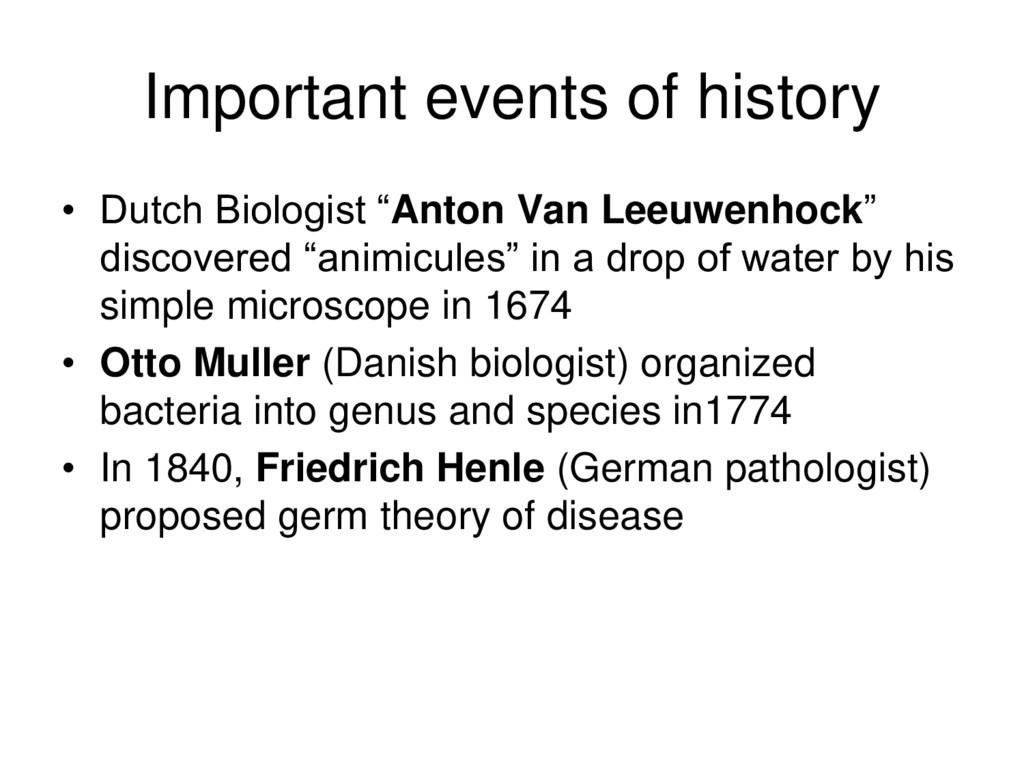
    discovered “animicules” in a drop of water by his simple microscope in 1674 • Otto Muller (Danish biologist) organized bacteria into genus and species in1774 • In 1840, Friedrich Henle (German pathologist) proposed germ theory of disease
  4. Conti… • Robert Koch – Father of medical Microbiology •

    Luis Pasteur (Father of Modern Microbiology) – identified causative agent of anthrax, rabies, plague, cholera and tuberculosis • Discovery of penicillin – 1928 (Alexander Flemming) • Discovery of sulphanilamide – 1935 (Gerhard Domagk’s) • Discovery of streptomycin – 1928 (Selman Waksman)
  5. Koch’s postulates 1. The causative (etiological) agent must be present

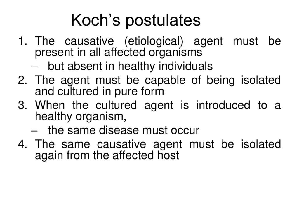
    in all affected organisms – but absent in healthy individuals 2. The agent must be capable of being isolated and cultured in pure form 3. When the cultured agent is introduced to a healthy organism, – the same disease must occur 4. The same causative agent must be isolated again from the affected host
  6. Branches of Medical Microbiology • Bacteriology – General – Systemic

    • Virology – General – Systemic • Parasitology – Protogoology – Helminthiology • Mycology • Immunology
  7. Difference between Virus And Bactria • Structure • Nucleic Acid

    • Type of Nucleus • Ribosomes • Nature of Outer Surface • Motility • Method of Replication
  8. What is bacterium? • Prokaryotic cells – Pro means primitive

    – Karyote means nuceus • Multiply by binary fission – 1divide to 2 • Devoid of chlorophil • ** single cell, living being, microscopic entity but fantastic in structural organization, reproduction, metabolism and function
  9. Eukaryotes • Eu means True (normal) • Karyote means nucleus

    • Much larger than most prokaryotes • Have subcellular, membrane-bound organelles • Includes all “higher" plants and animals • Microbiology includes – Fungi – Protozoa – Algae – Multicellular Organisms (helminthes)
  10. Fungus • Eukaryotic organism • cell walls contains Chitin •

    Molds are multicellular – masses of mycelia – composed of filaments called hyphae • Yeasts are unicellular
  11. Protozoa • Eukaryotes • No Cell Wall • Surround and

    absorb food • May be motile via – Pseudopods – cilia, or – flagella
  12. Algae • Eukaryotes • Cellulose cell walls • Use photosynthesis

    for energy • Produce molecular oxygen and organic compounds
  13. Virus • Acellular • Consist of DNA or RNA •

    Core is surrounded by a protein coat • Coat may be enclosed in a lipid envelope • Viruses are replicated only when they are in a living host cell • Living
  14. Multicellular- helminthes • Eukaryote • Multicellular animals • Parasitic flatworms

    and round worms are called helminths • Microscopic stages in life cycles
  15. Characteristics of Prokaryotes Nuclear level • Absence of – nuclear

    membrane – Nucleolie – Nucleoplasm – paired multiple chromosome – centriole
  16. Conti.. Cytoplasmic level • Absence of membrane bound organelles –

    Mitochondria – endoplasmic reticulum – golgi complex – Microtubules – Microfilaments
  17. Conti.. • Presence of – Ribosome – Plasmid • No

    protoplasmic streamming in the form of Brownian movement and Tyndall phenomena • No change in definite shape and formation of pseudopodia
  18. Characteristics of prokaryotes (Structural) Cell coverings (cell envelope) • Innermost

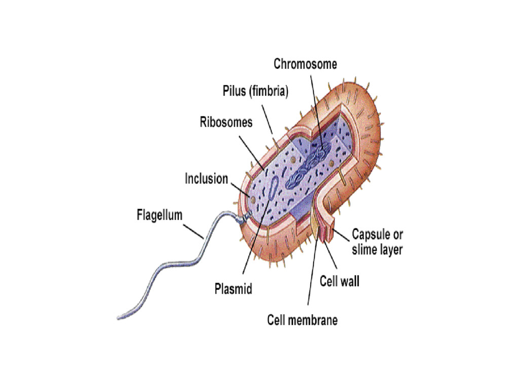
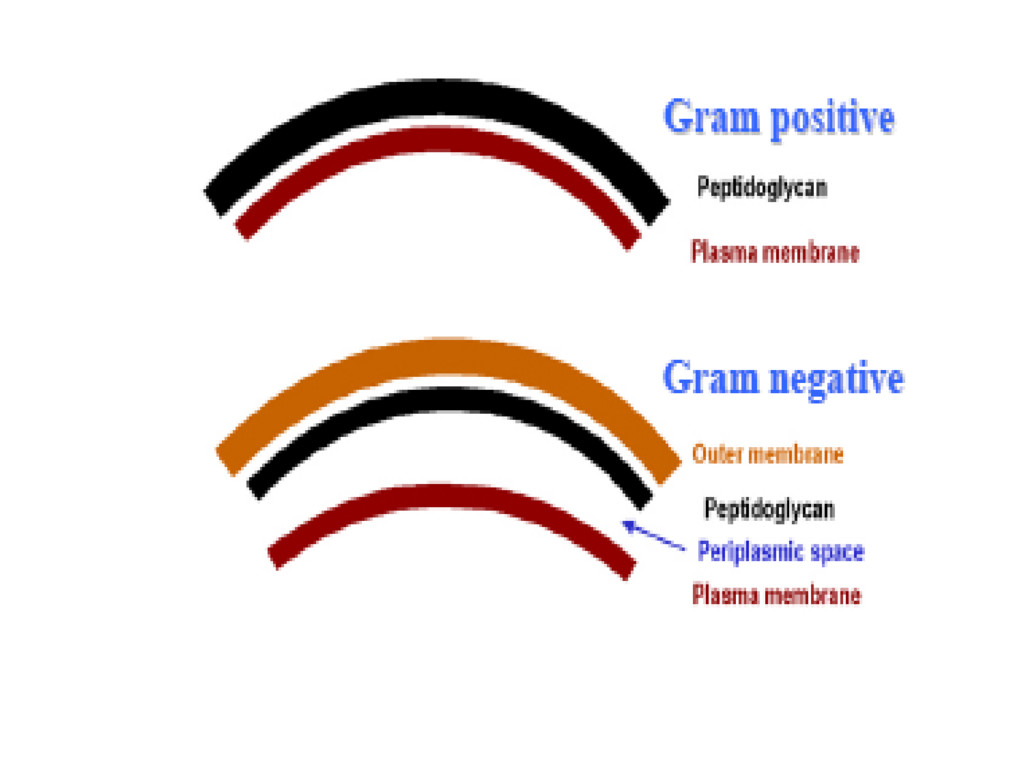
    layer – bilaminar phospholipid + Lipoprotein – cytoplasmic membrane (fluid stage) without any sterol • Two membrane in Gram negative bacteria • Condensation at some points forming mesosome
  19. Conti… 2) Outer to Cell Membrane – cell wall containing

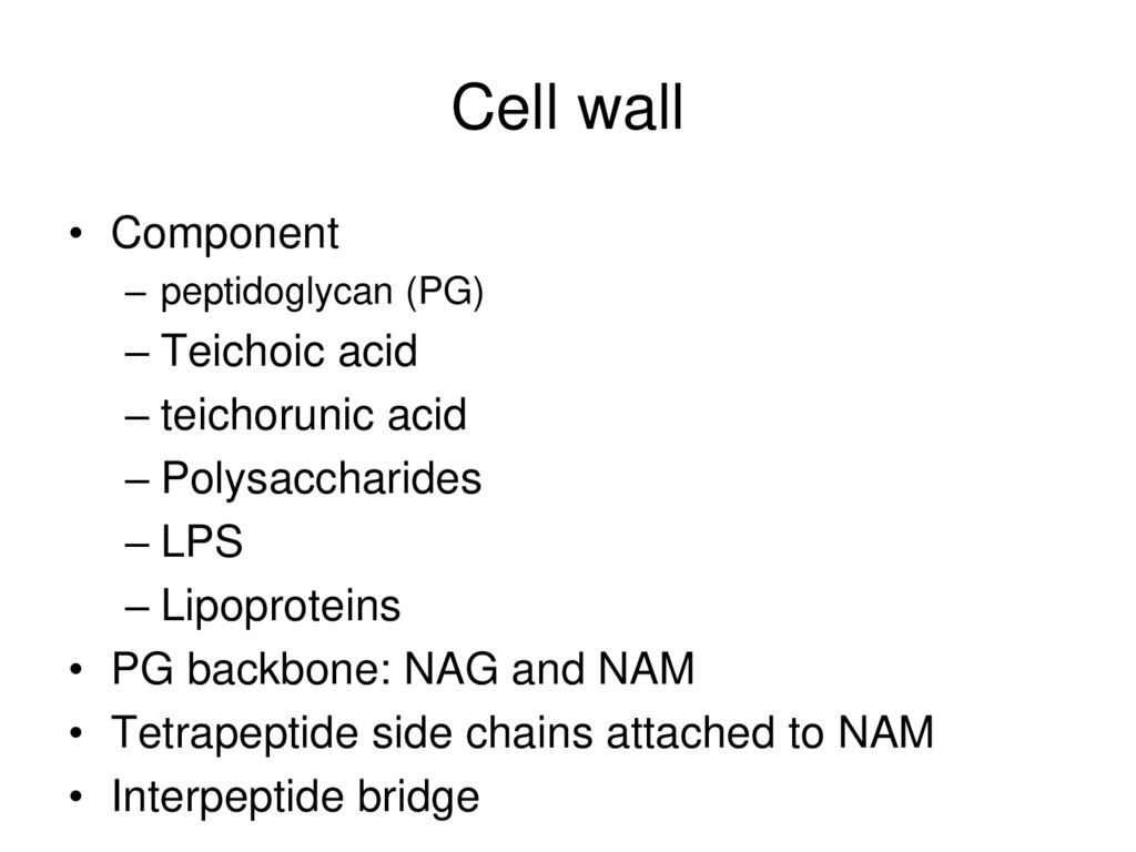
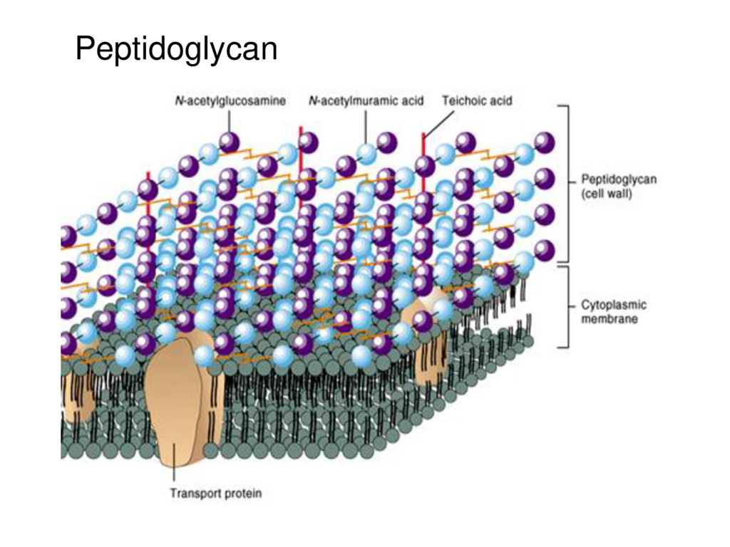
    specialized unique chemicals called peptidoglycan 3) Outer to Cell Wall – investing layer either capsule/glycocalyx/slime layer 4) S layer – Single type protein lattice outer to CW
  20. Characteristics of Prokaryotes • No genetic exchange during chromosome replication

    • Show secretory and excretory activities • Show chemical selectivity, motility and alteration of life stage (spore) • Wide range of temperature and pH tolerance • Heterogenous nutritional requirements and host specificity • Can produce self protective molecule bacteriocin • Become infected by particular virus • Can acquire extrachromosomal DNA (plasmid)
  21. Difference Between Eukaryotes and Prokaryotes • Nature of Nucleus •

    Mitotic Division • Presence of histones in DNA • Membrane bound Organelles • Size of Ribosomes • Peptidoglycan in Cell Wall
  22. Bacterial cells • essential structures – chemical composition – Organization

    – Functions • non-essential structures – chemical composition – Organization – Functions
  23. Structures of bacteria • Essential Structures (maintain viability) – Cell

    Wall – Cell Membrane – Mesosome – Ribosome – Nucleoid – Periplasm
  24. Structures of bacteria • Non-essential Structures (viability is not affected

    in their absence) – Flagella – fimbria (pili) – Capsule – Glycocalyx – slime layer – Plasmid – spore
  25. Cell wall • Component – peptidoglycan (PG) – Teichoic acid

    – teichorunic acid – Polysaccharides – LPS – Lipoproteins • PG backbone: NAG and NAM • Tetrapeptide side chains attached to NAM • Interpeptide bridge
  26. Conti… • Function – osmotic protection – integrity of cellular

    structures – virulence factors – Fully permeable to ions, aminoacids and sugars – makes it rigid – determines shape – Acts as antigen – Used as serological diagnosis
  27. Gram positive cell envelope • Covalently bound to the thick

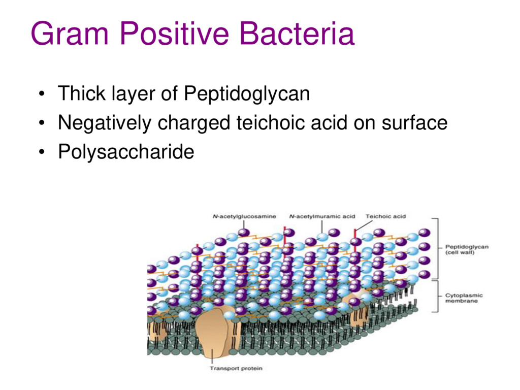
    peptidoglycan are – teichoic acid (their backbones are usually phosphorus containing polymers of ribitol or glycerol) or – teichuronic acid (glucuronic acid- containing polysaccharides • negatively charged molecules concentrate metal ions from the surroundings
  28. Conti… • Teichoic acids can also direct autolytic enzymes to

    sites of peptidoglycan digestion (autolysis). • This is needed to insert sections of cell wall for growth and division. • Lipoteichoic acid is primarily associated with the cell membrane.
  29. The Gram negative cell envelope • Covalently linked to the

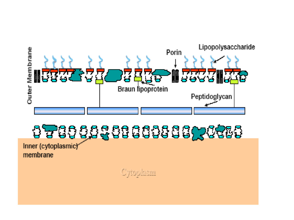
    thin peptidoglycan is the Braun lipoprotein which binds the outer membrane to the cell wall. • Like other membranes it contains proteins and phospholipids. • Unlike other membranes it contains lipopolysaccharide
  30. Conti… • Lipopolysaccharide – helps to provide a permeability barrier

    • LPS consists of three regions – an outer O antigen – a middle core – an inner lipid A region • core contains several sugars – lipid A contains β hydroxyfatty acids (uncommon in nature) – The molecule displays endotoxin activity
  31. Conti… • Porins in the outer membrane form channels to

    allow passage of small hydrophilic nutrients (such as sugars) through the outer membrane.
  32. Cell membrane • Phospholipid bilayer – Except triple layer in

    Mycoplasma • Interspersed with lipoprotein • Selectively permeable
  33. Cell Membrane • Functions – Transport – Synthesis – Excretion

    – Secretion – Metabolism – division by mesosome
  34. Nucleoid • Double stranded coiled helical DNA molecule – remain

    in a single chromosome • no introns • no non-coding sequences • no regulatory sequence • no long terminal repeats (LTRs) • Operon present
  35. Non-essential structures • Flagellum/-a (long whip like) – Filamentous Protein

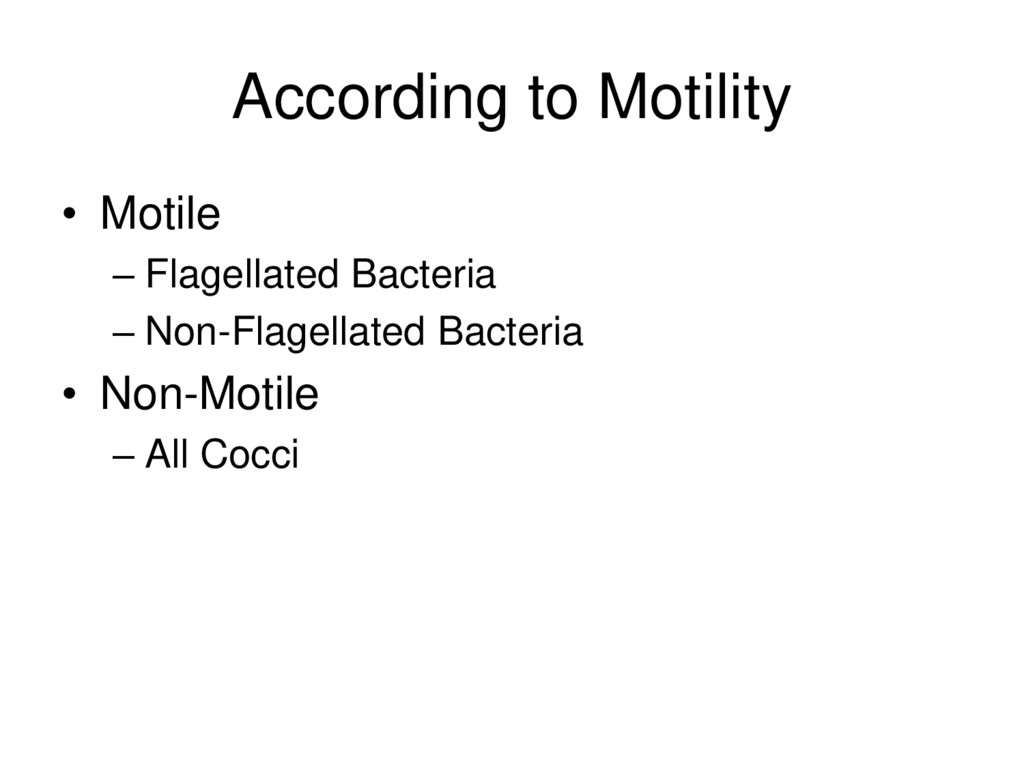
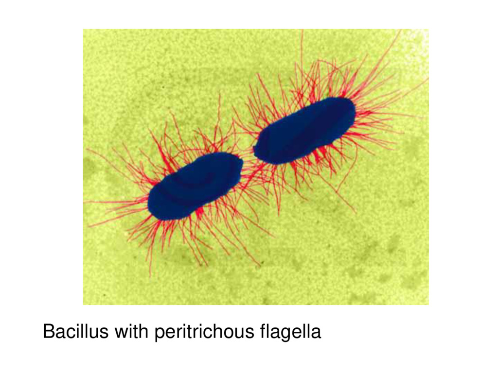
    Appendages – Composed of protein as flagellin unit – Arise from cytoplasmic membrane – Account for most bacterial motility – “Run and tumble” – Chemotaxis, phototaxis, aerotaxis, and magnetotaxis – Antigenic structure Fig 3.42
  36. Non-essential structures Pilus/-i • Thin hair like appendages • Composed

    of protein as pillin subunit • Arise from cytoplasmic membrane • Function – Attachment – Conjugation – Acts as virulence factors – Antigenic
  37. Capsule/Glycocalyx • External surface layer composed of polysaccharides Except –

    Polypeptide in Bacillus – Hyaluronic acid for Streptococci • Gel like • Forming either capsule (compact, complete and tight investing) or slime layer (loose meshwork) • Functions – prevent phagocytosis – Attachment of bacteria – Acts as Antigen
  38. Plasmid • Extra-chromosomal Double stranded Circular DNA • Independent of

    replication • Types – Transmissible – non-transmissible • Function – Carries gene for its own replication – Carries gene antibiotic resistance factors – Carries gene Proteins & toxin
  39. Endospores • Highly resistant structures formed at adverse environment •

    Composition – Bacterial DNA – small cytoplasm – cell membrane – Peptidoglycan – very little water – thick keratin like coat
  40. Endospores • Highly resistant to – heat, moisture, chemicals, radiation

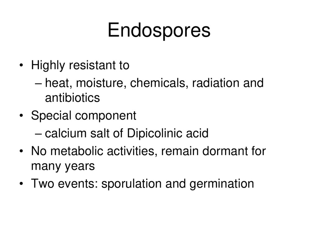
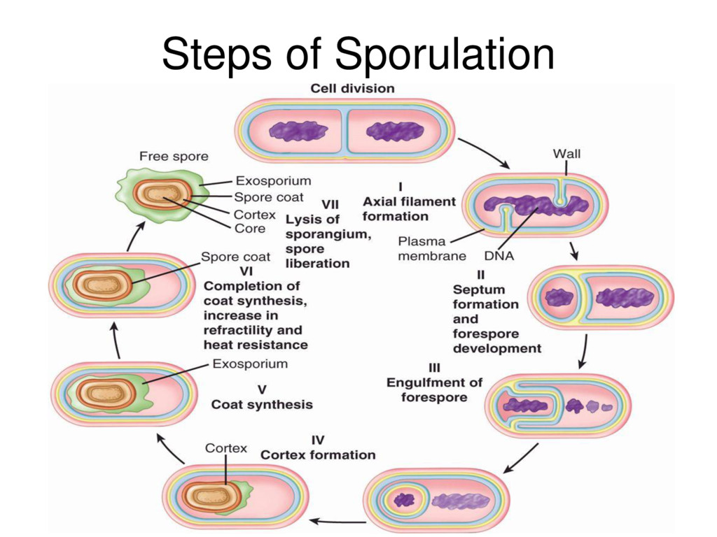
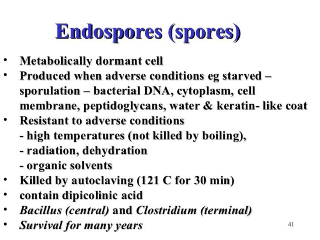
    and antibiotics • Special component – calcium salt of Dipicolinic acid • No metabolic activities, remain dormant for many years • Two events: sporulation and germination
  41. Classification of bacteria • Basis of Classification • differences between

    Gram positive and Gram negative bacteria • atypical characters of bacteria in relation to staining
  42. Morphological Classification Morphology of Bacteria • Cocci – rounded or

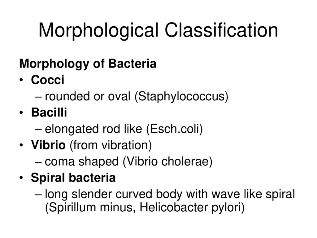
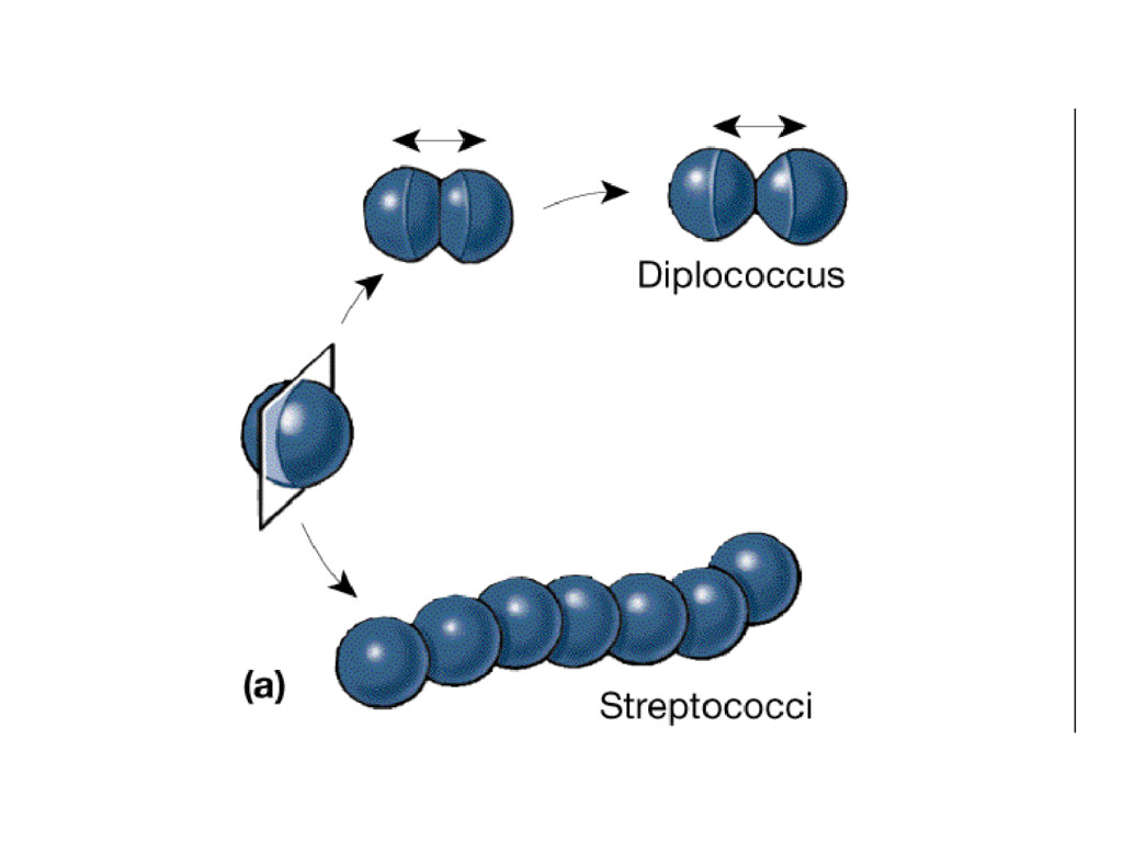
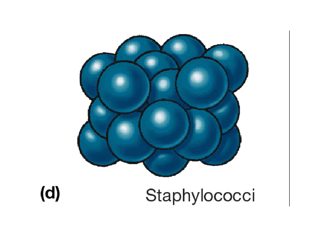
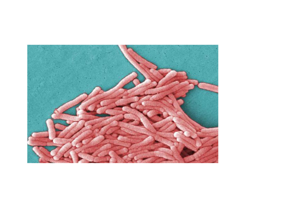
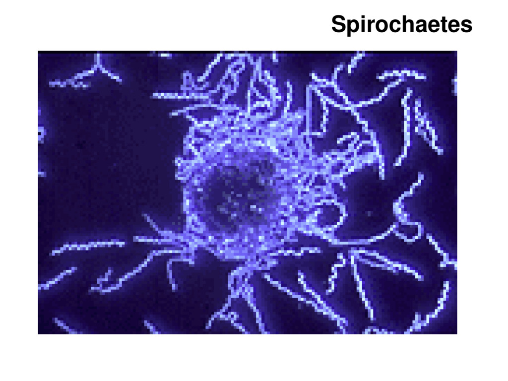
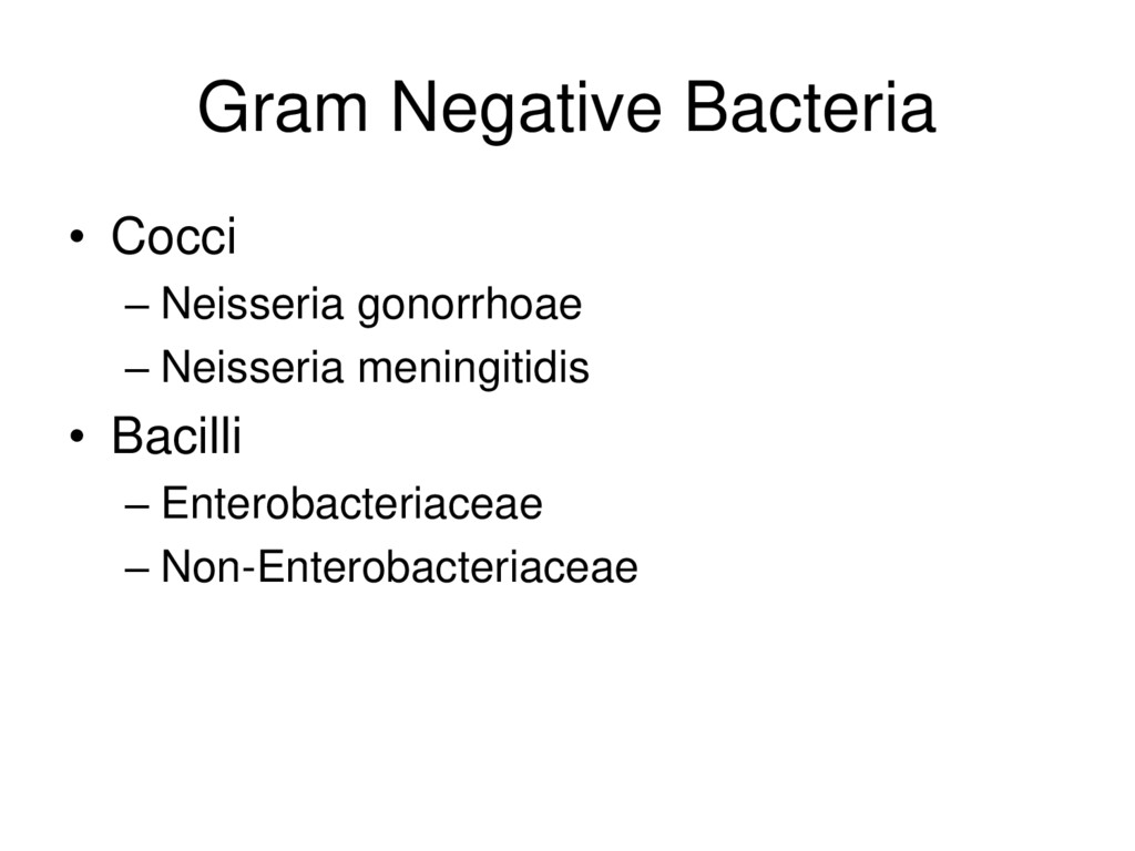
    oval (Staphylococcus) • Bacilli – elongated rod like (Esch.coli) • Vibrio (from vibration) – coma shaped (Vibrio cholerae) • Spiral bacteria – long slender curved body with wave like spiral (Spirillum minus, Helicobacter pylori)
  43. Others Shapes • Cocco-bacillus – longer than coccus, shorter than

    bacillus (Bordetella, Brucella) • Filamentous – long curved body like ribbon with branching (Actinomycetes, Nocardia)
  44. Thickness of Wall • Rigid thick wall Bacteria – Free-Living

    (Extracellular) – Non-Free Living (Obligate Intracellular) • Flexible thin walled Bacteria – Treponema – Borrela – Leptospira • Wall-less Bacteria – Mycoplasma – Ureaplasma
  45. Rigid thick wall Bacteria • Free-Living (Extracellular) – Gram Positive

    – Gram Negative – Acid-Fast • Non-Free Living (Obligate Intracellular) – Rickettsia – Chlamydia
  46. Gram Positive Bacteria • Cocci – Streptococcus – Staphylococcus •

    Bacilli – Spore Forming • Bacillus – Non-spore forming • Clostridium • Corynebacterium • Listeria
  47. Gram Negative Bacteria • Cocci – Neisseria gonorrhoae – Neisseria

    meningitidis • Bacilli – Enterobacteriaceae – Non-Enterobacteriaceae
  48. Staining Theoretical approach • Types • Laboratory procedures • findings

    of – Gram’s – Zeihl-neelsen (Z-N) – Albert’s staining
  49. Types of Staining Differential Staining • using primary and counter

    dye • performing a step of decolourisation • to see differential characters of bacteria – Gram’s stain – Ziehl-Neelsen stain – Albert stain
  50. Types of Staining Special Staining • to see unique property

    of particular bacteria – Fluorescense stain
  51. Gram’s staining • Named according to inventor Christian Gram •

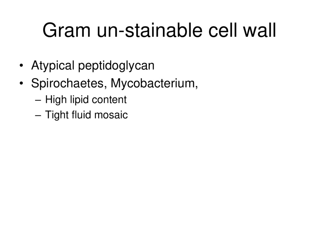
    Based on the property of bacterial cell envelope • stained at first by a primary dye & mordanted • Decolorized by acetone or alcohol • Counter stain is added • Gram positive bacteria – can resist decolourisation and retain the primary dye • Gram negative bacteria – can not resist decolourisation and take the counter dye
  52. Gram Positive Bacteria • Thick layer of Peptidoglycan • Negatively

    charged teichoic acid on surface • Polysaccharide
  53. Gram Negative Bacteria • Cell wall much more complex •

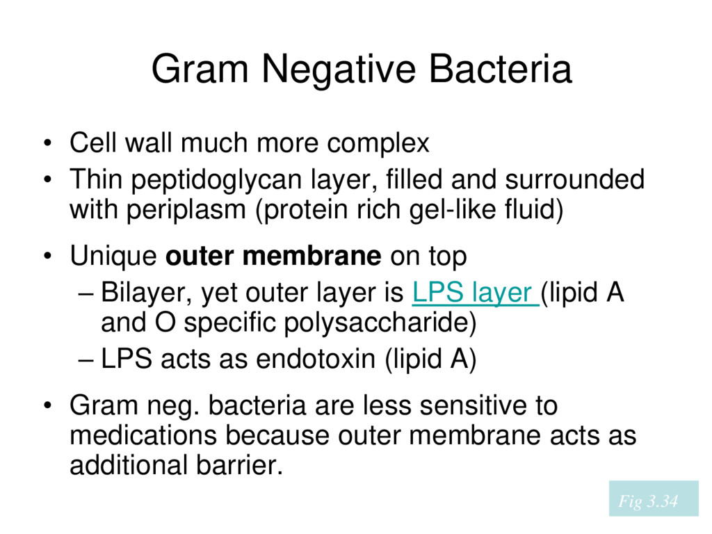
    Thin peptidoglycan layer, filled and surrounded with periplasm (protein rich gel-like fluid) • Unique outer membrane on top – Bilayer, yet outer layer is LPS layer (lipid A and O specific polysaccharide) – LPS acts as endotoxin (lipid A) • Gram neg. bacteria are less sensitive to medications because outer membrane acts as additional barrier. Fig 3.34
  54. Atypical cell envelope • Acid-fast and related bacteria – Mycobacteria

    – Nocardia • Presence of Mycolic acids – long, branch chained fatty acids