Upgrade to Pro — share decks privately, control downloads, hide ads and more …

Understanding the normal and abnormal post-operative MRI appearances after meniscal allograft transplant

Understanding the normal and abnormal post-operative MRI appearances after meniscal allograft transplant

Learning Objectives:

To know the current indications for meniscal allograft transplantation (MAT)
To become familiar with the different techniques used in MAT.
Expected post-operative MRI appearances and common complications after MAT
To know the MRI techniques best used for followup of meniscal allografts.

Timothy Woo

May 23, 2023
Tweet

Other Decks in Science

Transcript

  1. Understanding the normal and
    abnormal post-operative
    appearances after meniscal
    allograft transplant TIMOTHY WOO
    NATHAN JENKO
    NICOLE BAUSCH
    NICHOLAS SMITH
    UNIVERSITY HOSPITAL COVENTRY AND WARWICKSHIRE
    CLIFFORD BRIDGE ROAD, COVENTRY, UK
    [email protected]

    View Slide

  2. Introduction
    • The menisci are a crucial load bearing structure of the knee helping
    to decrease peak contact pressure and removal and debridement of
    irreparable (“white-zone”) meniscal tears greatly accelerate
    development of knee cartilage damage and osteoarthritis1,2
    • The first human meniscal allograft transplant was performed in
    19843 and since then thousands of procedures have been
    performed worldwide
    • Timing of transplant and the patient groups likely to benefit are still
    debated as well as whether it is truly chondroprotective
    • It is well-tolerated and has proven short to medium term
    symptomatic benefit
    Image credit Wikimedia commons: https://commons.wikimedia.org/wiki/File:Meniscus_tear_2010_-_closeup.JPG

    View Slide

  3. Learning Objectives
    • To know the current indications for meniscal allograft transplantation (MAT)
    • To become familiar with the different techniques used in MAT and the
    different graft types.
    • Expected post-operative MRI appearances and common complications after
    MAT
    • To know the MRI techniques best used for follow-up of meniscal allografts.

    View Slide

  4. Indications
    1. Unicompartmental pain in the
    presence of a total or subtotal
    meniscectomy
    2. As a concomitant procedure to ACL
    reconstruction if meniscus deficiency is
    believed to be a contributing factor to
    failure
    3. As a concomitant procedure to
    cartilage repair in a meniscus deficient
    compartment.
    1. Articular cartilage loss Outerbridge grade 3
    or above
    2. Any ligamentous deficiency or
    malalignment not surgically treatable.
    3. Inflammatory arthritis or previous septic
    arthritis
    4. Severe obesity (although no specific BMI
    threshold has been agreed on)
    5. Paediatric patient/immature skeleton
    Contraindications
    No age limit above which MAT not thought to be beneficial
    Consensus statement from the International Meniscus Reconstruction Experts Forum in 20154

    View Slide

  5. Evidence
    Several large case studies explore the outcomes following MAT, but the majority of these are
    only Level IV evidence (One long term level III study5)
    Heterogenous in outcome measures, exclusion criteria and patient cohort selection.
    1. Well-tolerated
    procedure with low
    failure rate (~10%)
    and low rate of
    complication
    (~14%6)
    2. All studies
    demonstrate increase
    in Patient Reported
    Outcome Measures
    (e.g. 55.7(poor) →
    81.3(good) mean
    improvement on
    Lysholm scale)7 and
    benefits maintained
    at 14 years
    3. No difference in
    PROMs or
    complication rates
    between operative
    techniques
    4. Uncertainty as to whether there is a chondroprotective
    effect:
    2 studies using contralateral
    knee control showed no
    radiographic progression8,9
    Other studies show progression
    of OA but often without
    adequate controls10,11.
    Probably there is a small chondroprotective effect but
    masked by a heterogenous cohort

    View Slide

  6. Operative Techniques
    BONE BRIDGE TECHNIQUE
    • Meniscus is harvested together with
    the meniscal roots attached to a
    “bone bridge” from the donor tibia.
    • An identical receptor site is cut into
    the patient’s native tibia and the
    bone bridge attached with the
    meniscal roots using suture anchors
    or interference screws.
    • Can be used for both medial and
    lateral meniscus
    BONE PLUG TECHNIQUE
    • Meniscus is harvested together with small bone plugs
    from the donor tibia, one attached to each root by long
    sutures
    • Suture tunnels are drilled from the root attachments to
    the anterior tibial cortex and the root sutures with the
    bone plugs are pulled through, tensioned and fixed –
    usually with a device such an endobutton
    SOFT TISSUE TECHNIQUE
    • No bone graft is used – Suture
    tunnels are drilled and used as
    for the bone plug technique
    • Some authors believe that the
    suture tunnel technique is not
    suitable for affixing the lateral
    meniscus due to the root
    proximity and risk of tunnel
    coalescence although there is
    no proven increased risk
    After root fixation, the remainder of
    the donor meniscus is attached to the
    recipient’s residual meniscal rim
    (if any) and/or joint capsule.

    View Slide

  7. Graft types
    Allografts currently available are either fresh frozen
    or cryopreserved with fresh frozen generally being
    preferred but there is no difference in outcome
    measures between groups. Rarely, fresh grafts are
    also used although these have logistical challenges.
    Fresh frozen soft
    tissue meniscal
    allograft:
    Figure from
    Spalding T. et al.
    (2015)12
    Previously, lyophilised grafts
    were also used, but these
    demonstrated graft
    shrinkage at second look
    arthroscopy and outcomes
    similar to meniscectomy
    groups in early case series13
    Grafts are stored in antibiotic
    solution while frozen to
    prevent pathogen transfer.
    Previously, some grafts were
    irradiated, but this also was
    found to compromise graft
    quality and this is no longer
    common practice.
    Artificially engineered graft material is much less commonly
    used with available products such as Collagen Meniscal
    Implant (Ivy Sports Medicine, Germany) which is composed
    of Achilles tendon Type I collagen and acellular polyurethane
    polymer scaffold (Actifit, Orteq Bioengineering, UK), but
    these are only usable for partial meniscal defects and require
    a viable meniscal rim and at least one root attachment.

    View Slide

  8. MRI technique
    ROUTINE IMAGING
    • Most of the fixation used in meniscal
    allograft transplant is non-metal except
    for the endobutton which is distant from
    the graft fixation
    • 3 Tesla imaging with fluid sensitive fat
    saturated structural sequences such as
    fat-saturated proton-density images using
    a dedicated knee coil in three orthogonal
    planes
    Proton-density weighted coronal MRI of the knee
    with a lateral meniscal transplant showing peripheral
    suture artefact
    ARTHROGRAPHY
    • Some authors advocate the use of
    arthrography to investigate graft/capsule
    healing as this is obscured by suture
    artefact. However, the clinical benefit of
    this is debated.
    • Arthrography can be used to tell
    between non-displaced graft tears and
    healing tissue and to investigate
    arthrofibrosis.

    View Slide

  9. Normal post-operative appearances
    SIGNAL
    • Similar to ACL graft
    • Initial T1/T2 high signal
    peaking at 6m then declining
    to T1/T2 low signal over the
    next 18 months
    SHRINKAGE/EXTRUSION
    • Commonly seen and not
    associated with worse PROMs15
    • However, thought undesirable
    as represents incompetence of
    graft
    GRAFT/CAPSULE HEALING
    • Assessed by fluid/contrast tracking
    between graft and capsule
    • Thought desirable – especially in soft
    tissue only fixations.
    • However, even arthrography
    underestimates healing14
    LINEAR DEFECTS
    • Not of clinical consequence
    unless16:
    • Increasing on serial imaging
    • True fluid signal
    • Painful
    Normal MRI of soft tissue only fixation lateral meniscus allograft
    Top: Normal appearances of the two suture tunnels on sagittal PDFS
    and T1-weighted images (arrowheads)
    Bottom: linear high signal in posterior horn found at arthroscopy to be
    unresorbed suture material

    View Slide

  10. Complications
    INFECTION
    • As with other joint procedures,
    superficial and deep joint
    infections are non-specific on
    imaging presenting early with
    synovitis and effusion
    SUTURE FAILURE
    • Root suture fixation sites are a potential site of failure in
    soft tissue fixation
    • Sutures should run continuously between the suture tunnel
    and the donor root
    • Anterior horn suture can often be poorly defined and
    detecting tears/suture failure here has low specificity
    Sagittal and coronal PD-weighted MRI of lateral meniscal allograft
    showing apparent absent anterior horn with no meniscal material visible
    continuous with the suture tunnel suggestive of a suture failure/tear
    The allograft was normal on arthroscopy.
    Early post-operative MRI of a patient with
    lateral meniscal allograft after re-presenting
    with tibial wound infection. PD-weighted
    sagittal images showing infrapatellar fat pad
    oedema, synovial thickening and effusion
    suspicious for joint infection

    View Slide

  11. NON-INCORPORATION OF BONEPLUGS/BONE-BRIDGE
    • The bonegraft should be visible on immediate and
    sometimes at 3 month post-operative imaging but
    should be completely incorporated by 6 months.
    • Signs of failure are as elsewhere with progressive
    sclerosis and cortication and persistent fluid cleft at
    MRI.
    GRAFT TEAR
    • Graft tear is the most common indication for non-
    routine followup MRI.
    • Linear high signal is normal in the graft meniscus
    usually representing suture material or
    granulation/epithelisation but tear is favoured if:
    • Displaced fragment
    • Increasing fluid signal over serial MRI scans esp if new
    cartilage defect
    • Imbibition of contrast on MRI arthrogram
    Sagittal PD-weighted MRI of a
    lateral meniscal allograft showing
    flipped tear of the posterior horn
    of the graft with empty posterior
    compartment (arrowhead)
    Immediate and 3 month postoperative radiographs showing normal
    incorporation of tibial bone-bridge of a lateral meniscal allograft

    View Slide

  12. ARTHROFIBROSIS
    • As with ACL graft reconstruction,
    meniscus transplant can rarely result in
    intra-articular scar mass formation or
    arthrofibrosis which usually presents as
    progressive restriction of range of
    movement.
    • Low signal soft tissue foci – often more
    evident on non-fat-saturated images are
    sometimes seen and MRI arthrography
    can sometimes also be helpful but most
    patients in which this is suspected
    progress to arthroscopy.
    PROGRESSION OF OSTEOARTHRITIS
    • In the medium to long term, progression
    of cartilage loss is important to note as
    this can indicate a functionally
    incompetent graft and can relate to
    recurrent patient pain (imaging after two
    years is often only undertaken in the
    context of new symptoms).
    • This can sometimes necessitate graft
    revision or in some cases joint
    replacement.
    Left: 1 year postoperative and
    Right: 3 year postoperative
    coronal PD-weighted MRI studies
    of a patient with lateral meniscal
    allograft and new symptoms
    showing definite interval cartilage
    degeneration despite meniscal
    allograft
    T1-weighted MRI through the intercondylar notch
    of a patient with lateral meniscal allograft with
    bone-bridge technique showing low signal scar in
    the infrapatellar fatpad and notch in keeping
    with arthrofibrosis

    View Slide

  13. Conclusion
    Meniscal allograft transplant is an increasingly
    popular choice for young to middle aged patients
    with significant meniscal injuries and is proven to
    have at least short to medium term symptomatic
    benefits.
    There is an increasing chance of coming across a
    patient who has undergone one of these
    procedures in the past and it is important that
    radiologists know the details of current and past
    surgical techniques and expected post-operative
    MRI appearances of these grafts to accurately
    assess re-injury. Lateral meniscal allograft in situ postfixation arthroscopic image
    From Spalding et al.12

    View Slide

  14. References
    1. Roos H, Laurén M, Adalberth T, Roos EM, Jonsson K, Lohmander LS. Knee osteoarthritis after meniscectomy: prevalence of radiographic changes after twenty-one years, compared with matched
    controls. Arthritis Rheum. 1998 Apr;41(4):687-93.
    2. Papalia R, Del Buono A, Osti L, Denaro V, Maffulli N. Meniscectomy as a risk factor for knee osteoarthritis: a systematic review. Br Med Bull. 2011;99:89-106.
    3. Milachowski KA, Weismeier K, Wirth CJ. Homologous meniscus transplantation. Experimental and clinical results. Int Orthop. 1989;13(1):1-11.
    4. Getgood A, LaPrade RF, Verdonk P, Gersoff W, Cole B, Spalding T; IMREF Group. International Meniscus Reconstruction Experts Forum (IMREF) 2015 Consensus Statement on the Practice of
    Meniscal Allograft Transplantation. Am J Sports Med. 2017 May;45(5):1195-1205.
    5. van der Wal RJP, Nieuwenhuijse MJ, Spek RWA, Thomassen BJW, van Arkel ERA, Nelissen RGHH. Meniscal allograft transplantation in The Netherlands: long-term survival, patient-reported
    outcomes, and their association with preoperative complaints and interventions. Knee Surg Sports Traumatol Arthrosc. 2020 Nov;28(11):3551-3560.
    6. Smith NA, MacKay N, Costa M, Spalding T. Meniscal allograft transplantation in a symptomatic meniscal deficient knee: a systematic review. Knee Surg Sports Traumatol Arthrosc. 2015
    Jan;23(1):270-9.
    7. Samitier G, Alentorn-Geli E, Taylor DC, Rill B, Lock T, Moutzouros V, Kolowich P. Meniscal allograft transplantation. Part 2: systematic review of transplant timing, outcomes, return to competition,
    associated procedures, and prevention of osteoarthritis. Knee Surg Sports Traumatol Arthrosc. 2015 Jan;23(1):323-33.
    8. Ha JK, Shim JC, Kim DW, Lee YS, Ra HJ, Kim JG. Relationship between meniscal extrusion and various clinical findings after meniscus allograft transplantation. Am J Sports Med. 2010
    Dec;38(12):2448-55.
    9. Sekiya JK, Giffin JR, Irrgang JJ, Fu FH, Harner CD. Clinical outcomes after combined meniscal allograft transplantation and anterior cruciate ligament reconstruction. Am J Sports Med. 2003 Nov-
    Dec;31(6):896-906.
    10. Hommen JP, Applegate GR, Del Pizzo W. Meniscus allograft transplantation: ten-year results of cryopreserved allografts. Arthroscopy. 2007 Apr;23(4):388-93
    11. Vundelinckx B, Bellemans J, Vanlauwe J. Arthroscopically assisted meniscal allograft transplantation in the knee: a medium-term subjective, clinical, and radiographical outcome evaluation. Am J
    Sports Med. 2010 Nov;38(11):2240-7.
    12. Spalding T, Parkinson B, Smith NA, Verdonk P. Arthroscopic Meniscal Allograft Transplantation With Soft-Tissue Fixation Through Bone Tunnels. Arthroscopy Techniques. 2015 Oct;4(5):e559–63.
    13. Wirth CJ, Peters G, Milachowski KA, Weismeier KG, Kohn D. Long-term results of meniscal allograft transplantation. Am J Sports Med. 2002 Mar-Apr;30(2):174-81.
    14. van Arkel ER, Goei R, de Ploeg I, de Boer HH. Meniscal allografts: evaluation with magnetic resonance imaging and correlation with arthroscopy. Arthroscopy. 2000 Jul-Aug;16(5):517-21.
    15. Boutin RD, Fritz RC, Marder RA. Magnetic resonance imaging of the postoperative meniscus: resection, repair, and replacement. Magn Reson Imaging Clin N Am. 2014 Nov;22(4):517-55.
    16. Lee DH, Lee BS, Chung JW, Kim JM, Yang KS, Cha EJ, Bin SI. Changes in magnetic resonance imaging signal intensity of transplanted meniscus allografts are not associated with clinical outcomes.
    Arthroscopy. 2011 Sep;27(9):1211-8.
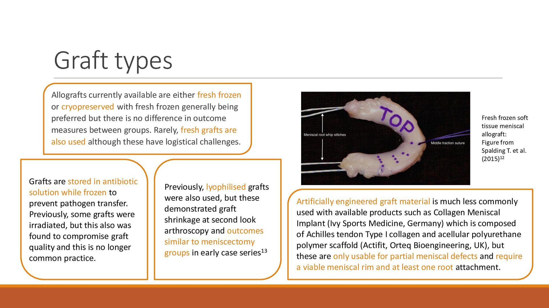
    View Slide