projects is increasing steadily and the number of cells that are being derived develops at a remarkable pace. However, stem cells around the world are vastly different in their provenance, programming and potentials. Furthermore, knowledge on the actual number of cell types, their derivation, availability and characteristics is rather sparse. Usually, “colleague-supply” avenues constantly furnish cells to laboratories around the world without ensuring their correct identity, characterization and quality. These parameters are critical if the cells will be eventually used in toxicology studies and drug discovery. Here we outline some basic principles in establishing a stem cell specific bank. 2
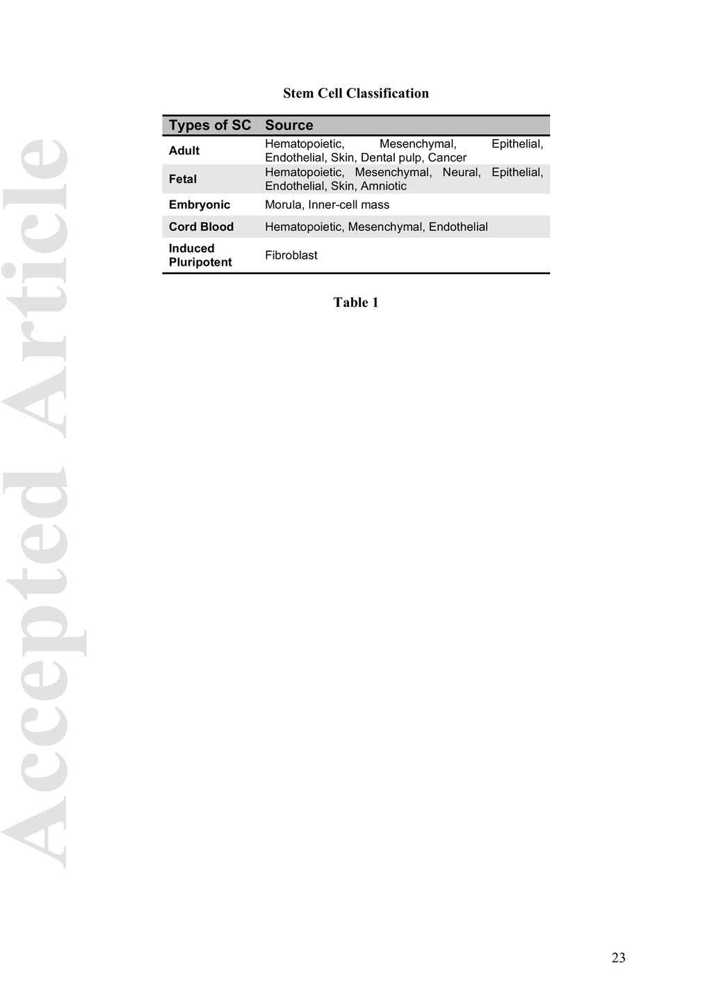
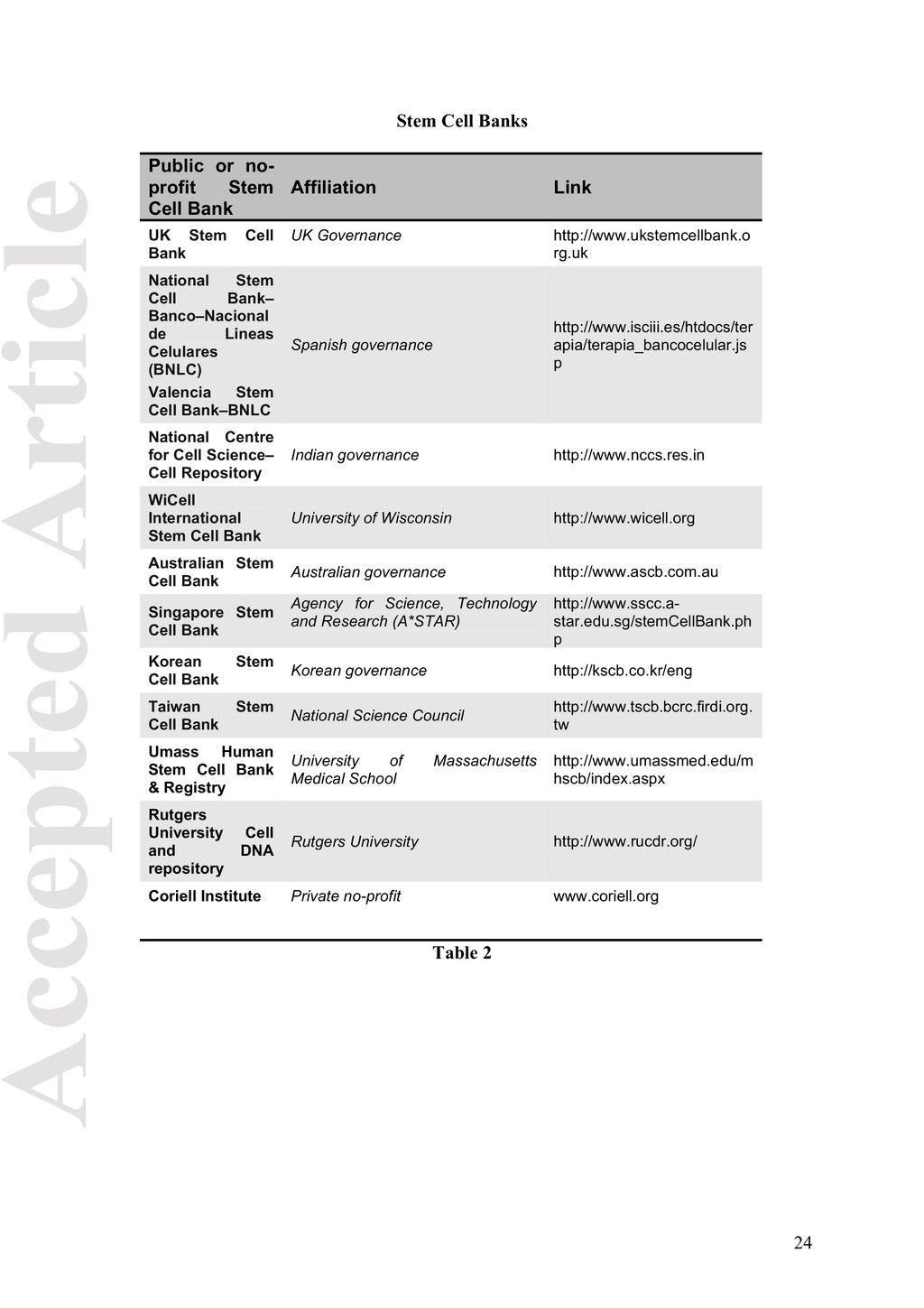
generating and intensively studying stem cells is partly due to their emerging potentials in cell-based medicine. Stem cell medicine is a dynamic field rapidly moving from basic research to clinical therapies. Many different stem cell types, to date, have been derived as listed in Table 1. The advent of disease-specific stem cells and stem cell-based therapies necessitates the implementation of accurate procedures, not only to guarantee the continuous supply of each cell line over long periods of time, but also to produce them with an identity and integrity as close as possible to the original cells. Since this bioproduct is becoming very demanding, the workload cannot be handled by small hospital based institutions, but by specialized and certified biobanks. These dedicated infrastructures can actively work towards establishing expedient procedures to promote prompt access to the cell lines and minimize the need for ‘colleague-supply’ routes, which over the years has promoted the widespread use of cross- contaminated and mycoplasma infected cells jeopardizing the validity of data in the literature (Chatterjee et al., 2007; NIH, 2007). Several specialized and specific repository laboratories, called “Stem Cell Bio-Bank” (listed in table 2) arose in the last decade to meet International Healthcare requests. The International Stem Cell Banking Initiative (ISCBI) committed to create a global network of banks that operate according to unified principles and practices for cell banking, testing and distribution. To this end, ISCBI has produced a consensus guidance document establishing the best practice in banking and supply of research-grade stem cells applicable to all stems cells including human embryonic (hES) and induced pluripotent (iPS) cells (ISCBI, 2009). High-throughput molecular technology platforms (“omics”) require the use of high quality bio-samples collected and managed following biosafety requirements and well-standardized procedures. A strong information technology (IT) structure, a robust tracking system linked 3
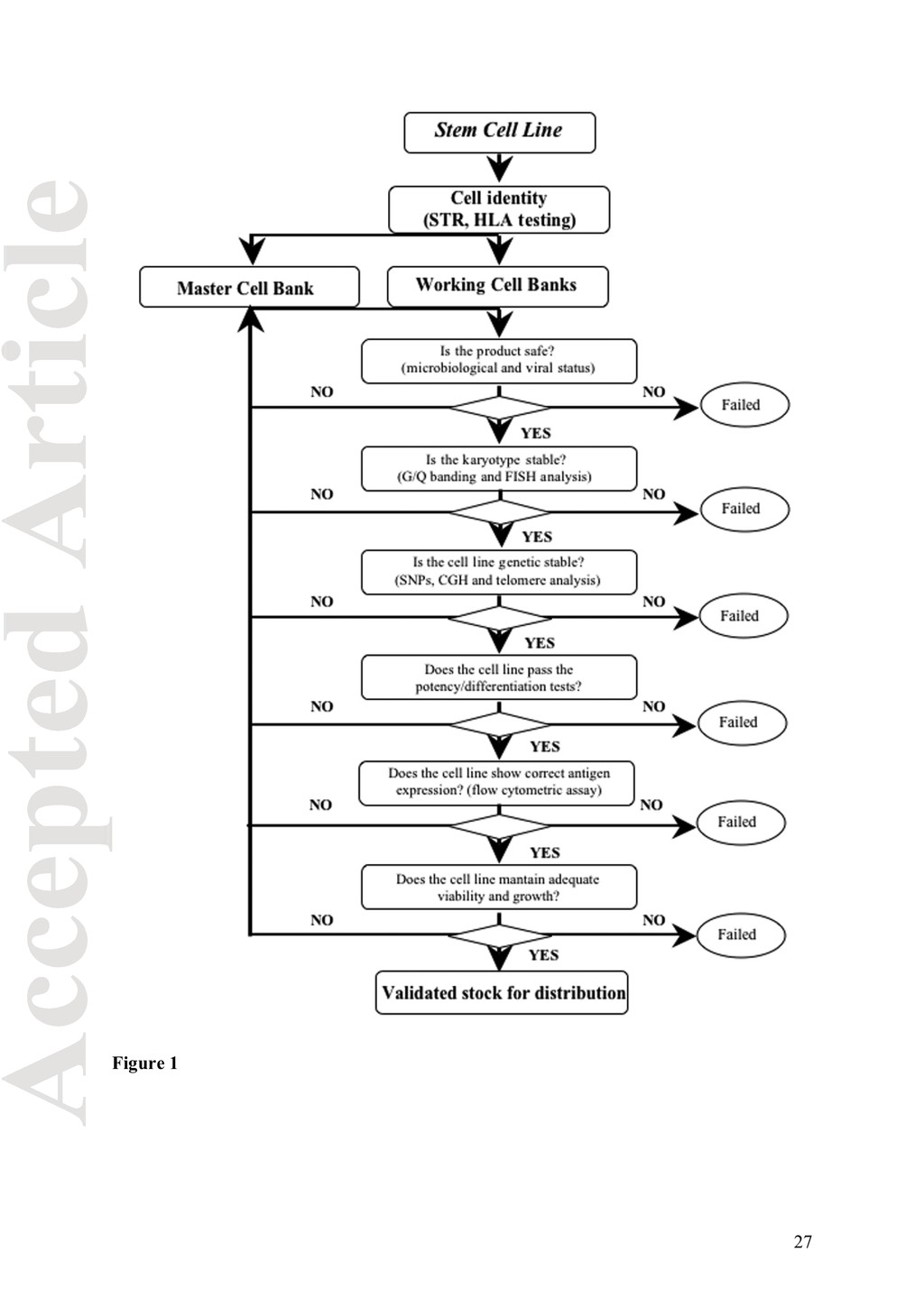
(SOPs) ensure the traceability of each cell line deposited from consent through distribution. The flow-chart in figure 1 schematizes the rules that a dedicated stem cell bank should follow within its infrastructure before making their cells a pharmaceutical product. Steps of the flow-chart will be discussed as specific topic. Mis-authentication and mis-identification of stem cell lines Biomedical researchers that use living cells in culture dishes, must cope with several risks: (i) misauthentication or misidentification of the cell lines (due to cross-contamination) particularly important for long term cultures (Drexler et al., 2003); (ii) contamination with bacteria, mycoplasma, viruses or other fast growing cells of undefined nature and (iii) cytogenetic status. Stringent control of these parameters requires trained personnel, systematic methods of analysis and rigorous application of well-standardized procedures (SOPs). When a cell line enters the biobank infrastructure, being it a primary or derived line, the first thing that must be done is its identification through HLA typing or short tandem repeats (STR or microsatellite) analysis (Masters et al., 2001). Then create a Master Cell Bank (MCB) consisting of at least 20 ampoules immediately stored at -196˚C and a Working Cell Bank (WCB) constituting the distribution line. The ethical committee approval from the donor‘s institution must be included in the IT database, certifying that all legal and ethical requirements have been met and proper informed consent has been signed from the donor. Furthermore, this database must contain all the information regarding the cell lines currently available and ethically approved for distribution. Although each institute has its own database, several efforts have been made to establish a centralized registry of public available stem cells (Table 3). For researchers seeking to work 4
find them; a good stem cell investment could be to create a centralized registry containing each stem cell background and their obtainability. Sterility QC testing of stem cell lines for basic and clinical research Ensuring sterility of MCB and WCB lines is fundamental since microbial contamination can change the characteristics of the cells without obvious cytopathic effects, but the use of such culture can damage the quality of research and/or expose researchers to infectious agents. However, cells with established contamination can be used in several research applications, but must be handled separately from the other cultures. The current testing standard for mycoplasma contamination simply relies on bacterial growth in nutrient media (broth and agar) (Barbara et al., 2008) for three-weeks (United States Food and Drug Administration, 2008a); polymerase chain reaction (PCR)-based mycoplasma detection methods expedite the results (Venor GeM kit, Minerva Biolabs, Berlin, Germany). Bacteria borne-disease contaminations can be detected by seeding cell culture medium on bacteriology plates containing selective growth medium specific for aerobic or anaerobic contaminants. After different incubation times (from 7 to 20 days), the plates are inspected for bacteria growth. The 16S DNA amplification assay can assess the nature of the putative bacterial colony (Gonzales and McDonough, 1998). Clinically significant transmissible infections which need to be tested before releasing the cells are: (i) parasitic protozoa (malaria, Chagas’disease, Leishmania, Plasmodium falciparum, etc.); (ii) bacteria borne-diseases such as: Chlamydia trachomatis (CT), Neisseria gonorrhoeae (GC) and Mycobacteria tuberculosis (MTB); (iii) viral borne diseases such as: Human Immunodeficiency Virus (HIV), Hepatitis C Virus (HCV), Hepatitis B Virus (HBV), Human Papilloma Virus (HPV), parvovarius and the prion variant VCJD (ISCB, 2009). Nucleic acids Amplification Technology (NAT) reflects a rapid means of detection and monitoring of the infective agents as well as assessing the clinical disease associated with the 5
Diagnostics, Abbott Diagnostics, Siemens Medical Solutions, Biomerieux, Becton, Dickinson and Gen-Probe, basic to these station is nucleic acids extraction protocols (SOPs) and a real-time PCR instrument which enhances the accuracy (highly specific detection and quantification of extremely low levels of disease agents), rapidity and ability to quantitate viral target sequences (Logna et al., 2009). In order to decrease human errors, robotized stations can be used. Currently there are no available validated sensitive detection methods for prions and transmissible spongiform encephalopathies (TSEs) agents. However, a constant traceability of the geographic origin of donor animals must be included in the database avoiding products derive from countries classified as geographical bovine spongiform encephalopathy (BSE)- risk I and II (Heim and Mumford, 2005). Genetic Stability Extensive in vitro culturing over large periods can also lead to cellular senescence, as well as genetic and epigenetic changes. Spectral karyotyping (to detect gross chromosomal alterations) must be performed regularly to ensure chromosomal stability as a function of culturing, freezing and thawing. Even if human stem cell lines have shown an overall preservation of euploidy during long term culturing (Hoffman and Carpenter, 2005), recurrent abnormalities have been reported (Narva et al., 2010; Lefort et al., 2008; Buzzard et al., 2004). Studies on mouse stem cell lines have in fact described that many chromosomal aberrations did occur during in vitro culturing and genetic engineering procedures, but such genetic changes did not affect the overall biological properties of the cells (Diaferia et al, 2011). Notably, propensity to karyotype instability can vary between different lines, regardless of the culturing conditions, duration or splitting techniques; it is therefore encouraged to regularly perform regular high-resolution molecular and cytogenetic studies to 6
disease or transformation. Small genomic variations and submicroscopic DNA alterations that can potentially affect cell phenotype, may be evaluated by performing comparative genomic hybridisation (CGH) where the use of fluorescent dyes that bind to specific regions of the chromosomes, allows the acquisition of full digital coloured images of the chromosomes. DNA gain or loss can be identified as well as cell characteristic patterns including mutations at chromosomal or sub- chromosomal levels (Stephenson et al., 2010). CGH is a powerful molecular genetic tool to screen genomes for chromosomal imbalances but has some limitations: the technique does not detect balanced chromosomal translocations or inversion nor small interstitial deletions. Other methods such as “spectral karyotyping” (SKY) and multiple single nucleotide polymorphism (SNP) analysis could provide useful and specific information about copy number changes (gains/losses) and/or nucleotide variation in the DNA content (International Stem Cell Banking Initiative, 2009). However, despite these standardized definitions and criteria that aid the reproducibility of stem cell cultures, little is known about their long-term safety, stability and differentiated derivates. Furthermore, the extension in telomere length that occurs with the increasing number of stem cells passages and the reduced rate of spontaneous differentiation, suggest that a standardized and validated procedure for characterizing stem cells at a specific passage number is essential for their potential use in therapeutic applications. Thus, further analysis can be required to determine whether telomere length stabilizes or shortens during cell culture. The polymerase chain reaction (PCR) based telomeric repeat amplification protocol (TRAP) assay can be used to detect telomerase activity and the terminal restriction fragment (TRF) analysis to evaluate the length of telomere (Herbert et al., 2003). 7
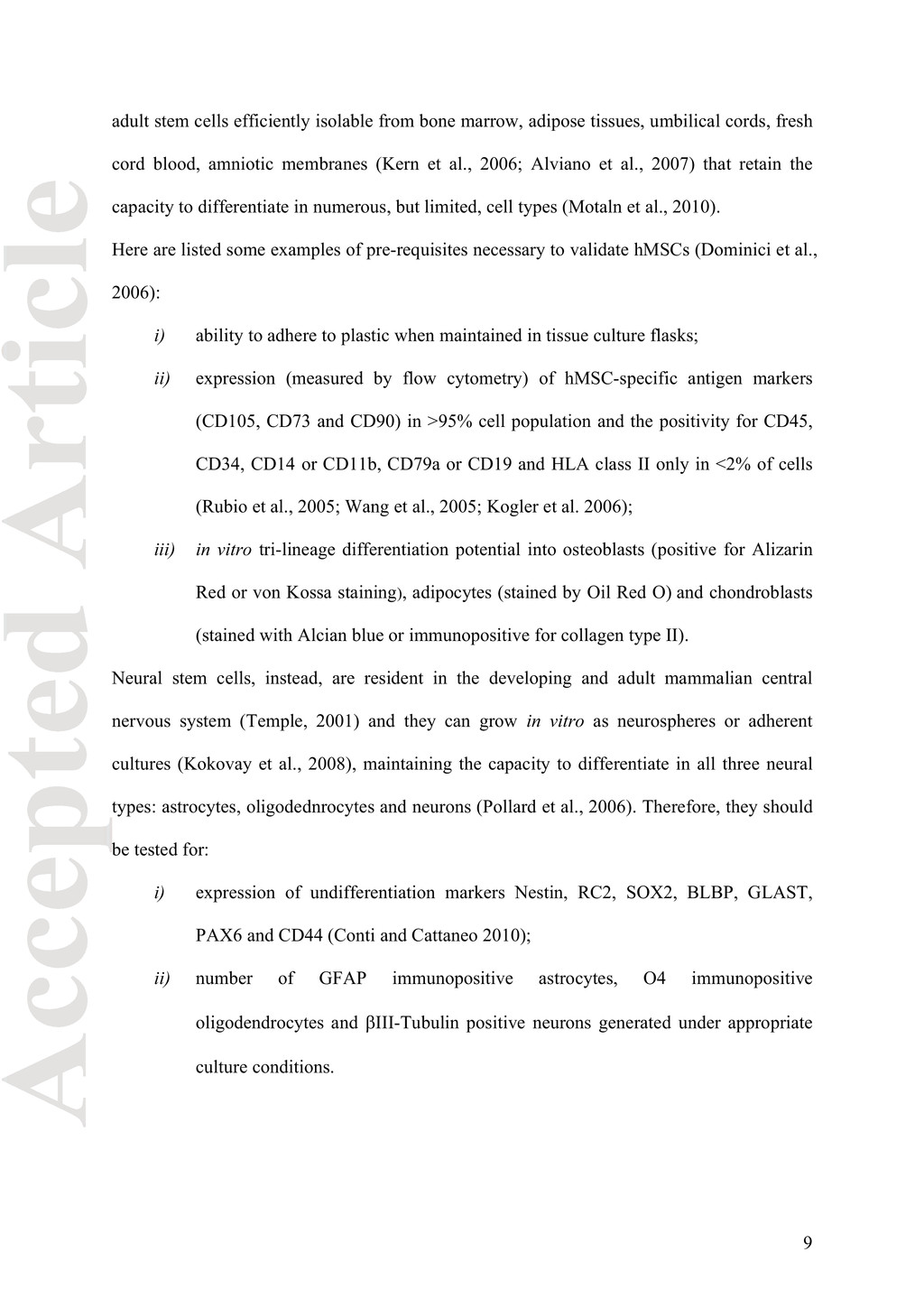
The self-renewal and the capacity to differentiate into multiple cell types (pluripotency) make stem cells attractive candidates for the functional regeneration of damaged tissues. Once assured the sterility from microbial, fungal, endotoxin, mycoplasma and viral contamination, each cell line must be controlled for its “potency”; the US Food and Drug Administration (FDA) regulates clinical trials to ensure that subjects enrolled in a study involving stem cell- based products are not exposed to significant and unreasonable risks (United States Food and Drug Administration, 2008b). The only way to attest the physical nature of stem cells is to evaluate their potency as the capacities to self-renew and differentiate in appropriate conditions. Obviously substantial differences in term of potency exist among the various types of stem cells: the pluripotency characterizes only embryonic stem cells (ESCs) that have the virtual ability to generate any adult cell type, while the embryonic germ cells (EGCs) can ultimately produce only gametes. The limitless self-renewal potential and the capacity to give rise to all differentiated cell types make ESCs able to spontaneously form teratoma-like masses after injection into immunodeficient mice (Thomson et al, 1998; Brivanlou et al., 2003). Biobanks should verify that this characteristic is preserved in their cell collections both in the MCB and in the WCB. Alternatively, the capacities of ESCs to form embryoid bodies or to differentiate under specific conditions in vitro could also be evaluated (ISCBI 2009). Since these biobanks are stem cell dedicated, specific tests should be set up to verify surface antigen marker profiles: SSEA-1 negative or very low, SSEA-3 positive, SSEA-4 positive, TRA-1-60 positive and TRA- 1-81 positive. A more complete analysis should include the evaluation of the expression of six other genes particularly linked to the ESC phenotype: Nanog, Oct 4, DNMT 3B, TDGF, GABRB3, GDF3 (International Stem Cell Initiative, 2007). Differently from ESCs, the fetal or adult stem cells, which are capable of producing a limited pool of different mature cell types. For example, human mesenchymal stem cells (hMSCs) are 8
umbilical cords, fresh cord blood, amniotic membranes (Kern et al., 2006; Alviano et al., 2007) that retain the capacity to differentiate in numerous, but limited, cell types (Motaln et al., 2010). Here are listed some examples of pre-requisites necessary to validate hMSCs (Dominici et al., 2006): i) ability to adhere to plastic when maintained in tissue culture flasks; ii) expression (measured by flow cytometry) of hMSC-specific antigen markers (CD105, CD73 and CD90) in >95% cell population and the positivity for CD45, CD34, CD14 or CD11b, CD79a or CD19 and HLA class II only in <2% of cells (Rubio et al., 2005; Wang et al., 2005; Kogler et al. 2006); iii) in vitro tri-lineage differentiation potential into osteoblasts (positive for Alizarin Red or von Kossa staining), adipocytes (stained by Oil Red O) and chondroblasts (stained with Alcian blue or immunopositive for collagen type II). Neural stem cells, instead, are resident in the developing and adult mammalian central nervous system (Temple, 2001) and they can grow in vitro as neurospheres or adherent cultures (Kokovay et al., 2008), maintaining the capacity to differentiate in all three neural types: astrocytes, oligodednrocytes and neurons (Pollard et al., 2006). Therefore, they should be tested for: i) expression of undifferentiation markers Nestin, RC2, SOX2, BLBP, GLAST, PAX6 and CD44 (Conti and Cattaneo 2010); ii) number of GFAP immunopositive astrocytes, O4 immunopositive oligodendrocytes and EIII-Tubulin positive neurons generated under appropriate culture conditions. 9
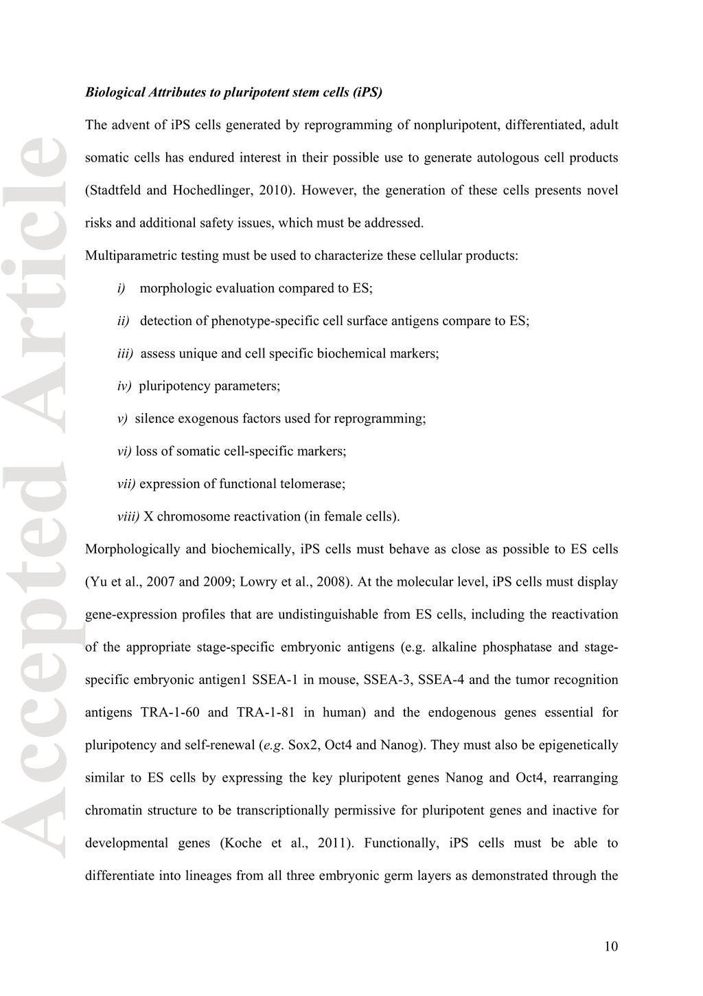
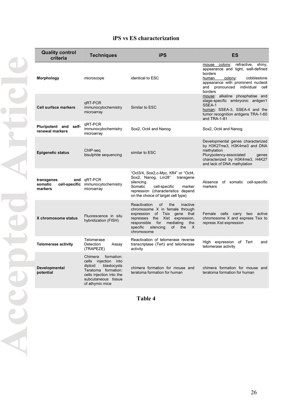
iPS cells generated by reprogramming of nonpluripotent, differentiated, adult somatic cells has endured interest in their possible use to generate autologous cell products (Stadtfeld and Hochedlinger, 2010). However, the generation of these cells presents novel risks and additional safety issues, which must be addressed. Multiparametric testing must be used to characterize these cellular products: i) morphologic evaluation compared to ES; ii) detection of phenotype-specific cell surface antigens compare to ES; iii) assess unique and cell specific biochemical markers; iv) pluripotency parameters; v) silence exogenous factors used for reprogramming; vi) loss of somatic cell-specific markers; vii) expression of functional telomerase; viii) X chromosome reactivation (in female cells). Morphologically and biochemically, iPS cells must behave as close as possible to ES cells (Yu et al., 2007 and 2009; Lowry et al., 2008). At the molecular level, iPS cells must display gene-expression profiles that are undistinguishable from ES cells, including the reactivation of the appropriate stage-specific embryonic antigens (e.g. alkaline phosphatase and stage- specific embryonic antigen1 SSEA-1 in mouse, SSEA-3, SSEA-4 and the tumor recognition antigens TRA-1-60 and TRA-1-81 in human) and the endogenous genes essential for pluripotency and self-renewal (e.g. Sox2, Oct4 and Nanog). They must also be epigenetically similar to ES cells by expressing the key pluripotent genes Nanog and Oct4, rearranging chromatin structure to be transcriptionally permissive for pluripotent genes and inactive for developmental genes (Koche et al., 2011). Functionally, iPS cells must be able to differentiate into lineages from all three embryonic germ layers as demonstrated through the 10
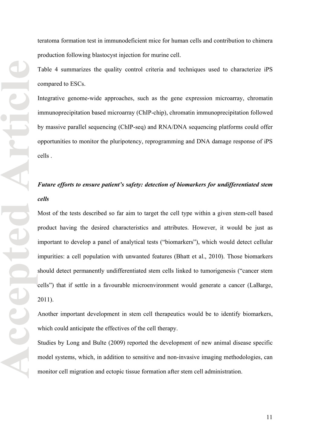
contribution to chimera production following blastocyst injection for murine cell. Table 4 summarizes the quality control criteria and techniques used to characterize iPS compared to ESCs. Integrative genome-wide approaches, such as the gene expression microarray, chromatin immunoprecipitation based microarray (ChIP-chip), chromatin immunoprecipitation followed by massive parallel sequencing (ChIP-seq) and RNA/DNA sequencing platforms could offer opportunities to monitor the pluripotency, reprogramming and DNA damage response of iPS cells . Future efforts to ensure patient’s safety: detection of biomarkers for undifferentiated stem cells Most of the tests described so far aim to target the cell type within a given stem-cell based product having the desired characteristics and attributes. However, it would be just as important to develop a panel of analytical tests (“biomarkers”), which would detect cellular impurities: a cell population with unwanted features (Bhatt et al., 2010). Those biomarkers should detect permanently undifferentiated stem cells linked to tumorigenesis (“cancer stem cells”) that if settle in a favourable microenvironment would generate a cancer (LaBarge, 2011). Another important development in stem cell therapeutics would be to identify biomarkers, which could anticipate the effectives of the cell therapy. Studies by Long and Bulte (2009) reported the development of new animal disease specific model systems, which, in addition to sensitive and non-invasive imaging methodologies, can monitor cell migration and ectopic tissue formation after stem cell administration. 11
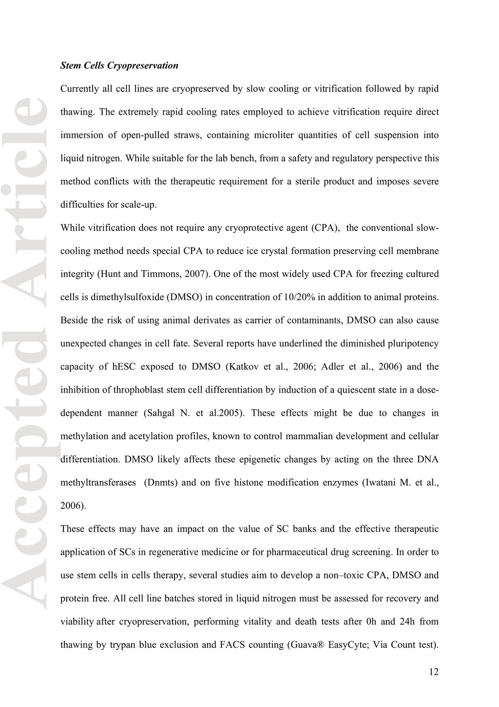
slow cooling or vitrification followed by rapid thawing. The extremely rapid cooling rates employed to achieve vitrification require direct immersion of open-pulled straws, containing microliter quantities of cell suspension into liquid nitrogen. While suitable for the lab bench, from a safety and regulatory perspective this method conflicts with the therapeutic requirement for a sterile product and imposes severe difficulties for scale-up. While vitrification does not require any cryoprotective agent (CPA), the conventional slow- cooling method needs special CPA to reduce ice crystal formation preserving cell membrane integrity (Hunt and Timmons, 2007). One of the most widely used CPA for freezing cultured cells is dimethylsulfoxide (DMSO) in concentration of 10/20% in addition to animal proteins. Beside the risk of using animal derivates as carrier of contaminants, DMSO can also cause unexpected changes in cell fate. Several reports have underlined the diminished pluripotency capacity of hESC exposed to DMSO (Katkov et al., 2006; Adler et al., 2006) and the inhibition of throphoblast stem cell differentiation by induction of a quiescent state in a dose- dependent manner (Sahgal N. et al.2005). These effects might be due to changes in methylation and acetylation profiles, known to control mammalian development and cellular differentiation. DMSO likely affects these epigenetic changes by acting on the three DNA methyltransferases (Dnmts) and on five histone modification enzymes (Iwatani M. et al., 2006). These effects may have an impact on the value of SC banks and the effective therapeutic application of SCs in regenerative medicine or for pharmaceutical drug screening. In order to use stem cells in cells therapy, several studies aim to develop a non–toxic CPA, DMSO and protein free. All cell line batches stored in liquid nitrogen must be assessed for recovery and viability after cryopreservation, performing vitality and death tests after 0h and 24h from thawing by trypan blue exclusion and FACS counting (Guava® EasyCyte; Via Count test). 12
confirm the integrity of liquid nitrogen storage long-term. Administrative Requirements The science of stem cell banking must also meet all the ethical and legal requirements to be able to demonstrate that it has followed all the appropriate regulatory standards. Documentation of banking procedures is vital and it should include: (i) informed consent from the donor for the procurement of the tissue and derivation of the cell lines, (ii) all relevant data from quality controls and characterization, (iii) a material safety datasheet on hazards associated with the cells, (iv) a material transfer agreement (MTA) to guarantee ownership and avoid third party distribution and (v) all necessary declarations and labels to meet local import regulations (ISCBI, 2009). For these reasons a stem cell bank should be accredited or authorized by appropriate authorities for the purpose of their activities and should have an independent and transparent governance structure which reviews the ethical and legal requirements. Even with such structured organization the terms of the informed consent, national laws on stem cell research and the continuous improvement in stem cell culture protocols may complicate the establishment of one uniform set of ethics and standards by which cells should be handled and used (Healy et al., 2008; Crook et al., 2010). The review process of these dynamic landscape, spanning from ethic to law, science and management needs to be continuously performed and made available to the international scientific community to improve the generation of high-quality biomaterial and facilitate its accessibility. Discussion Creating standards for the characterizing of all stem cell types is a big challenge. Pluripotent stem cells tend to be genetically unstable, particularly if less than optimal culture techniques 13
needed to make high quality pluripotent stem cells available to the broad research community are beyond their means. Academics sharing is a nice idea, but unrealistic given the subject matter, times and law. University administrations will put strings on stem cell distributions by their scientists due to intellectual property rights and potential source of revenue. On the other hand, National Stem Cell Bank, funded by the government and making high quality cells available to all researchers at reasonable cost, would actually be quite ideal and desirable. The exponential generation and use of disease- and patient-specific stem cell lines will require proper storage facilities to handle high quality and precious material. But even if the number of cell lines grow, the number of banks will probably level off in the near future due to extensive quality controls and excellence of the banking effort in the long term. That's especially true for embryonic stem cells and ultimately for iPS. While the establishment of iPS cell lines is conceptually and technically simple, direct reprogramming is a slow and inefficient process consisting of largely unknown events. A single reprogramming experiment usually generates multiple iPS cell lines that are not always identical. Each individual iPS cell line needs to be fully characterized to ensure safety and pluripotency capacity. Several variables must be considered in order to reproducibly obtain iPS, which include (i) the choice of factors used to reprogram cells; (ii) the methods used to deliver these factors; (iii) the choice of target cell type; (iv) the parameters of factor expression, such as timing and levels; (v) the culture conditions used to derive iPSCs; and the methods of (vi) identifying and (vii) characterizing reprogrammed cells. The fact that iPS can be maintained in vitro indefinitely makes the establishment of cell banking facilities attractive and distribution of high-quality iPS lines to interested parties would facilities further development of clinical regenerative therapies and toxicological tests. We should not forget that the biggest obstacle in every aspect of stem cell study is still the 14
diseases. Because the biology is so tough everything else should be made accessible. Acknowledgments We acknowledge the support of NeuroStemcell (European Community's Seventh Framework Programme grant agreement nr. HEALTH-2008-B-222943). We also are grateful to Marble Arch working Group on International Biobanking and the European Middle Eastern and African Society for Biopreservation and Biobanking (ESSB) as knowledge provider. 15
S. 2006. The effects of solvents on embryonic stem cell differentiation. Toxicol In Vitro. 20(3):265-71. Alviano F, Fossati V, Marchionni C, Arpinati M, Bonsi L, Franchina M, Lanzoni G, Cantoni S, Cavallini C, Bianchi F, Tazzari PL, Pasquinelli G, Foroni L, Ventura C, Grossi A, Bagnara GP. 2007. Term amniotic membrane is a high throughput source for multipotent mesenchymal stem cells with the ability to differentiate into endothelial cells in vitro. BMC Dev Biol. 1:11. Barbara JAJ, Regan FAM, Contreras MC. 2008. Transfusion Microbiology. Cambridge University press. 390p. Benjamin RJ. 2001. Nucleic acid testing: update and applications. Semin Hematol 38(4 Suppl 9):11-6. Review. Bhatt AN, Mathur R, Farooque A, Verma A, Dwarakanath BS. 2010. Cancer biomarkers - current perspectives. Indian J Med Res. 132:129-49. Brivanlou AH, Gage FH, Jaenisch R, Jessell T, Melton D, Rossant J. 2003. Stem cells. Setting standards for human embryonic stem cells. Science 300(5621):913-916. Buzzard JJ, Gough NM, Crook JM, Colman A. 2004. Karyotype of human ES cells during extended culture. Nat Biotechnol 22:381-2. Chatterjee R. 2007. Cell biology: cases of mistaken identity Science. 315:928–931. Conti L, Cattaneo E, 2010. Neural stem cell systems: physiological players or in vitro entities? Nat Rev Neurosci 11(3):176-187. 16
Cell Banking Initiative (ISCBI): raising standards to bank on. In Vitro Cell Dev Biol Anim. 46(3-4):169-72. Diaferia GR, Conti L, Redaelli S, Cattaneo M, Mutti C, DeBlasio P, Dalprà L, Cattaneo E, Biunno I. 2011. Systematic chromosomal analysis of cultured mouse neural stem cell lines. Stem Cell Dev, in press. Dominici M, LeBlanc K, Mueller I, Slaper-Cortenbach I, Marini F, Krause D, Deans R, Keating A, Prockop Dj, Horwitz E. 2006. Minimal criteria for defining multipotent mesenchymal stromal cells. The International Society for Cellular Therapy Position Statement.Cytotherapy 8:315-317. Drexler HG, Dirks WG, Matsuo Y and MacLeod RA. 2003. False leukemia-lymphoma cell lines: an update on over 500 cell lines. Leukemia. 17:416–426. Gonzales F and McDonough S. 1998. Applications of Transcription-Mediated Amplification to Quantification of Gene Sequences. In: Ferre F, editor. Gene Amplification. Birkhauser, Boston. p189-204. Healy LE, Ludwig TE, Choo A. 2008. International banking: checks, deposits, and withdrawals. Cell Stem Cell. 2(4):305-6. Heim D and Mumford E. 2005. The future of BSE from the global perspective. J Meat Sci 70: 555–562. Herbert BS, Shay JW, Wright WE. 2003. Analysis of telomeres and telomerase. Curr Protoc Cell Biol. Chapter 18:Unit 18.6 17
Stem Cell Rev 1:139-44 Review. Hunt CJ, Timmons PM. 2007. Cryopreservation of human embryonic stem cell lines. Methods Mol Biol. 368:261-70. International Stem Cell Initiative, Adewumi O, Aflatoonian B, Ahrlund-Richter L, Amit M, Andrews PW, Beighton G, Bello PA, Benvenisty N, Berry LS, Bevan S, Blum B, Brooking J, Chen KG, Choo AB, Churchill GA, Corbel M, Damjanov I, Draper JS, Dvorak P, Emanuelsson K, Fleck RA, Ford A, Gertow K, Gertsenstein M, Gokhale PJ, Hamilton RS, Hampl A, Healy LE, Hovatta O, Hyllner J, Imreh MP, Itskovitz-Eldor J, Jackson J, Johnson JL, Jones M, Kee K, King BL, Knowles BB, Lako M, Lebrin F, Mallon BS, Manning D, Mayshar Y, McKay RD, Michalska AE, Mikkola M, Mileikovsky M, Minger SL, Moore HD, Mummery CL, Nagy A, Nakatsuji N, O'Brien CM, Oh SK, Olsson C, Otonkoski T, Park KY, Passier R, Patel H, Patel M, Pedersen R, Pera MF, Piekarczyk MS, Pera RA, Reubinoff BE, Robins AJ, Rossant J, Rugg-Gunn P, Schulz TC, Semb H, Sherrer ES, Siemen H, Stacey GN, Stojkovic M, Suemori H, Szatkiewicz J, Turetsky T, Tuuri T, van den Brink S, Vintersten K, Vuoristo S, Ward D, Weaver TA, Young LA, Zhang W. 2007. Characterization of human embryonic stem cell lines by the International Stem Cell Initiative. Nat Biotechnol 25(7):803- 816. ISCBI (International Stem Cell Banking Initiative). 2009. Consensus guidance for banking and supply of human embryonic stem cell lines for research purposes. Stem Cell Rev and Rep. 5:301-314. 18
Yagi S, Shiota K. 2006. Dimethyl sulfoxide has an impact on epigenetic profile in mouse embryoid body. Stem Cells, 24: 2549- 2556. Katkov II, Kim MS, Bajpai R, Altman YS, Mercola M, Loring JF, Terskikh AV, Snyder EY, Levine F. 2006. Cryopreservation by slow cooling with DMSO diminished production of Oct-4 pluripotency marker in human embryonic stem cells. Cryobiology. 53(2):194-205. Kern S, Eichler H, Stoeve J, Kluter H, Bieback K. 2006. Comparative analysis of mesenchymal stem cells from bone marrow, umbilical cord blood, or adipose tissue. Stem Cells 24: 1294-1301. Koche RP, Smith ZD, Adli M, Gu H, Ku M, Gnirke A, Bernstein BE, Meissner. 2011. Reprogramming factor expression initiates widespread targeted chromatin remodeling. Cell Stem Cell. 8(1):96-105. Kogler G, Radke FT, Lefort A, Sensken S, Fischer J, Sorg RV, Wernet P. 2006. Cytokine production and hematopoiesis supporting activity of cord blood-derived unrestricted somatic stem cells. Exp Hematol. 33:573-583. Kokovay E, Shen Q, Temple S. 2008. The incredible elastic brain: how neural stem cells expand our minds. Neuron 60(3):420-429. LaBarge MA. 2010. The difficulty of targeting cancer stem cell niches. Clin Cancer Res. 16(12):3121-9. Lefort N, Feyeux M, Bas C, Féraud O, Bennaceur-Griscelli A, Tachdjian G, Peschanski M, Perrier AL. 2008. Human embryonic stem cells reveal recurrent genomic instability at 20q11.21. Nat Biotechnol 26(12):1364-6. 19
Current Technology and Applications. Caister Academic Press. 284 p. Long CM, Bulte JW. 2009. In vivo tracking of cellular therapeutics using magnetic resonance imaging. Expert Opin Biol Ther. 9(3):293-306. Lowry WE, Richter L, Yachechko R, Pyle AD, Tchieu J, Sridharan R, Clark AT, Plath K. 2008. Generation of human induced pluripotent stem cells from dermal fibroblasts. Proc Natl Acad Sci U S A. 105(8):2883-8. Masters JR, Thomson JA, Daly-Burns B, Reid YA, Dirks WG, Packer P, Toji LH, Ohno T, Tanabe H, Arlett CF et al. 2001. Short tandem repeat profiling provides an international reference standard for human cell lines. Proc. Natl Acad. Sci. USA. 98:8012–8017. Motaln H, Schichor C, Lah TT. 2010. Human mesenchymal stem cells and their use in cell- based therapies. Cancer. 116(11):2519-2530. Närvä E, Autio R, Rahkonen N, Kong L, Harrison N, Kitsberg D, Borghese L, Itskovitz- Eldor J, Rasool O, Dvorak P, Hovatta O, Otonkoski T, Tuuri T, Cui W, Brüstle O, Baker D, Maltby E, Moore HD, Benvenisty N, Andrews PW, Yli-Harja O, Lahesmaa R. 2010 High- resolution DNA analysis of human embryonic stem cell lines reveals culture-induced copy number changes and loss of heterozygosity. Nat Biotechnol 28(4):371-7 NIH. 2007. Notice regarding authentication of cultured cell lines, NOTod-08-017, Nov 28, NiH. Pollard SM, Conti L, Sun Y, Goffredo D, Smith A. 2006. Adherent neural stem (NS) cells from fetal and adult forebrain. Cereb Cortex. 16 Suppl 1:i112-20. 20
Cigudosa JC, Lloyd AC, Bernad A. 2005. Spontaneous human adult stem cell transformation. Cancer Res. 65: 3035-3039. Sahgal N, Canham LN, Konno T, Wolfe MW, Soares MJ. 2005. Modulation of trophoblast stem cell and giant cell phenotypes: analyses using the Rcho-1 cell model. Differentiation. 73(9-10):452-62 Stadtfeld M, Hochedlinger K. 2010. Induced pluripotency: history, mechanisms, and applications. Genes Dev. 24(20):2239-63. Stephenson E, Ogilvie CM, Patel H, Cornwell G, Jacquet L, Kadeva N, Braude P, Ilic D. 2010. Safety paradigm: genetic evaluation of therapeutic grade human embryonic stem cells. J R Soc Interface. 7 Suppl 6:S677-88. Temple S. 2001.The development of neural stem cells. Nature 414, 112–117. Thomson JA, Itskovitz-Eldor J, Shapiro SS, Waknitz MA, Swiergiel JJ, Marshall VS, Jones JM. 1998. Embryonic stem cell lines derived from human blastocysts. Science 282(5391):1145-1147. Erratum in: Science 282(5395):1827. United States Food and Drug Administration. 2008a. Validation of growth-based rapid microbiological methods for sterility testing of cellular and gene therapy products. Rockville: Guidance for Industry. United States Food and Drug Administration. 2008b. Potency Tests for Cellular and Gene Therapy Products. Rockville: Guidance for Industry. 21
Affiliation Link UK Stem Cell Bank UK Governance http://www.ukstemcellbank.o rg.uk National Stem Cell Bank– Banco–Nacional de Lineas Celulares (BNLC) Valencia Stem Cell Bank–BNLC Spanish governance http://www.isciii.es/htdocs/ter apia/terapia_bancocelular.js p National Centre for Cell Science– Cell Repository Indian governance http://www.nccs.res.in WiCell International Stem Cell Bank University of Wisconsin http://www.wicell.org Australian Stem Cell Bank Australian governance http://www.ascb.com.au Singapore Stem Cell Bank Agency for Science, Technology and Research (A*STAR) http://www.sscc.a- star.edu.sg/stemCellBank.ph p Korean Stem Cell Bank Korean governance http://kscb.co.kr/eng Taiwan Stem Cell Bank National Science Council http://www.tscb.bcrc.firdi.org. tw Umass Human Stem Cell Bank & Registry University of Massachusetts Medical School http://www.umassmed.edu/m hscb/index.aspx Rutgers University Cell and DNA repository Rutgers University http://www.rucdr.org/ Coriell Institute Private no-profit www.coriell.org Table 2 24
http://www.hescreg.eu/ StemDB EU http://www.stemdb.org US NIH US http://grants.nih.gov/stem_cells/registry/current.htm International Stem Cell Registry Massachusetts Life Sciences Center http://www.umassmed.edu/iscr/index.aspx Table 3 25
Morphology microscope identical to ESC mouse colony: refractive, shiny, appearance and tight, well-defined borders human colony: cobblestone appearance with prominent nucleoli and pronounced individual cell borders Cell surface markers qRT-PCR immunocytochemistry microarray Similar to ESC mouse: alkaline phosphatise and stage-specific embryonic antigen1 SSEA-1 human: SSEA-3, SSEA-4 and the tumor recognition antigens TRA-1-60 and TRA-1-81 Pluripotent and self- renewal markers qRT-PCR immunocytochemistry microarray Sox2, Oct4 and Nanog Sox2, Oct4 and Nanog Epigenetic status ChIP-seq bisulphite sequencing similar to ESC Developmental genes characterized by H3K27me3, H3K4me3 and DNA methylation Plurypotency-associated genes characterized by H3K4me3, H4K27 and lack of DNA methylation transgenes and somatic cell-specific markers qRT-PCR immunocytochemistry microarray “Oct3/4, Sox2,c-Myc, Kfl4” or “Oct4, Sox2, Nanog, Lin28” transgene silencing. Somatic cell-specific marker repression (characteristics depend on the choice of target cell type) Absence of somatic cell-specific markers X chromosome status Fluorescence in situ hybridization (FISH) Reactivation of the inactive chromosome X in female through expression of Tsix gene that represses the Xist expression, responsible for mediating the specific silencing of the X chromosome Female cells carry two active chromosome X and express Tsix to repress Xist expression Telomerase activity Telomerase Detection Assay (TRAPEZE) Reactivation of telomerase reverse transcriptase (Tert) and telomerase activity High expression of Tert and telomerase activity Developmental potential Chimera formation: cells injection into diploid blastocysts Teratoma formation: cells injection into the subcutaneous tissue of athymic mice chimera formation for mouse and teratoma formation for human chimera formation for mouse and teratoma formation for human Table 4 26