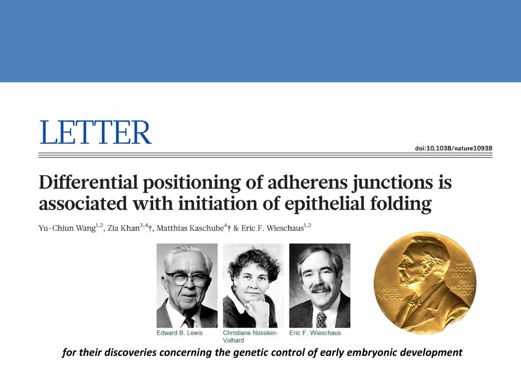
in physiology or medicine for discovering that ordinary cells of the body can be reprogrammed into stem cells, a discovery which may lead to new treatments. Nobel prize
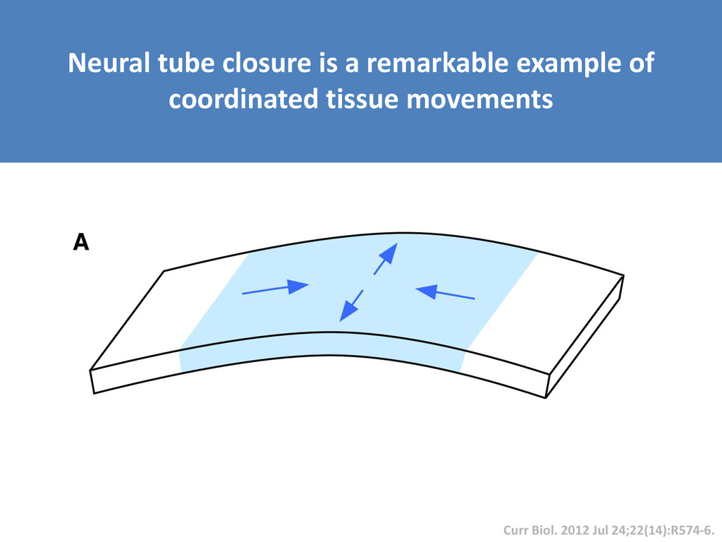
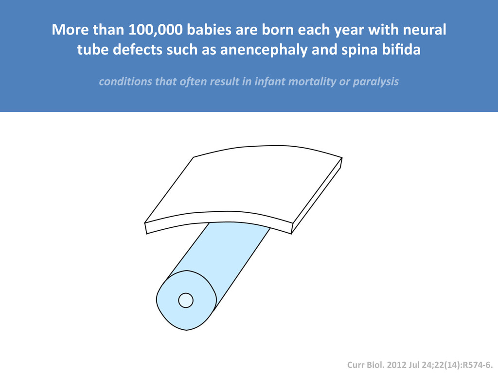
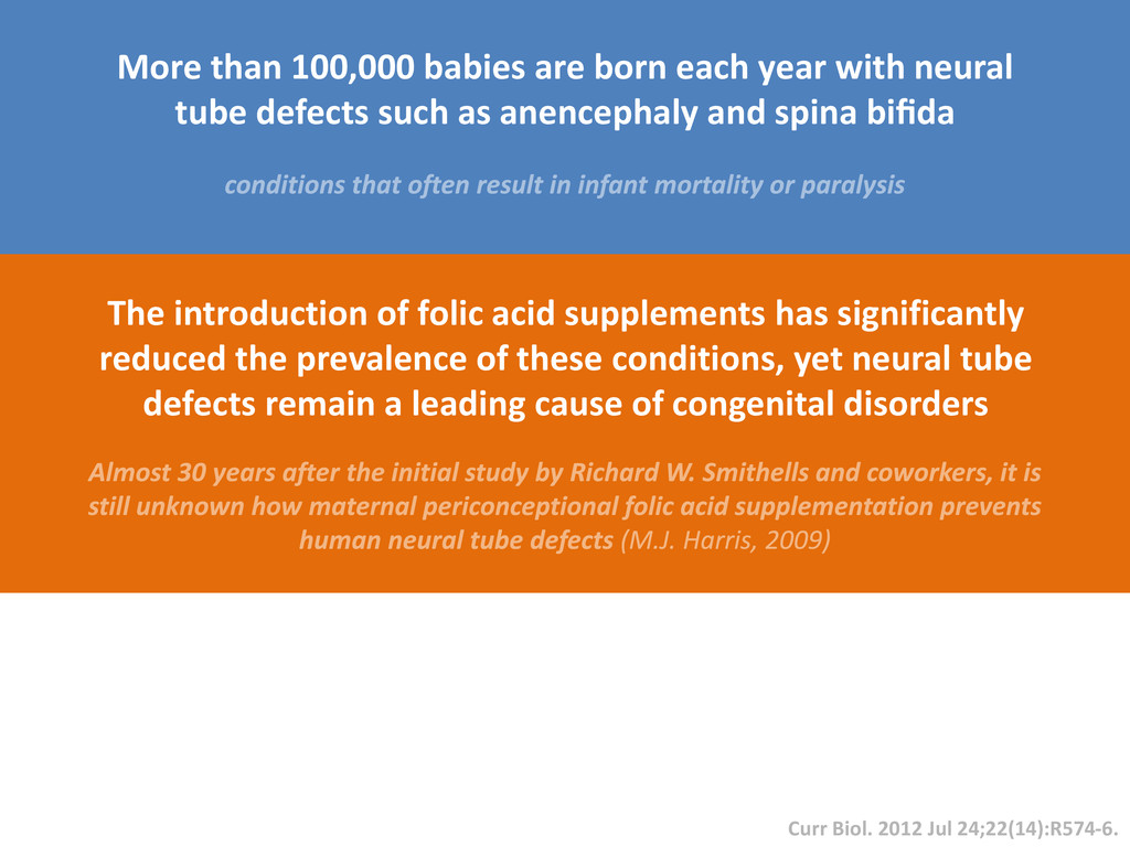
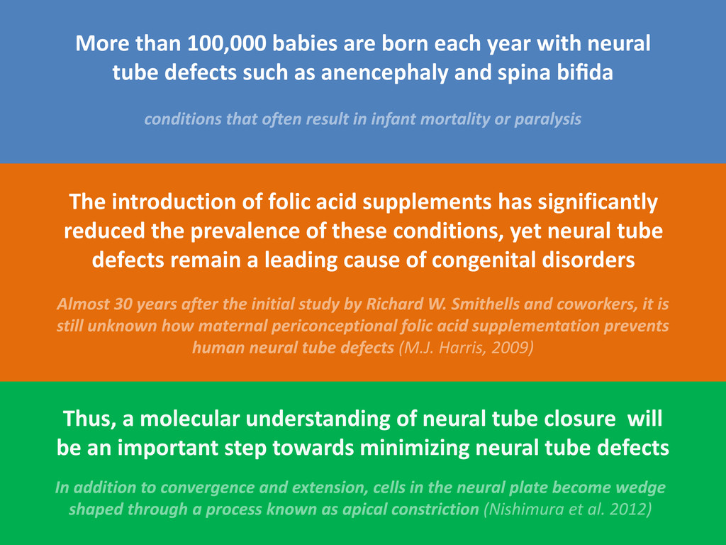
born each year with neural tube defects such as anencephaly and spina bifida The introduction of folic acid supplements has significantly reduced the prevalence of these conditions, yet neural tube defects remain a leading cause of congenital disorders
tube defects such as anencephaly and spina bifida Curr Biol. 2012 Jul 24;22(14):R574-6. The introduction of folic acid supplements has significantly reduced the prevalence of these conditions, yet neural tube defects remain a leading cause of congenital disorders
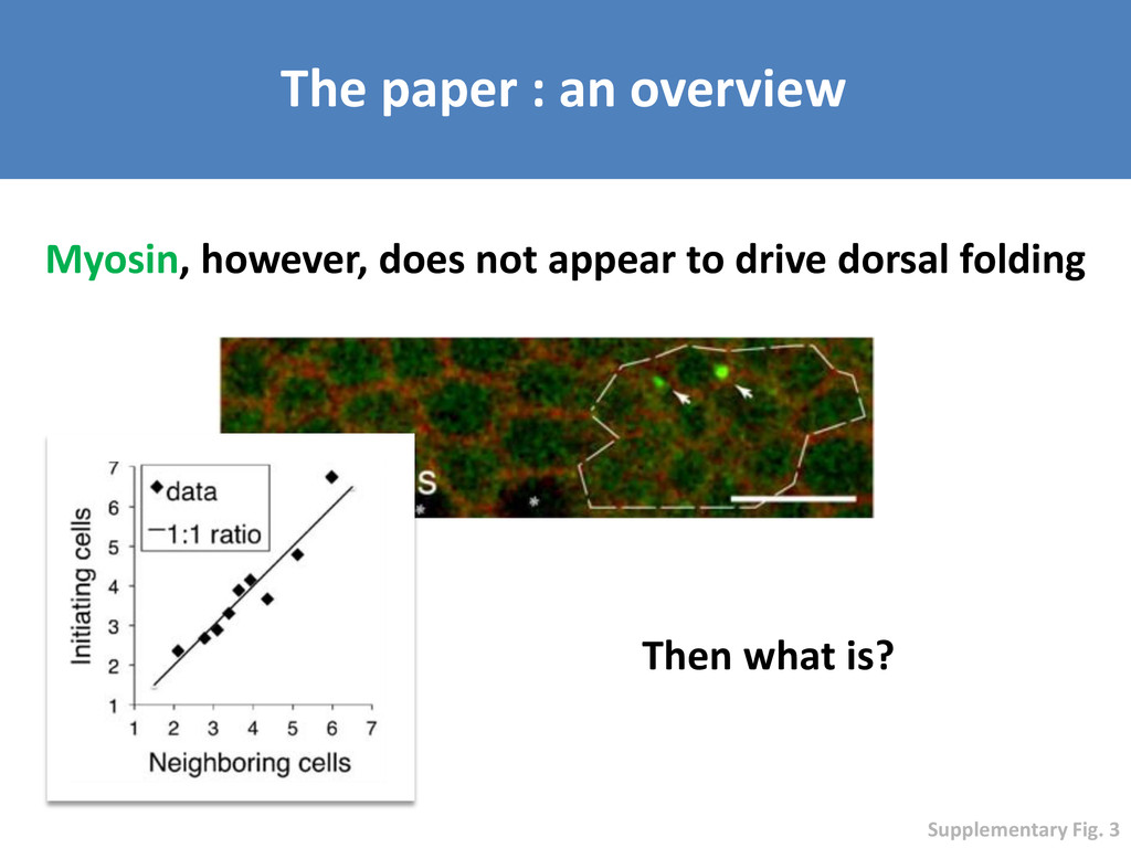
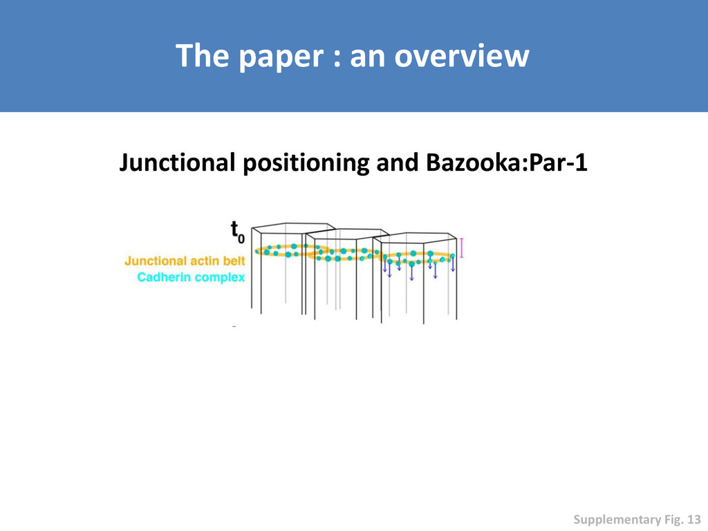
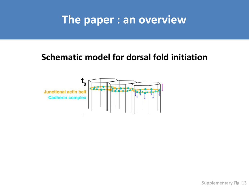
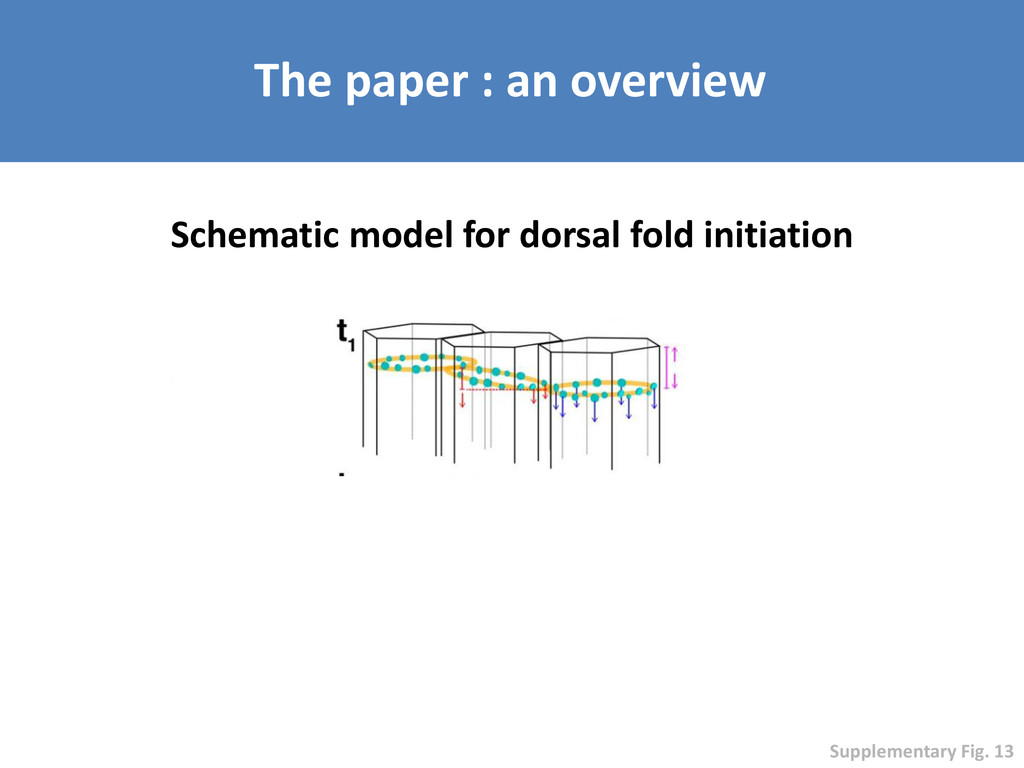
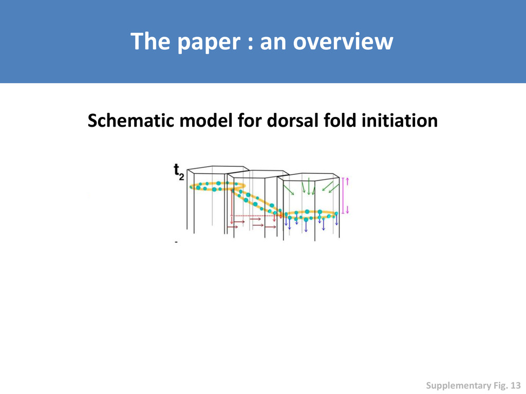
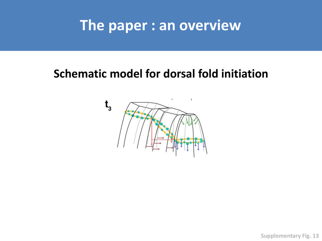
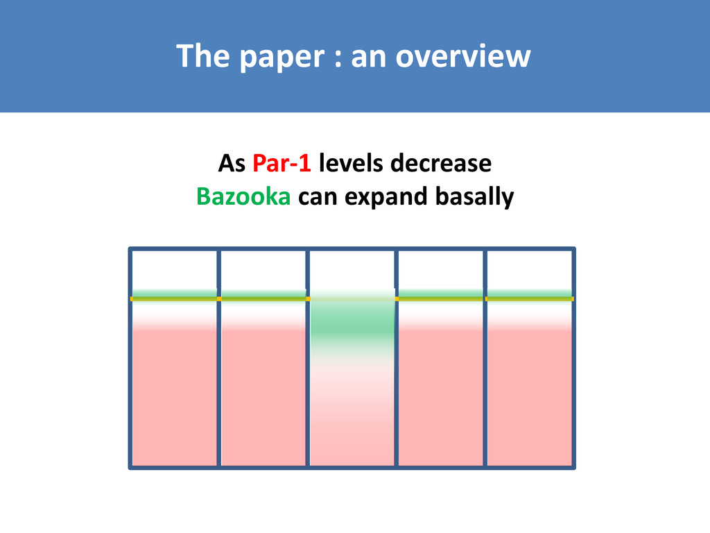
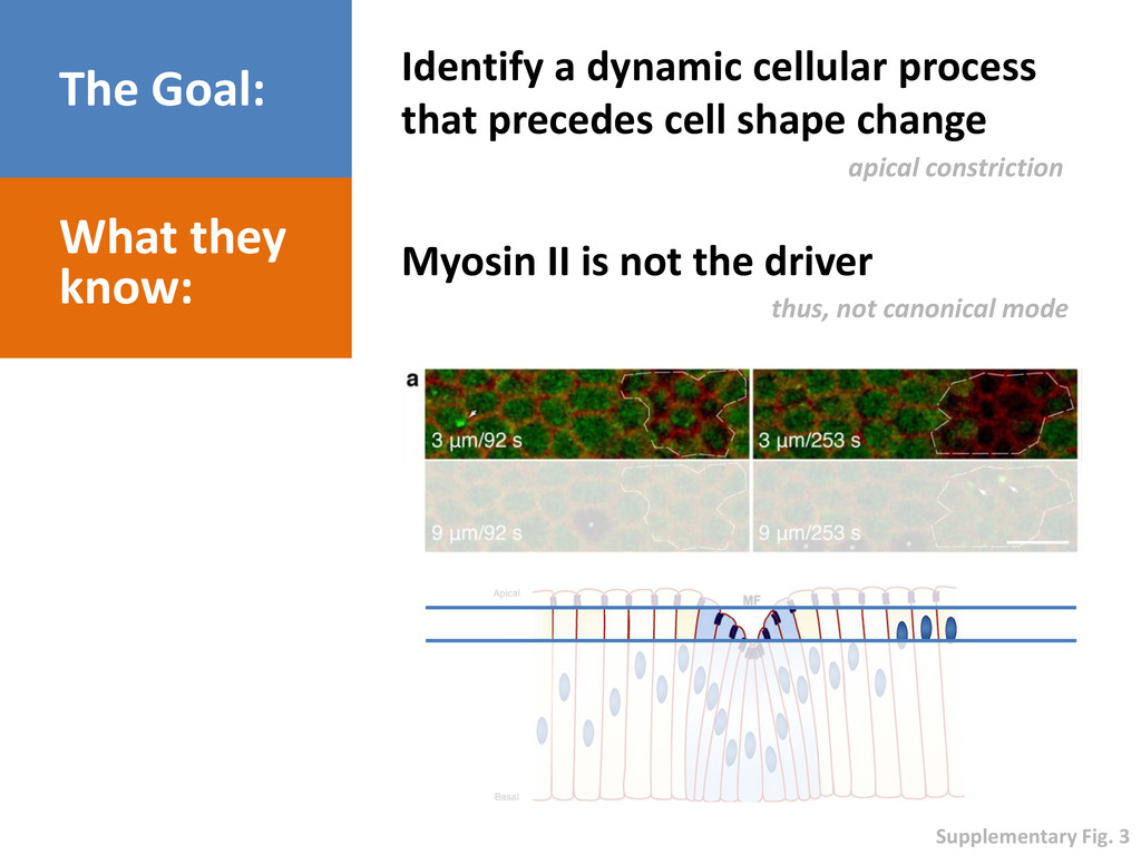
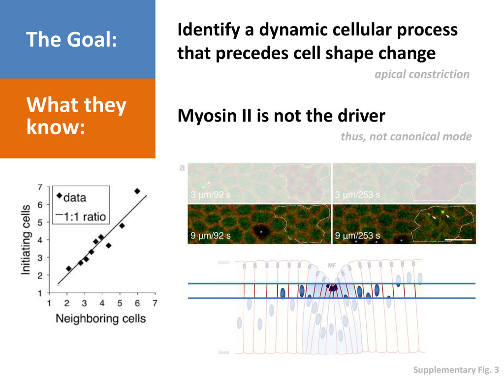
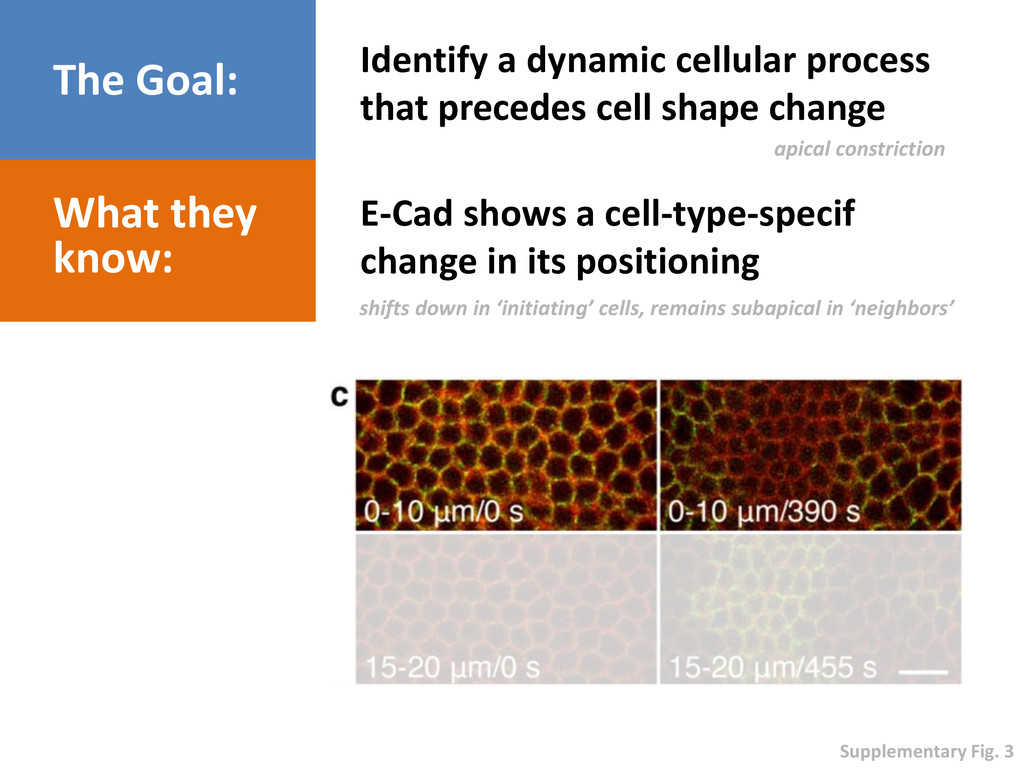
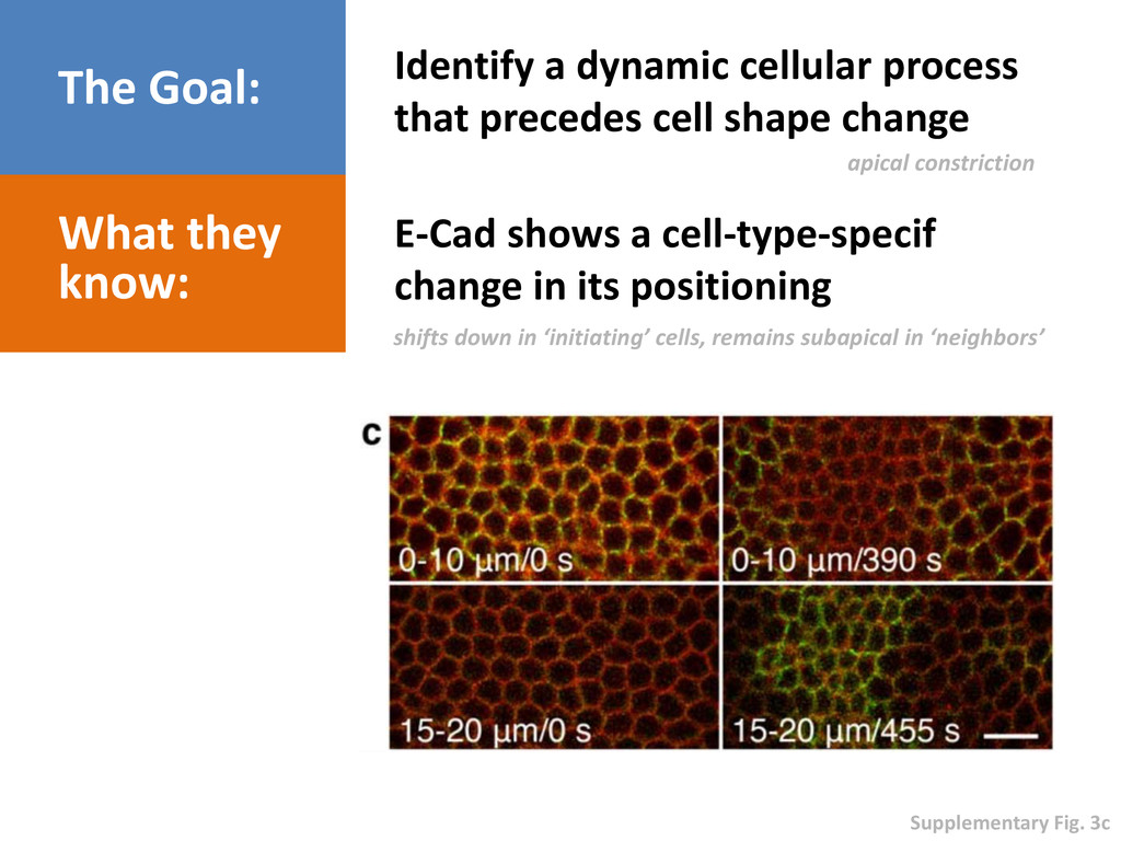
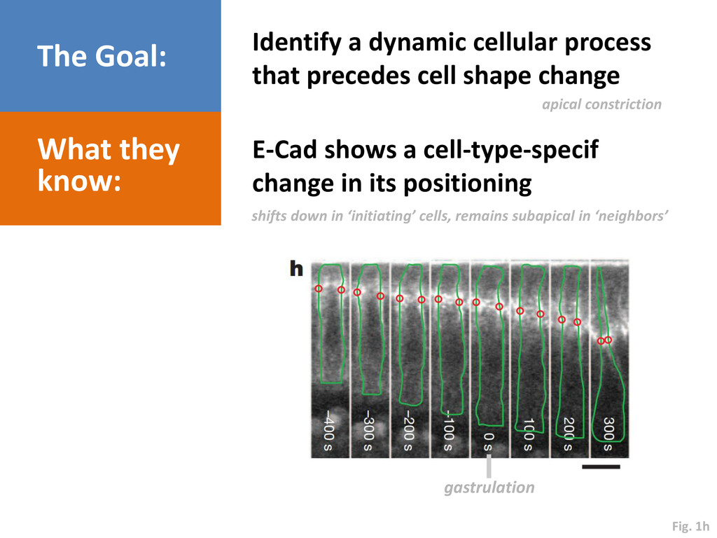
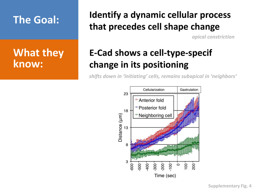
shape change What they know: E-Cad shows a cell-type-specif change in its positioning apical constriction shifts down in ‘initiating’ cells, remains subapical in ‘neighbors’
shape change What they know: E-Cad shows a cell-type-specif change in its positioning apical constriction shifts down in ‘initiating’ cells, remains subapical in ‘neighbors’ Supplementary Fig. 3
shape change What they know: E-Cad shows a cell-type-specif change in its positioning apical constriction shifts down in ‘initiating’ cells, remains subapical in ‘neighbors’ Supplementary Fig. 3c
shape change What they know: E-Cad shows a cell-type-specif change in its positioning apical constriction shifts down in ‘initiating’ cells, remains subapical in ‘neighbors’ gastrulation Fig. 1h
shape change What they know: E-Cad shows a cell-type-specif change in its positioning apical constriction shifts down in ‘initiating’ cells, remains subapical in ‘neighbors’ Supplementary Fig. 4
shape change What they know: E-Cad shows a cell-type-specif change in its positioning apical constriction shifts down in ‘initiating’ cells, remains subapical in ‘neighbors’ Supplementary Fig. 4
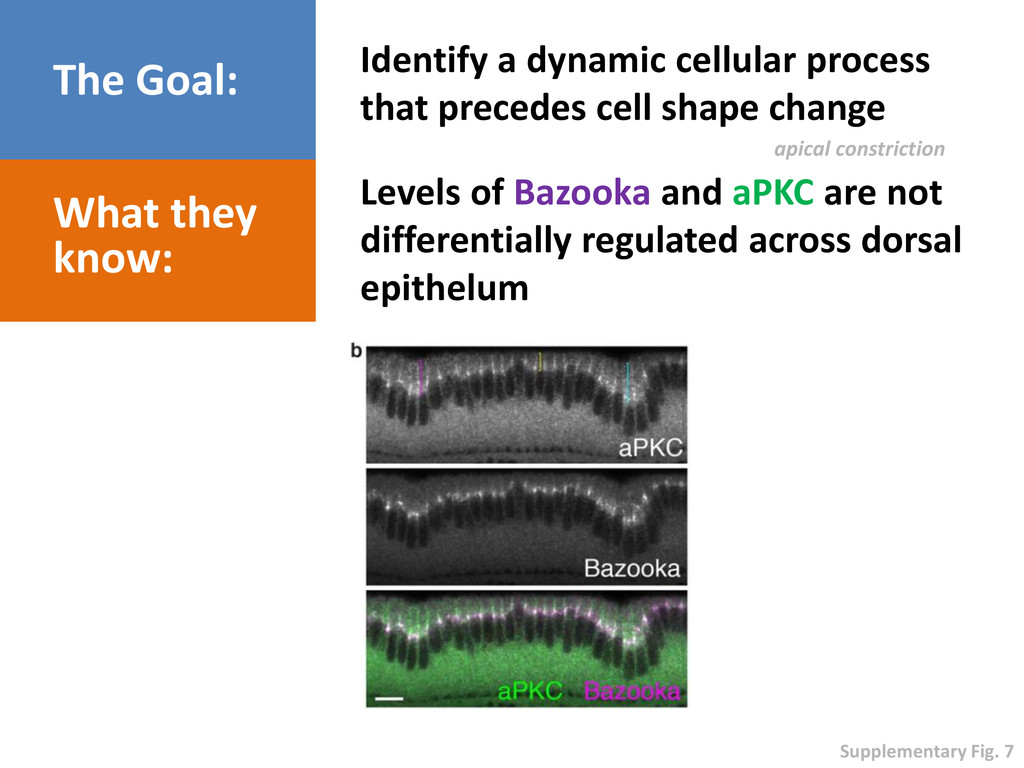
shape change What they know: Levels of Bazooka and aPKC are not differentially regulated across dorsal epithelum apical constriction Supplementary Fig. 7
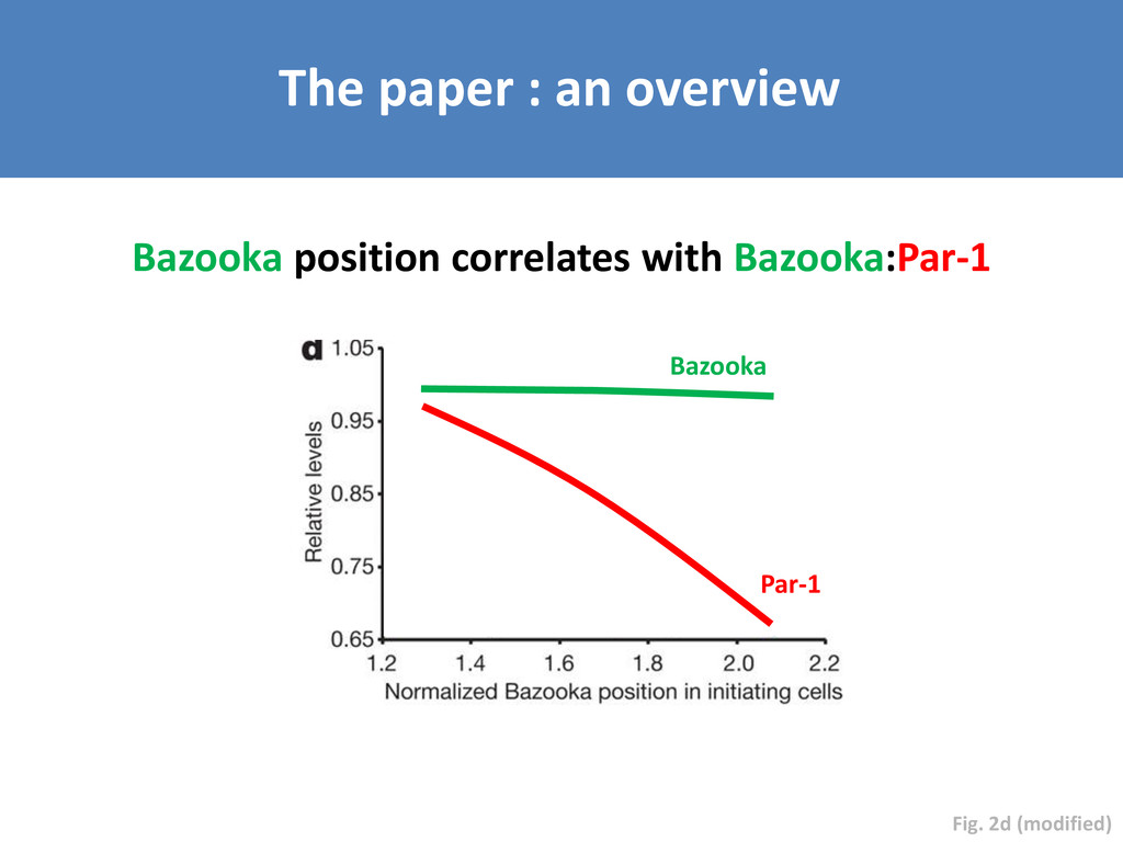
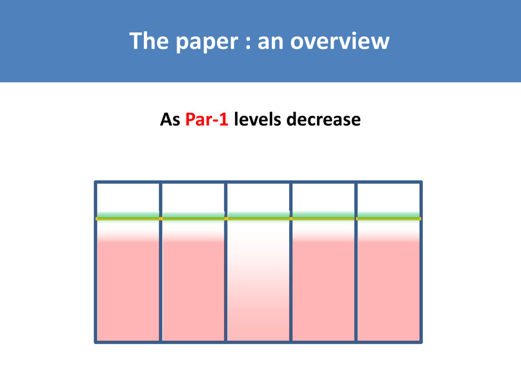
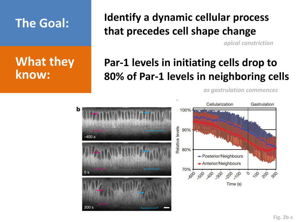
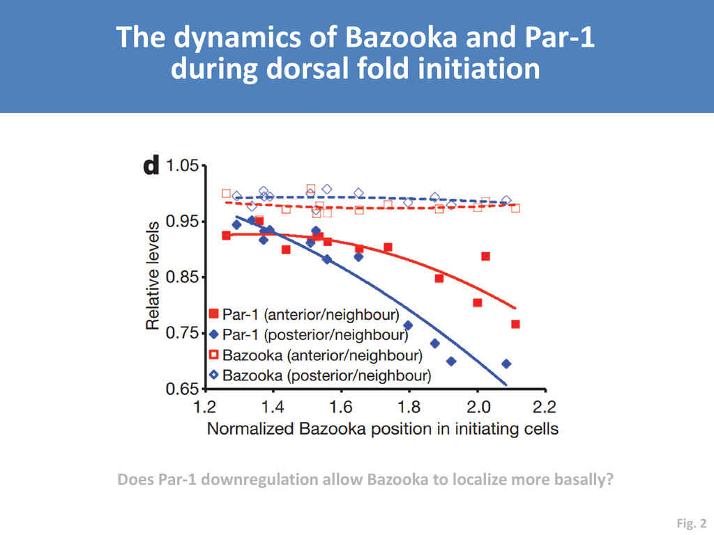
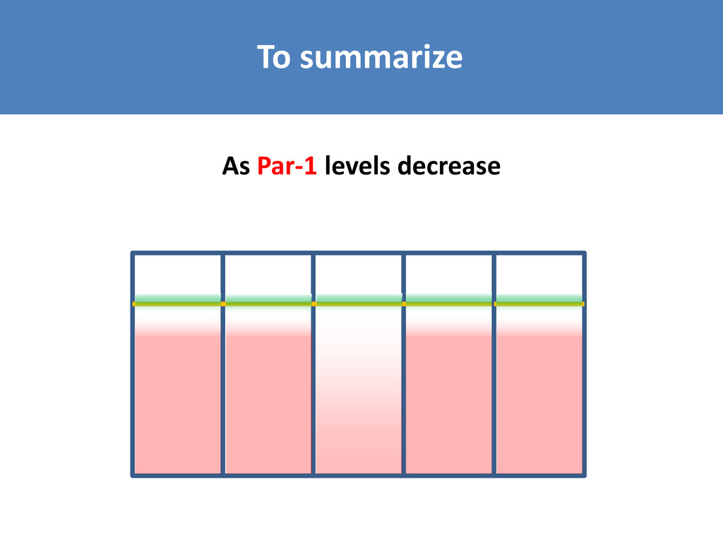
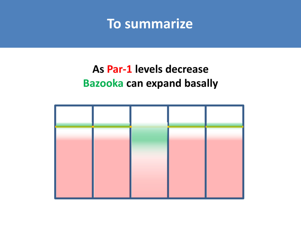
shape change What they know: Par-1 levels in initiating cells drop to 80% of Par-1 levels in neighboring cells apical constriction as gastrulation commences Fig. 2b-c
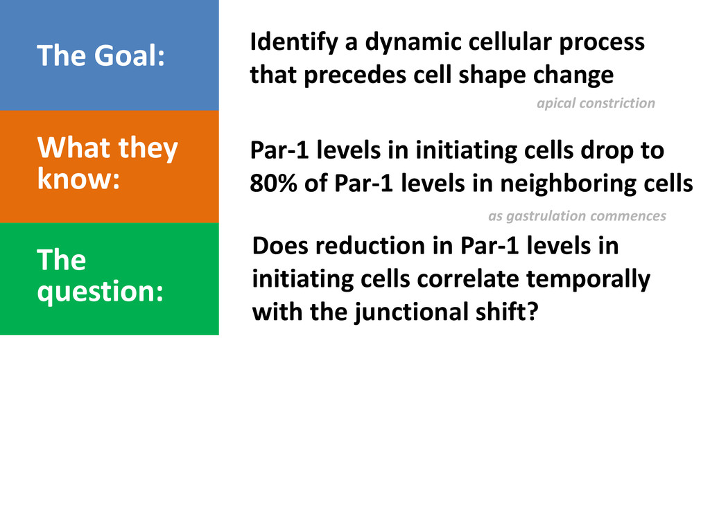
shape change What they know: Par-1 levels in initiating cells drop to 80% of Par-1 levels in neighboring cells apical constriction as gastrulation commences The question: Does reduction in Par-1 levels in initiating cells correlate temporally with the junctional shift?
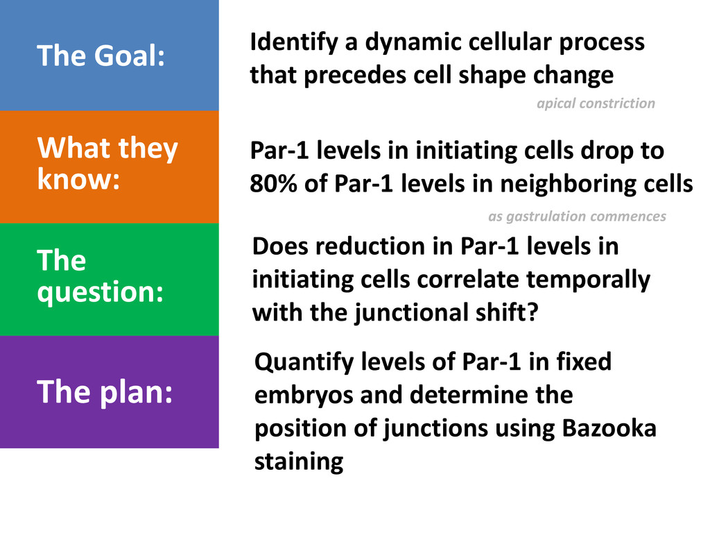
shape change What they know: Par-1 levels in initiating cells drop to 80% of Par-1 levels in neighboring cells apical constriction as gastrulation commences The question: Does reduction in Par-1 levels in initiating cells correlate temporally with the junctional shift? The plan: Quantify levels of Par-1 in fixed embryos and determine the position of junctions using Bazooka staining
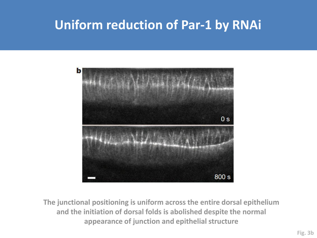
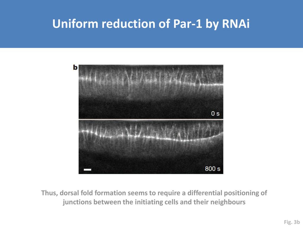
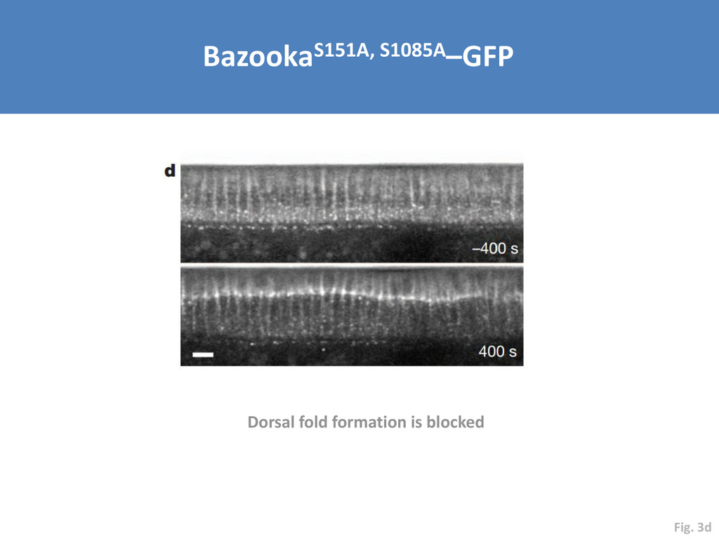
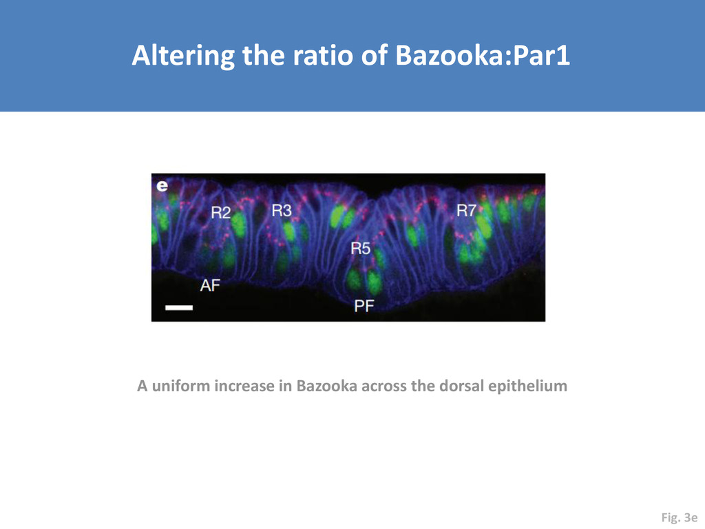
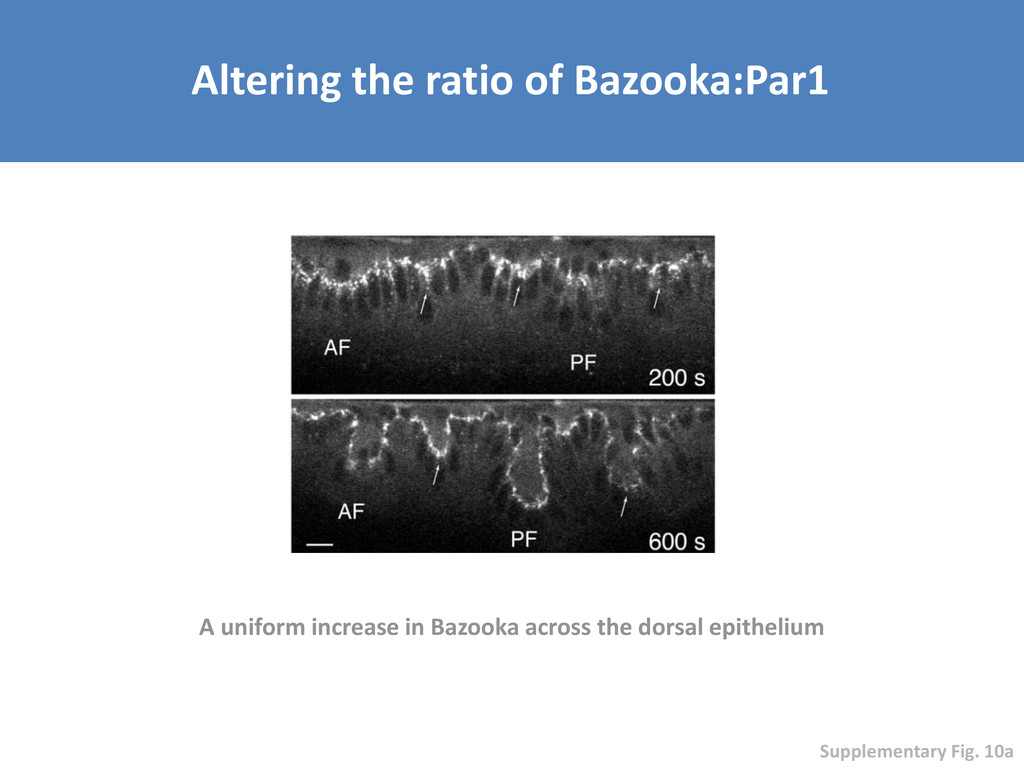
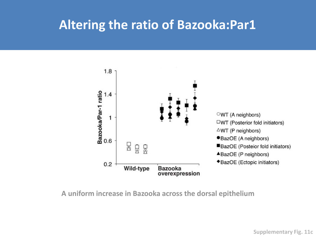
uniform across the entire dorsal epithelium and the initiation of dorsal folds is abolished despite the normal appearance of junction and epithelial structure Fig. 3b
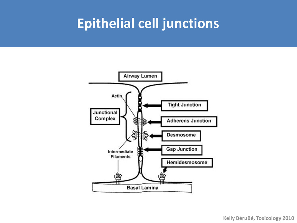
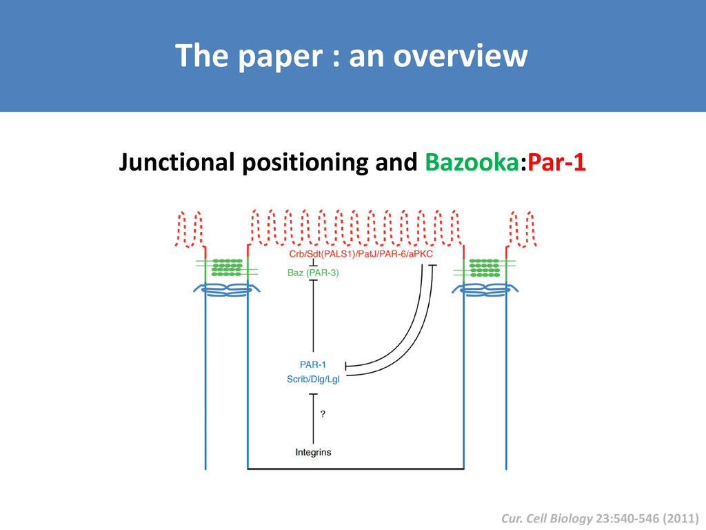
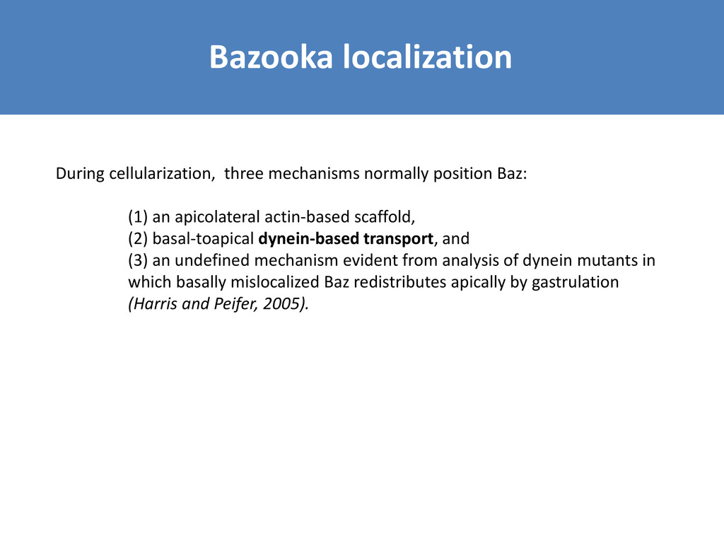
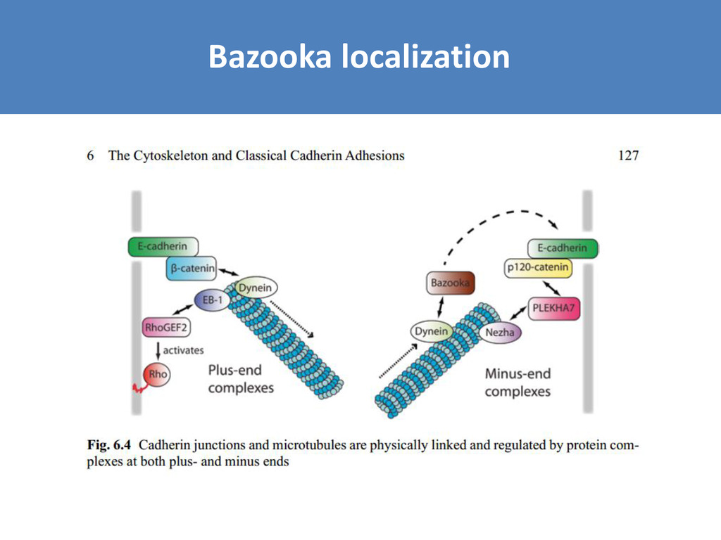
an apicolateral actin-based scaffold, (2) basal-toapical dynein-based transport, and (3) an undefined mechanism evident from analysis of dynein mutants in which basally mislocalized Baz redistributes apically by gastrulation (Harris and Peifer, 2005).


 rasagy
 baasie
 davidcarrasco
 axbom
 mayatellez
 aemeredith
 dougneiner
 aleyda
 reverentgeek
 keavy
 tammyeverts
 yeseniaperezcruz