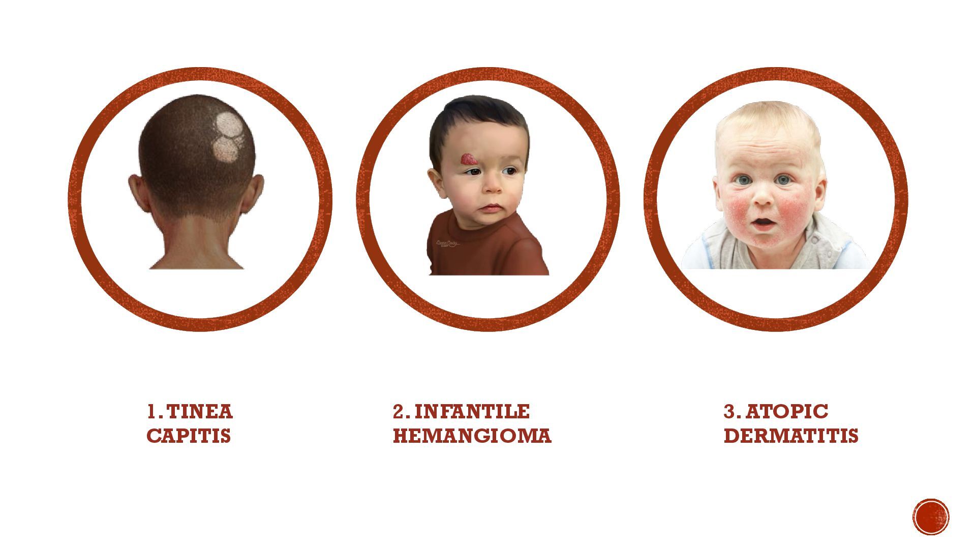
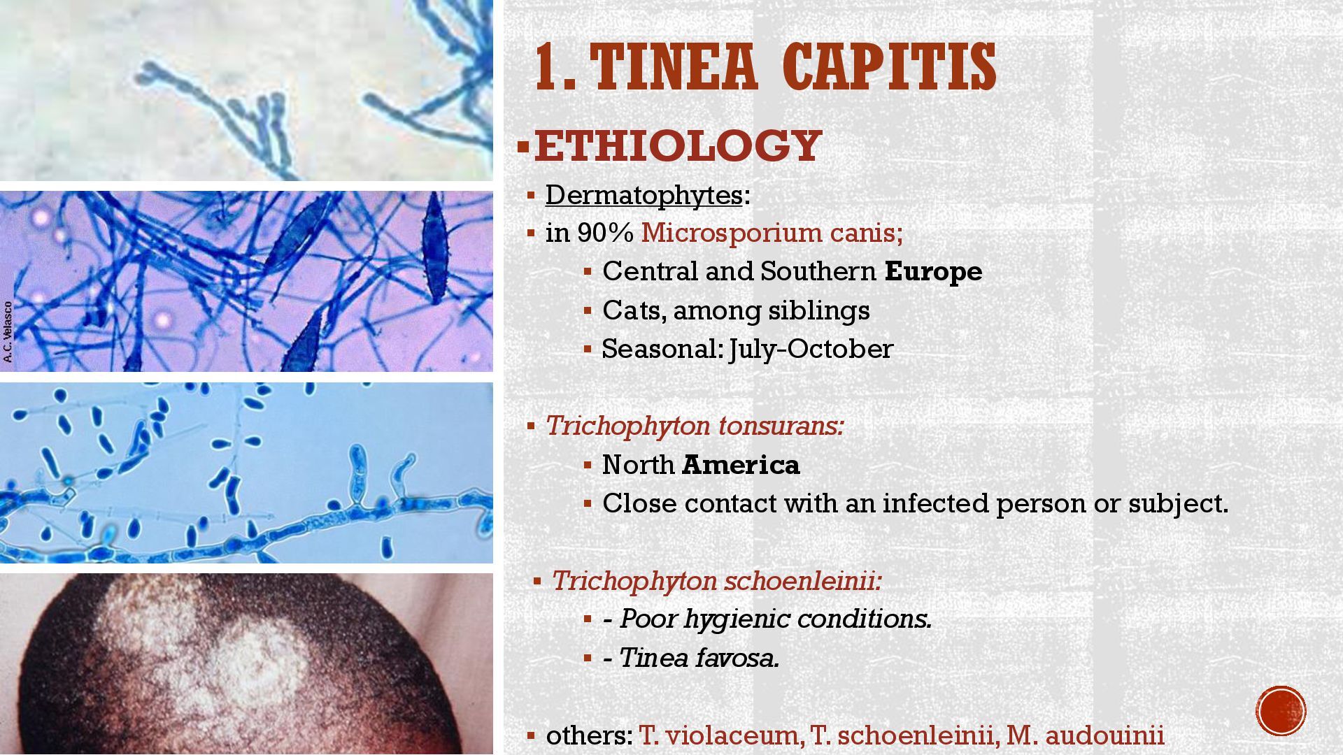
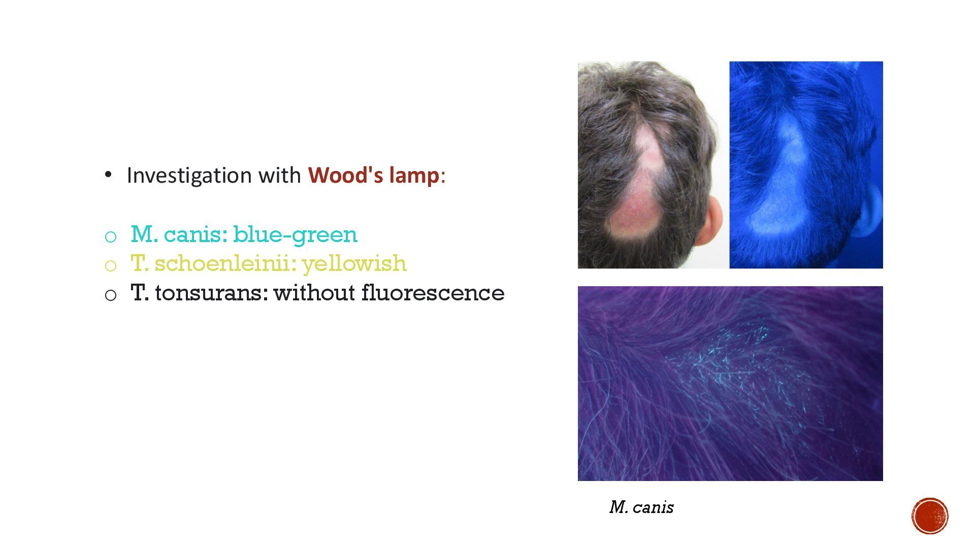
canis; ▪ Central and Southern Europe ▪ Cats, among siblings ▪ Seasonal: July-October ▪ Trichophyton tonsurans: ▪ North America ▪ Close contact with an infected person or subject. ▪ Trichophyton schoenleinii: ▪ - Poor hygienic conditions. ▪ - Tinea favosa. ▪ others: T. violaceum, T. schoenleinii, M. audouinii
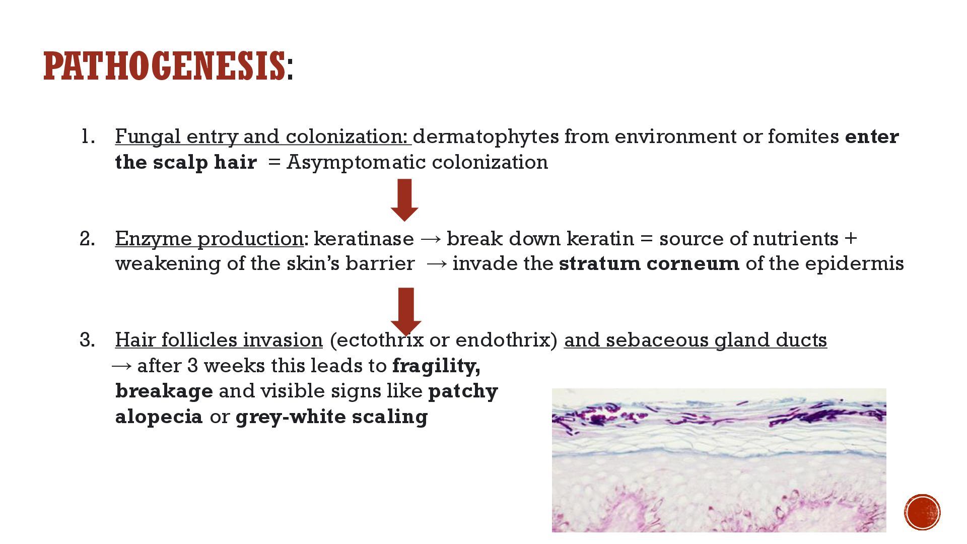
fomites enter the scalp hair = Asymptomatic colonization 2. Enzyme production: keratinase → break down keratin = source of nutrients + weakening of the skin’s barrier → invade the stratum corneum of the epidermis 3. Hair follicles invasion (ectothrix or endothrix) and sebaceous gland ducts → after 3 weeks this leads to fragility, breakage and visible signs like patchy alopecia or grey-white scaling
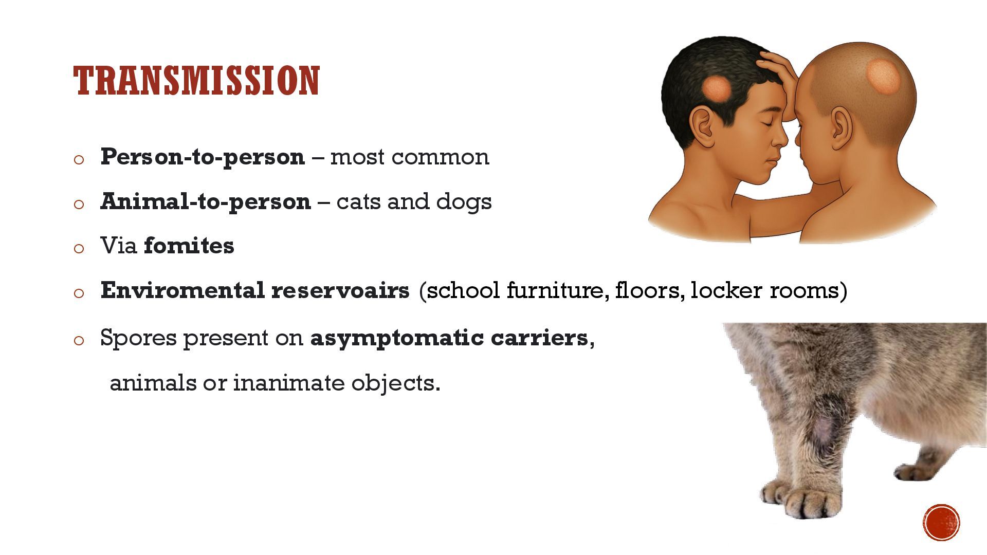
and dogs o Via fomites o Enviromental reservoairs (school furniture, floors, locker rooms) o Spores present on asymptomatic carriers, animals or inanimate objects.
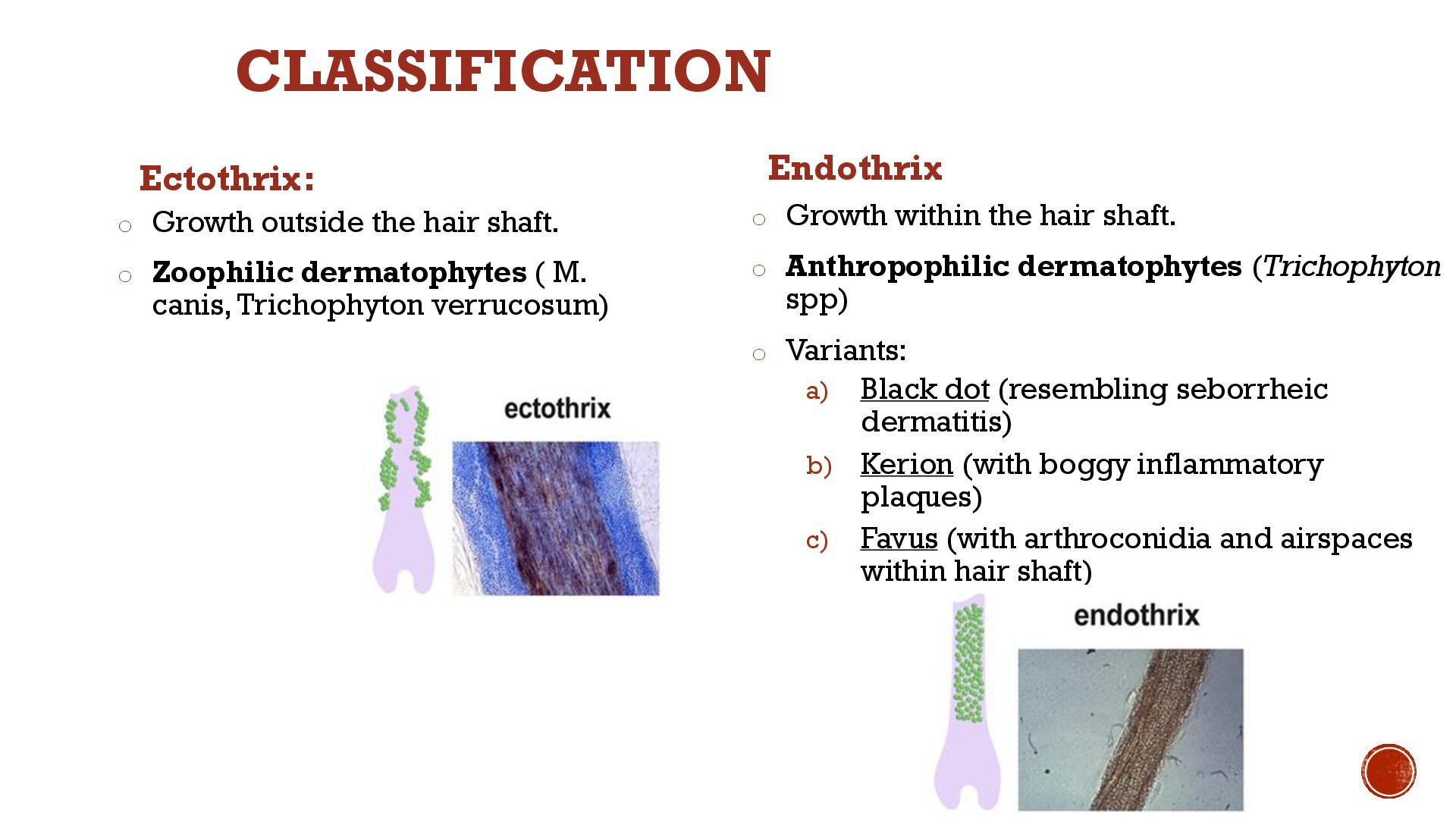
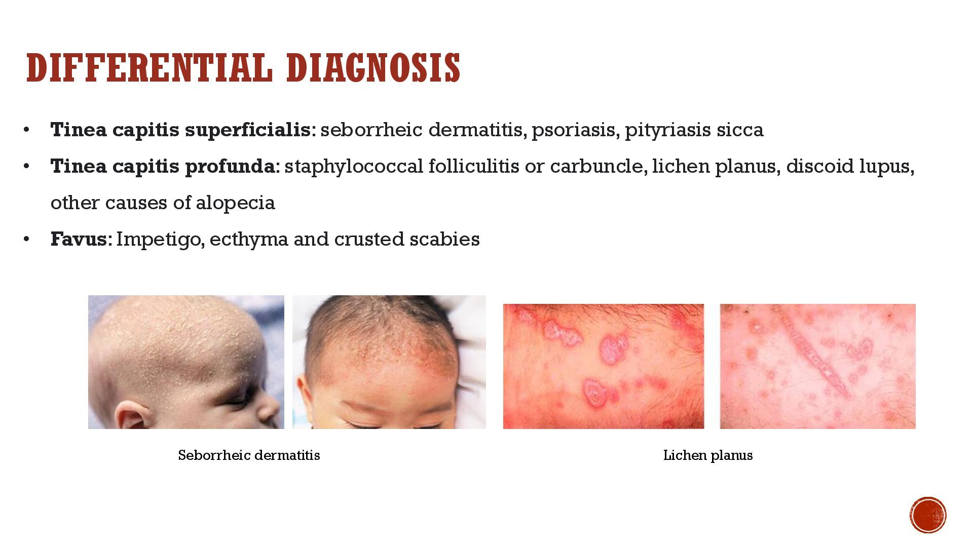
( M. canis, Trichophyton verrucosum) Endothrix o Growth within the hair shaft. o Anthropophilic dermatophytes (Trichophyton spp) o Variants: a) Black dot (resembling seborrheic dermatitis) b) Kerion (with boggy inflammatory plaques) c) Favus (with arthroconidia and airspaces within hair shaft) CLASSIFICATION
(more common in boys) ▪ Densely populated areas, kindergartens, schools ▪ Immunocompromised ▪ Contact with animals (dogs, cats), close contact with peers, through objects (toothbrushes, combs, towels) ▪ Asymptomatic vectors (humans or animals) ▪ Geographical distribution of each fungus ▪ North and Central America: 90% T. tonsurans. Less common: M. canis. ▪ Europe: M. canis, T, violaceum, M. audouinii, ▪ Asia: T. violaceum, M. canis. ▪ Africa: T. violaceium, T. schonleinii, M. canis.
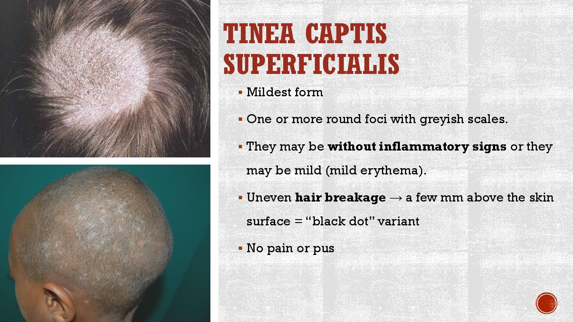
round foci with greyish scales. ▪ They may be without inflammatory signs or they may be mild (mild erythema). ▪ Uneven hair breakage → a few mm above the skin surface = “black dot” variant ▪ No pain or pus
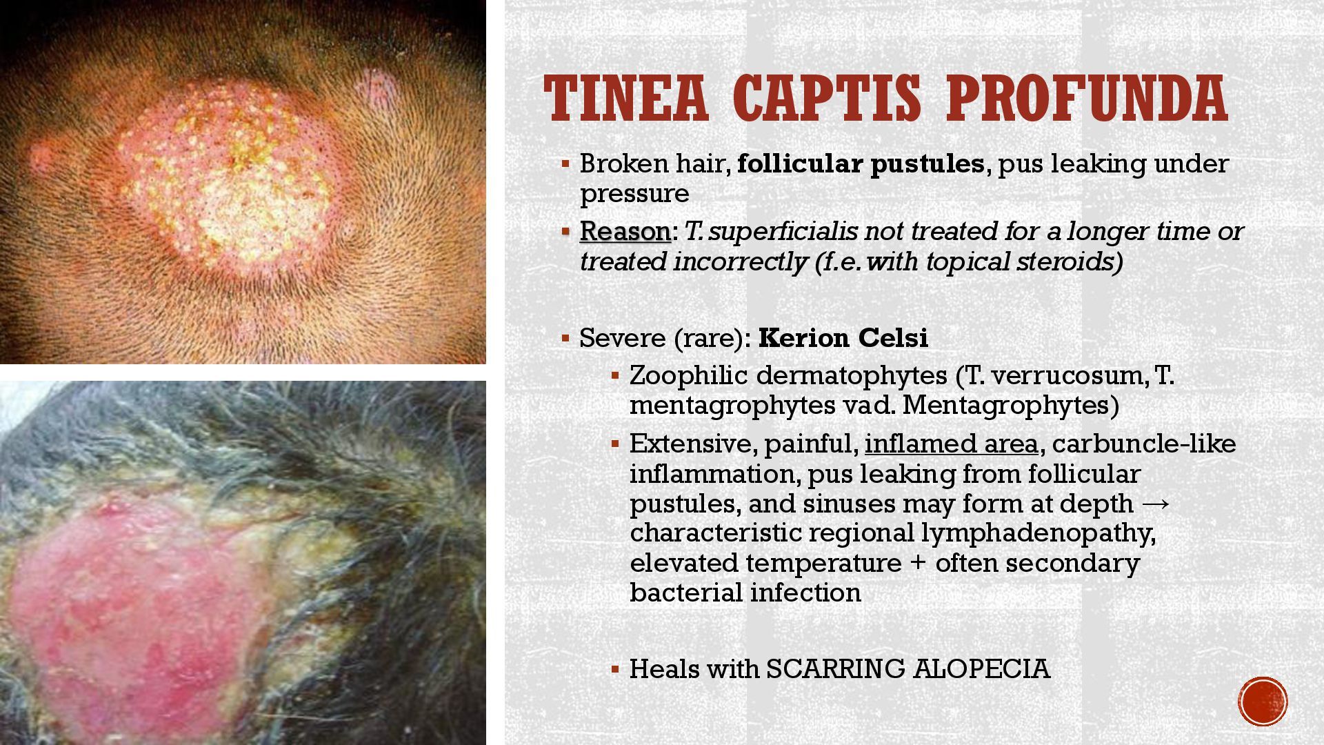
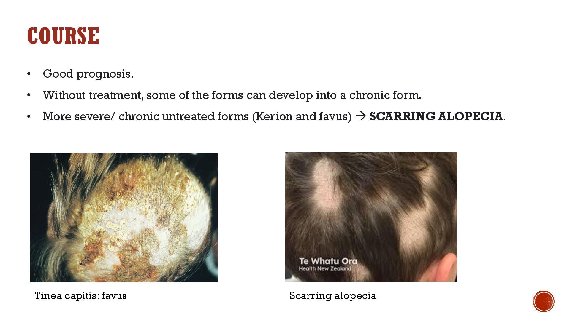
under pressure ▪ Reason: T. superficialis not treated for a longer time or treated incorrectly (f.e. with topical steroids) ▪ Severe (rare): Kerion Celsi ▪ Zoophilic dermatophytes (T. verrucosum, T. mentagrophytes vad. Mentagrophytes) ▪ Extensive, painful, inflamed area, carbuncle-like inflammation, pus leaking from follicular pustules, and sinuses may form at depth → characteristic regional lymphadenopathy, elevated temperature + often secondary bacterial infection ▪ Heals with SCARRING ALOPECIA
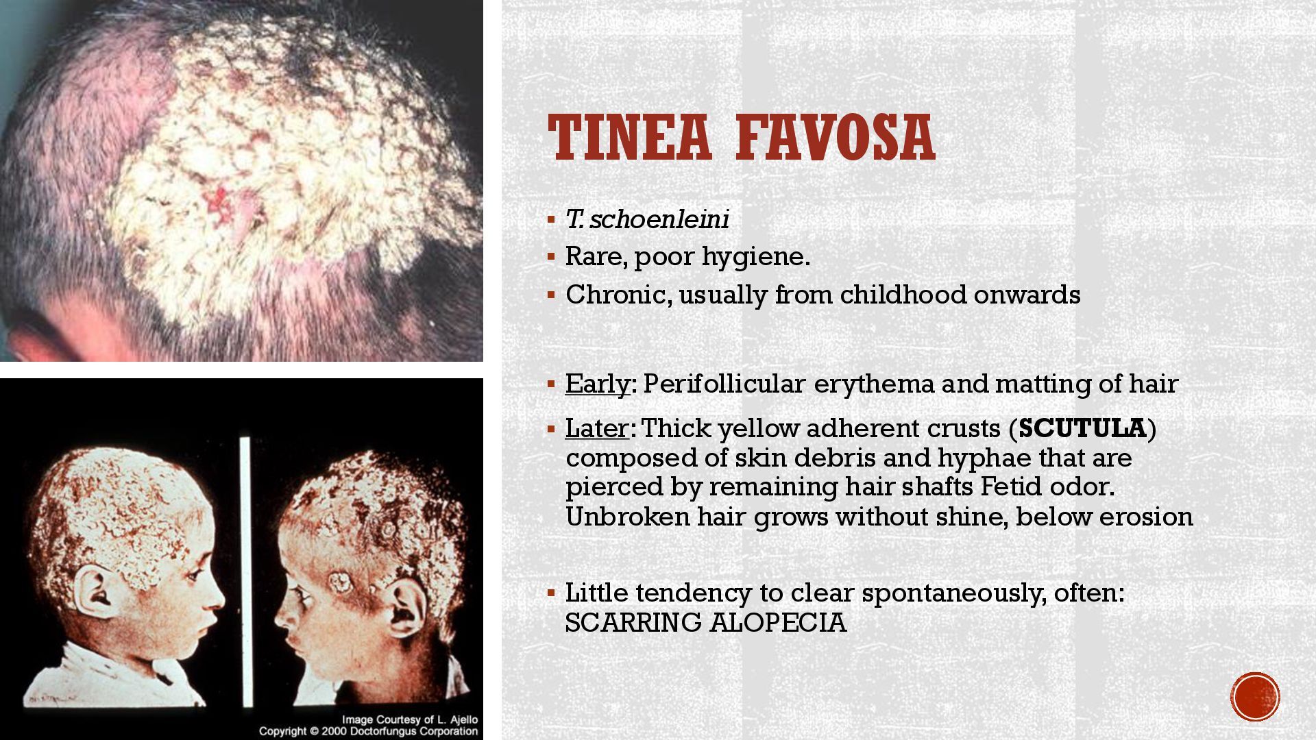
Chronic, usually from childhood onwards ▪ Early: Perifollicular erythema and matting of hair ▪ Later: Thick yellow adherent crusts (SCUTULA) composed of skin debris and hyphae that are pierced by remaining hair shafts Fetid odor. Unbroken hair grows without shine, below erosion ▪ Little tendency to clear spontaneously, often: SCARRING ALOPECIA
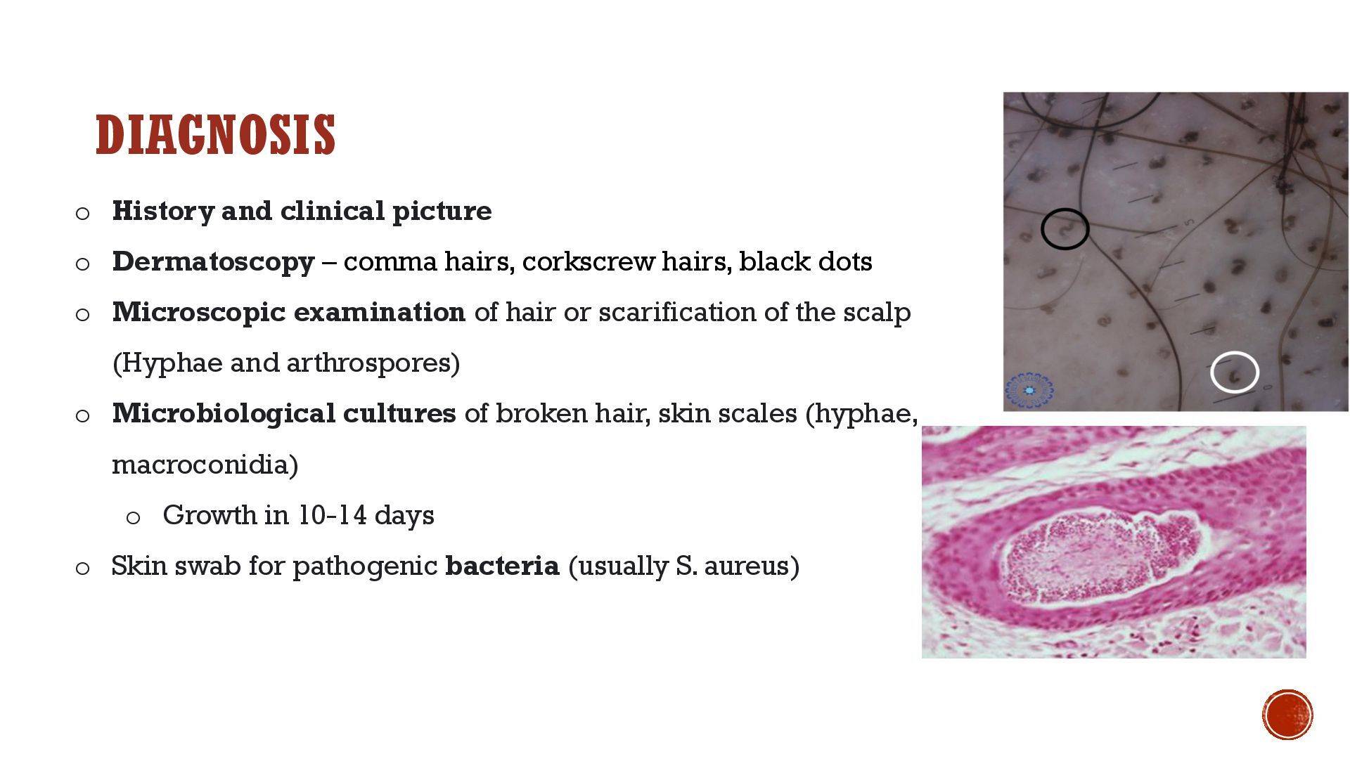
hairs, corkscrew hairs, black dots o Microscopic examination of hair or scarification of the scalp (Hyphae and arthrospores) o Microbiological cultures of broken hair, skin scales (hyphae, macroconidia) o Growth in 10-14 days o Skin swab for pathogenic bacteria (usually S. aureus)
- terbinafine NOT a good option for T. capitis caused by M. canis -> other systemic antimycotic, f.e. itraconazole! • Additionally LOCAL antifungal cream: o terbinafine or azole antifungal • We recommend washing the scalp with an antifungal shampoo (ketoconazole, selenium sulfide) o Reducing the transfer of spores o Cover infected areas – NO transmission • Control mycological results 1x / 4-6 weeks: o 1. negative report → we cancel the SYSTEMIC TH o 2. negative report → cancel LOCAL TH • Systemic antibiotic (if associated bacterial infection)
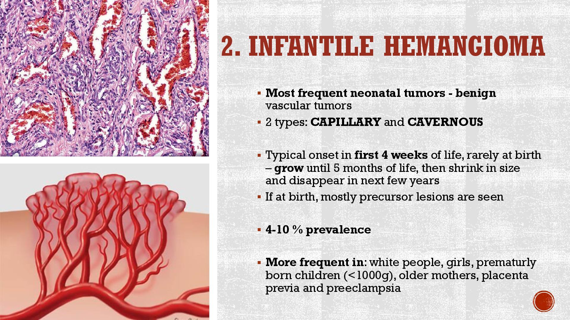
vascular tumors ▪ 2 types: CAPILLARY and CAVERNOUS ▪ Typical onset in first 4 weeks of life, rarely at birth – grow until 5 months of life, then shrink in size and disappear in next few years ▪ If at birth, mostly precursor lesions are seen ▪ 4-10 % prevalence ▪ More frequent in: white people, girls, prematurly born children (<1000g), older mothers, placenta previa and preeclampsia
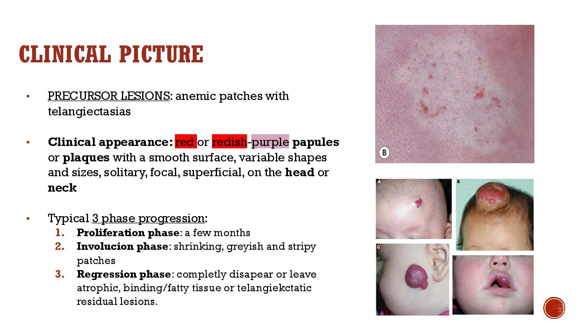
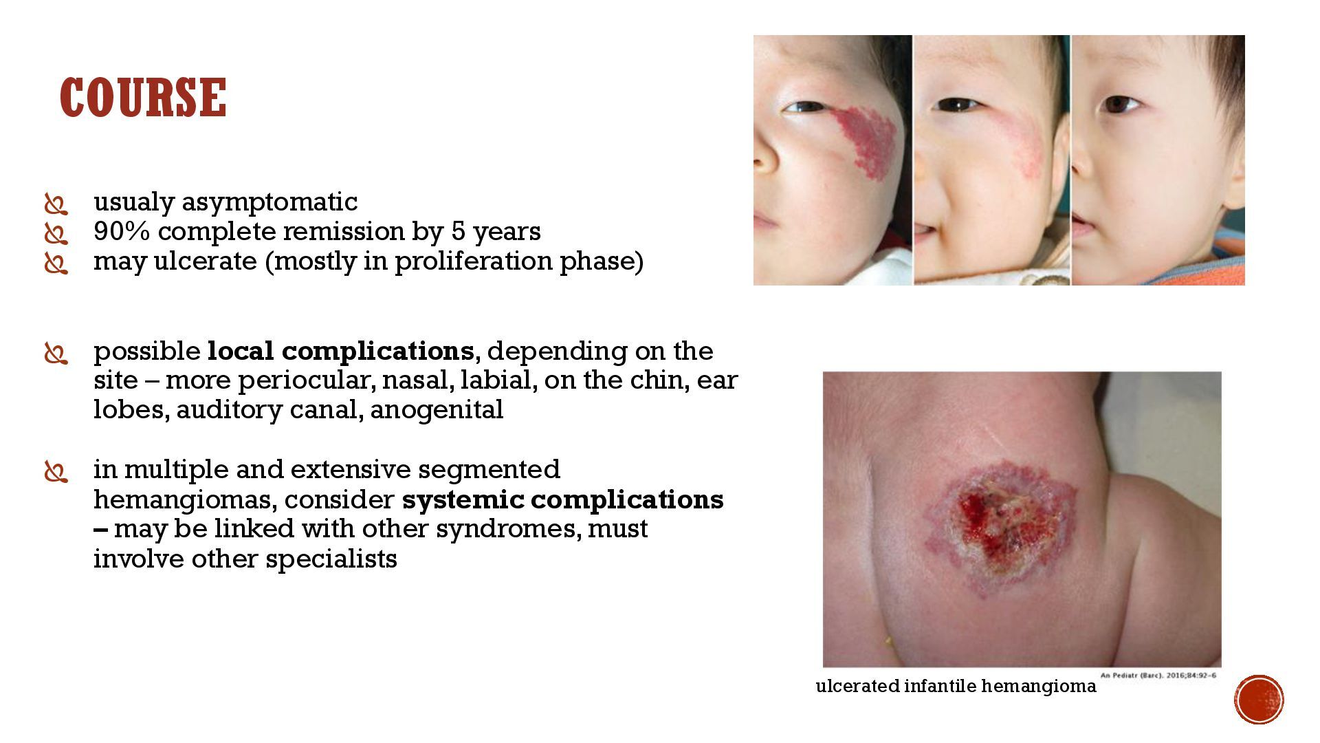
Clinical appearance: red or redish-purple papules or plaques with a smooth surface, variable shapes and sizes, solitary, focal, superficial, on the head or neck • Typical 3 phase progression: 1. Proliferation phase: a few months 2. Involucion phase: shrinking, greyish and stripy patches 3. Regression phase: completly disapear or leave atrophic, binding/fatty tissue or telangiekctatic residual lesions.
years may ulcerate (mostly in proliferation phase) possible local complications, depending on the site – more periocular, nasal, labial, on the chin, ear lobes, auditory canal, anogenital in multiple and extensive segmented hemangiomas, consider systemic complications – may be linked with other syndromes, must involve other specialists ulcerated infantile hemangioma
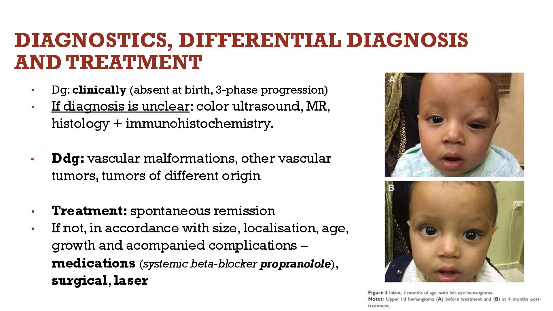
birth, 3-phase progression) • If diagnosis is unclear: color ultrasound, MR, histology + immunohistochemistry. • Ddg: vascular malformations, other vascular tumors, tumors of different origin • Treatment: spontaneous remission • If not, in accordance with size, localisation, age, growth and acompanied complications – medications (systemic beta-blocker propranolole), surgical, laser
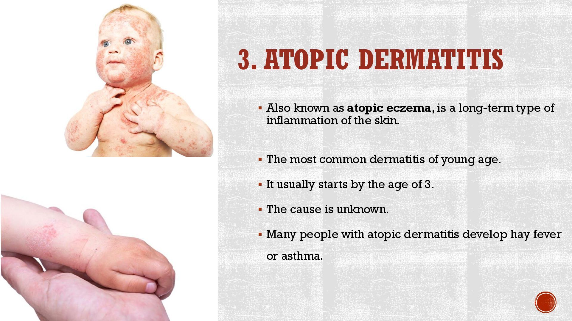
a long-term type of inflammation of the skin. ▪ The most common dermatitis of young age. ▪ It usually starts by the age of 3. ▪ The cause is unknown. ▪ Many people with atopic dermatitis develop hay fever or asthma.
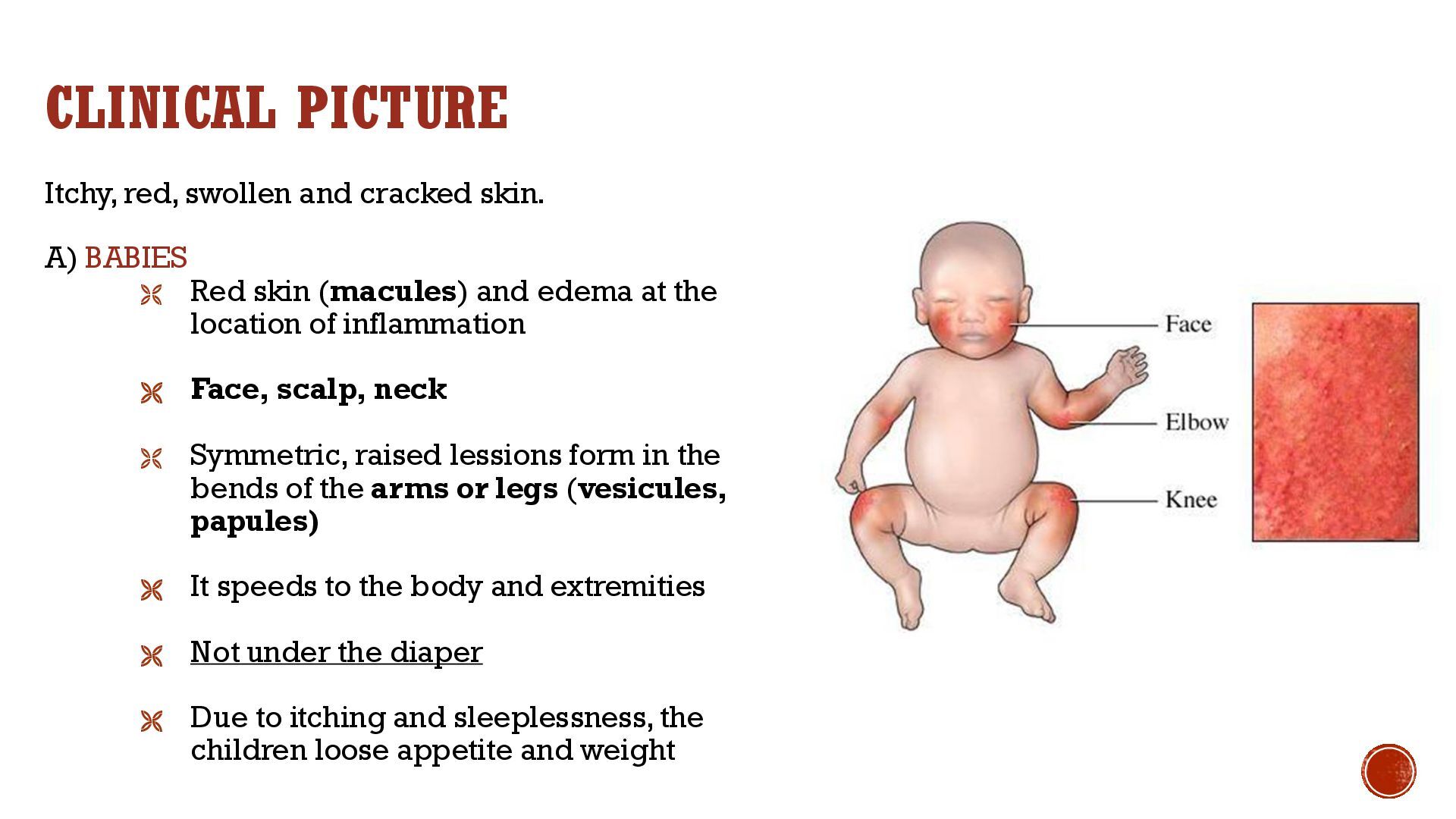
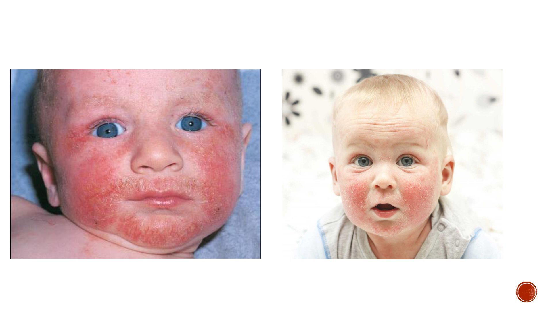
Red skin (macules) and edema at the location of inflammation Face, scalp, neck Symmetric, raised lessions form in the bends of the arms or legs (vesicules, papules) It speeds to the body and extremities Not under the diaper Due to itching and sleeplessness, the children loose appetite and weight
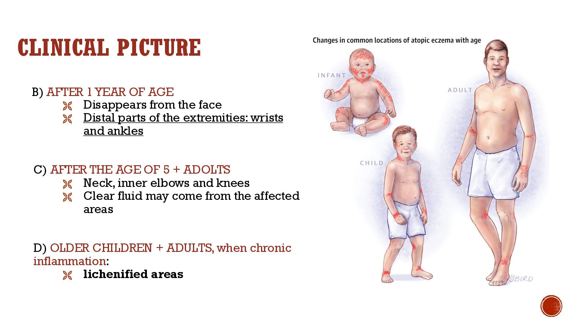
from the face Distal parts of the extremities: wrists and ankles C) AFTER THE AGE OF 5 + ADOLTS Neck, inner elbows and knees Clear fluid may come from the affected areas D) OLDER CHILDREN + ADULTS, when chronic inflammation: lichenified areas
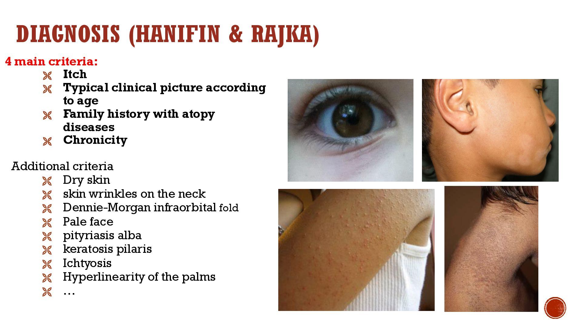
Typical clinical picture according to age Family history with atopy diseases Chronicity Additional criteria Dry skin skin wrinkles on the neck Dennie-Morgan infraorbital fold Pale face pityriasis alba keratosis pilaris Ichtyosis Hyperlinearity of the palms …


 codingconduct
 aleyda
 sarafernandez
 tmiket
 jonoalderson
 techseoconnect
 speakerdeck
 akademiya2063
 trishagee
 katarinadahlin
 geoffreycrofte