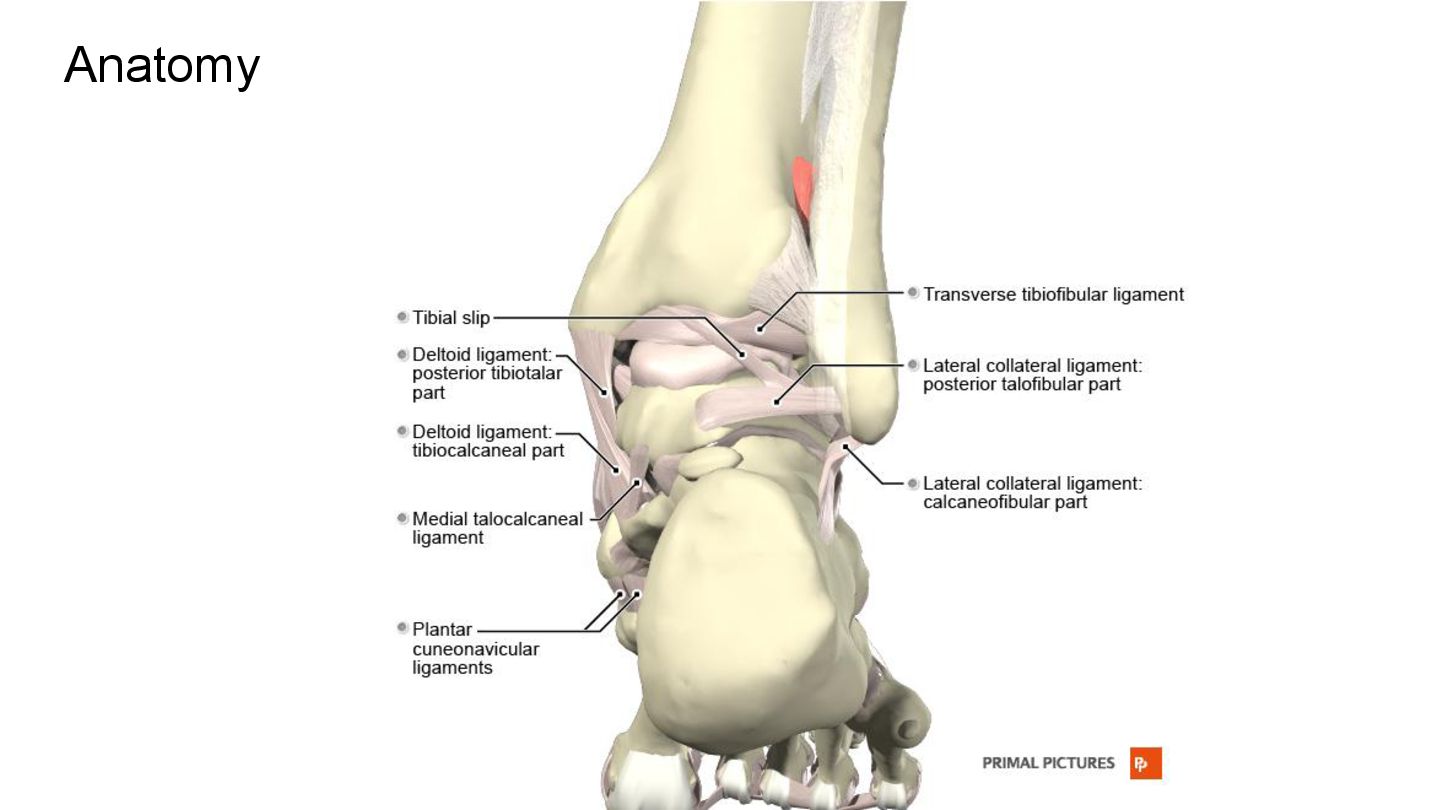
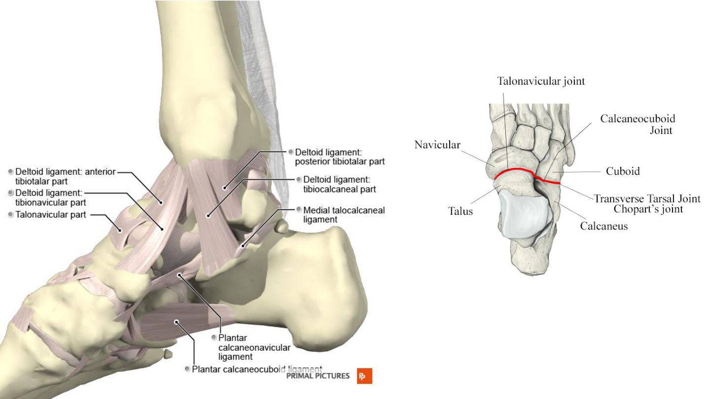
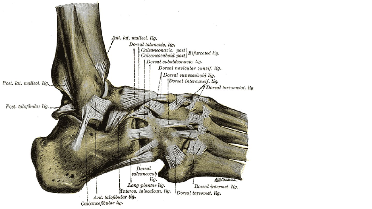
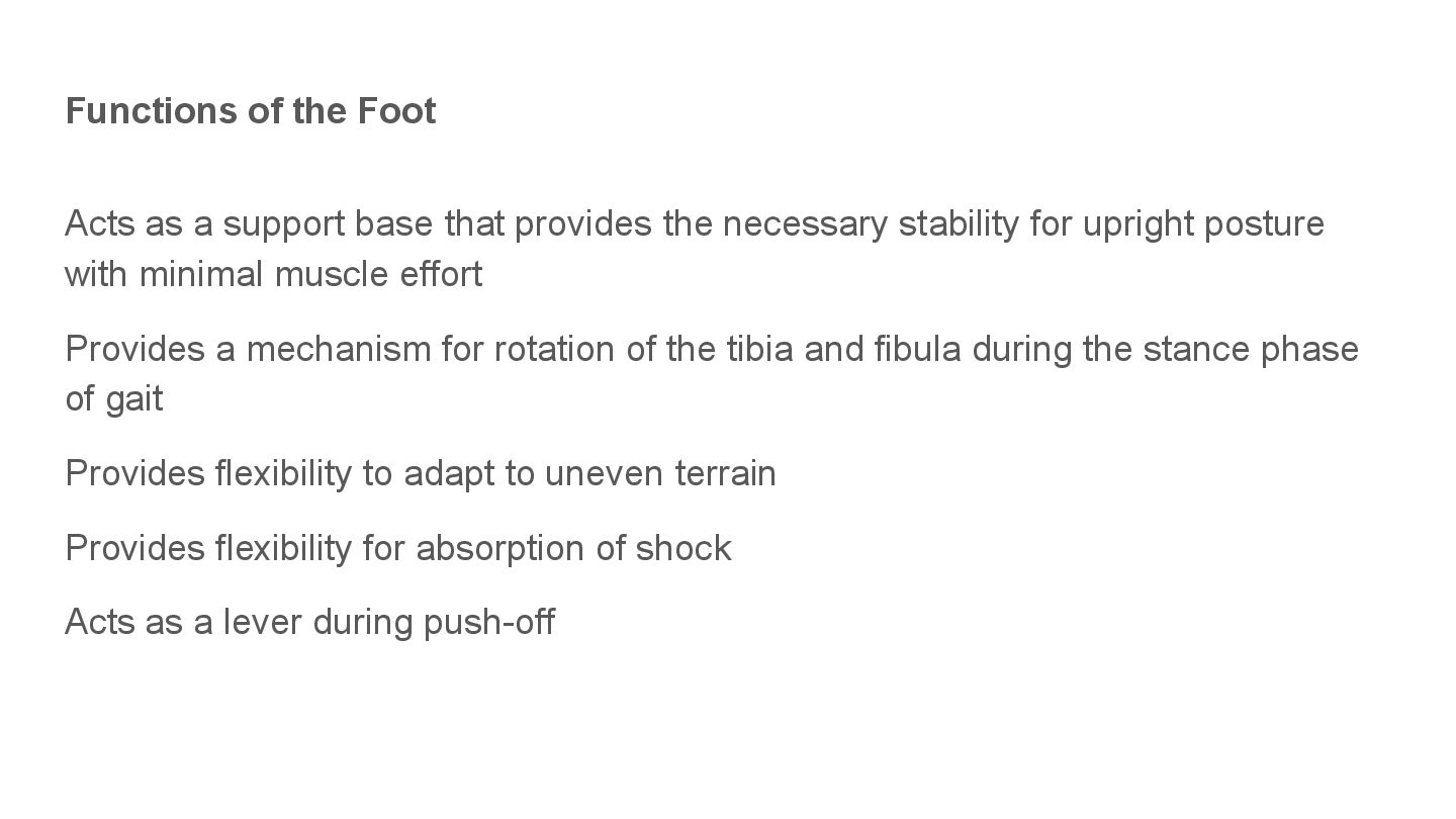
provides the necessary stability for upright posture with minimal muscle effort Provides a mechanism for rotation of the tibia and fibula during the stance phase of gait Provides flexibility to adapt to uneven terrain Provides flexibility for absorption of shock Acts as a lever during push-off
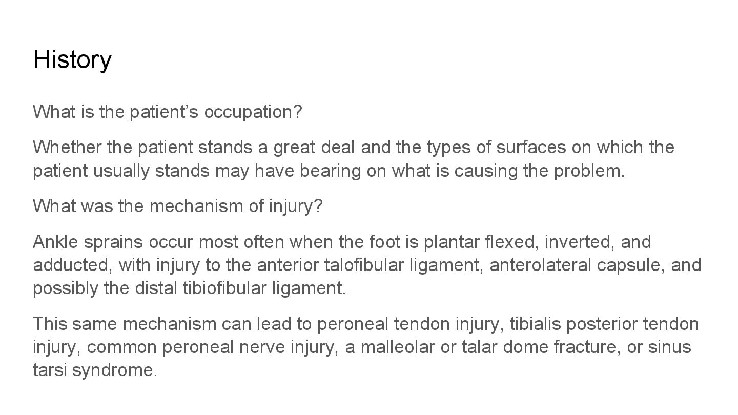
a great deal and the types of surfaces on which the patient usually stands may have bearing on what is causing the problem. What was the mechanism of injury? Ankle sprains occur most often when the foot is plantar flexed, inverted, and adducted, with injury to the anterior talofibular ligament, anterolateral capsule, and possibly the distal tibiofibular ligament. This same mechanism can lead to peroneal tendon injury, tibialis posterior tendon injury, common peroneal nerve injury, a malleolar or talar dome fracture, or sinus tarsi syndrome.
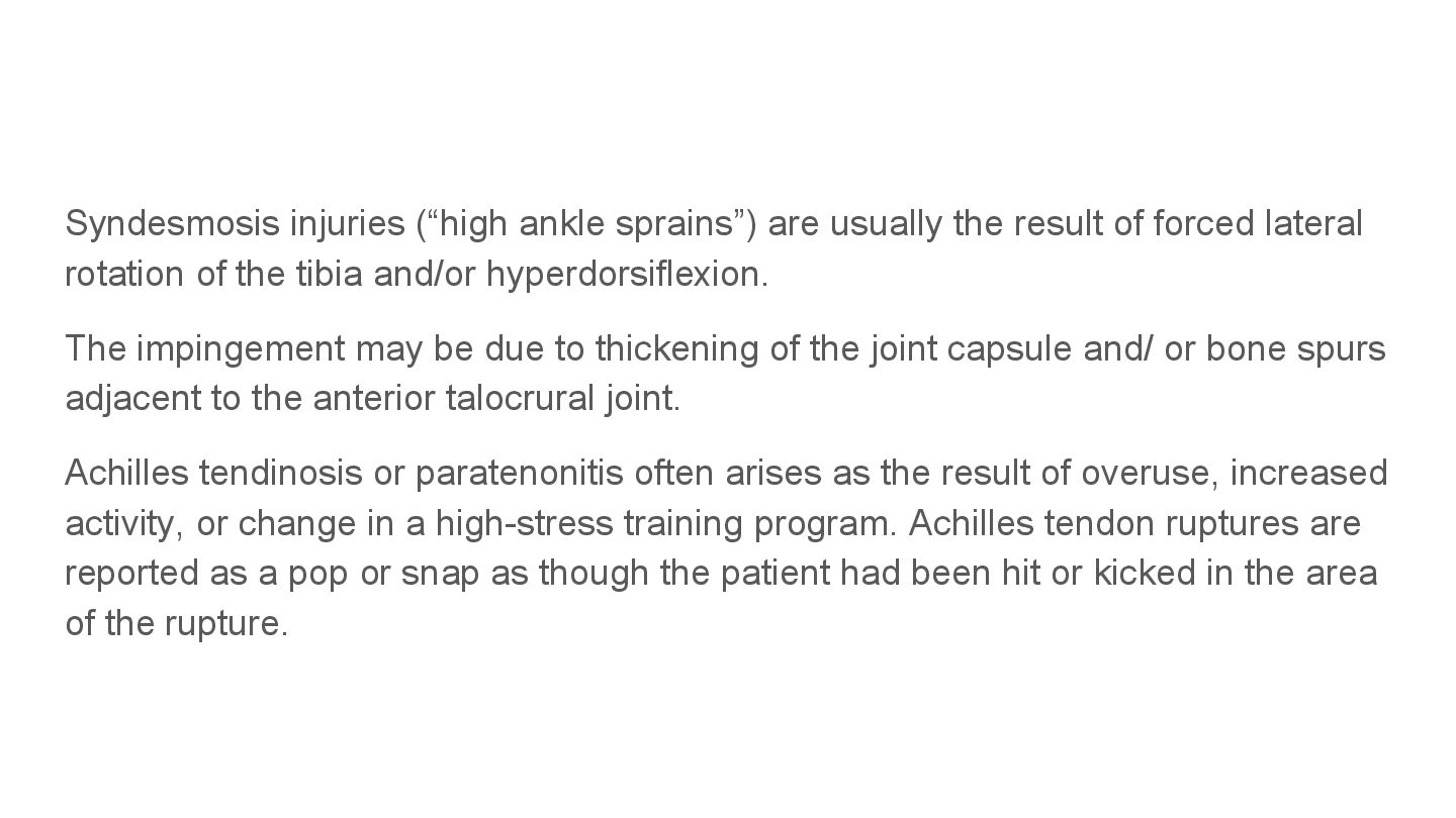
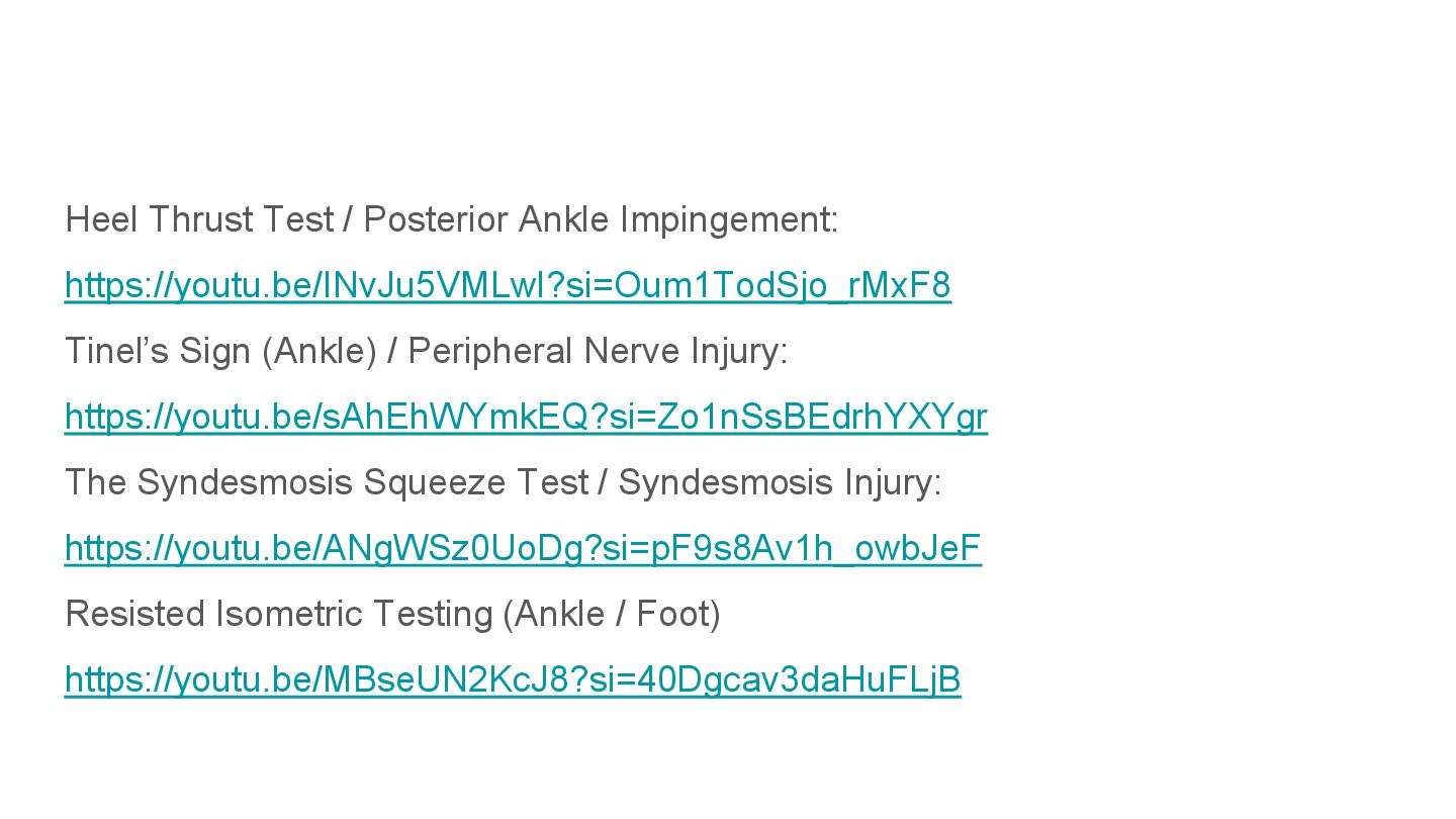
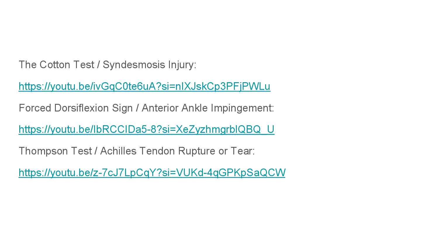
forced lateral rotation of the tibia and/or hyperdorsiflexion. The impingement may be due to thickening of the joint capsule and/ or bone spurs adjacent to the anterior talocrural joint. Achilles tendinosis or paratenonitis often arises as the result of overuse, increased activity, or change in a high-stress training program. Achilles tendon ruptures are reported as a pop or snap as though the patient had been hit or kicked in the area of the rupture.
the lateral aspect that rapidly diminishes, may indicate a tear of the peroneal retinaculum. Individuals such as dancers, soccer players, and track and field athletes may have posterior ankle impingement because of excessive repetitive plantar flexion of the foot. Did the patient notice a transient or fixed deformity of the foot or ankle at the time of injury? Was there any transitory locking (e.g., loose body, muscle spasm)? Positive answer may indicate a fracture causing immediate swelling that decreased as it spread into the surrounding tissue.
injury? If so, the injury is probably not too severe, provided there is no loss of stability. Inability to bear weight, severe pain, and rapid swelling indicate a severe injury. Walking is compatible with a second degree sprain; pain with running usually indicates a first-degree injury. Was there any swelling or bruising (ecchymosis)? How quickly and where did it develop? This question can elicit some idea of the type of swelling (e.g., blood, synovial, purulent) and whether it is intracapsular or extracapsular. Swelling over the extensor tendons of the foot caused by irritation from doing up (i.e., lacing up) stiff ice skates too tight.
recurrent ankle instability will be indicated by one or more significant lateral ankle sprains involving functional and mechanical instability, increased subtalar laxity and greater anterior laxity, or history of giving way usually during initial contact when walking, running, cutting, or rapidly decelerating in the last 6 months. What is the patient’s usual activity or pastime? Answers to this question should give some idea of the stresses placed on the lower leg, ankle, and foot; how frequently they are applied; and whether the patient is suffering from a repetitive stress injury. For example, tarsal navicular stress fractures may be seen in runners resulting in dorsal midfoot pain radiating into the medial arch with little swelling.
For example, with overuse injuries, pain initially comes on after the activity. As the injury progresses, pain or soreness is present at the beginning of the activity, and then it goes away during the activity only to return afterward. In later stages of the problem, the pain is constantly present. Pain during the activity suggests stress on the injured structure.
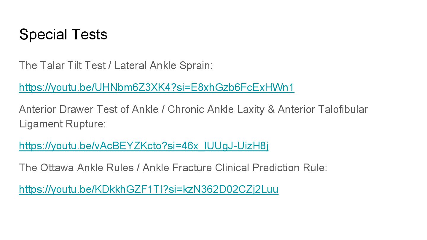
location or area? For example, with shin splints (medial tibial stress syndrome [MTSS]) or a compartment syndrome (acute or chronic exertional type), the patient usually indicates a diffuse area. With a stress fracture, the area of pain tends to be more specific. Anterolateral ankle impingement demonstrates anterolateral ankle joint tenderness, anterolateral ankle joint swelling (extracapsular), pain with force dorsiflexion and eversion, pain with single leg squat, pain with activities, and possible absence of ankle instability. Peroneal tendon problems show posterolateral pain and may be associated with lateral ankle instability. Plantar fasciitis is the most common cause of heel pain on the anteromedial aspect of the heel, it may be accompanied by a heel spur, which is the result of the plantar fasciitis.
of heel do the shoes have? Are the shoes in good condition? Does the patient make use of orthoses? If so, are they still functional? Poor footwear may contribute to patient pain and subsequent impairment. Is there a history of previous injury, affliction, or surgery? For example, poliomyelitis may lead to a pes cavus. Systemic conditions, such as diabetes, gout, psoriasis, and collagen diseases, may manifest themselves first in the foot. If there was previous surgery, did the pain resolve following surgery? Is the pain the same as before surgery? Is it new pain?
six symptoms must be positive. Anterolateral ankle joint tenderness Anterolateral ankle joint swelling Pain on forced dorsiflexion Pain on affected side with single leg squat Pain with activities Absence of ankle instability
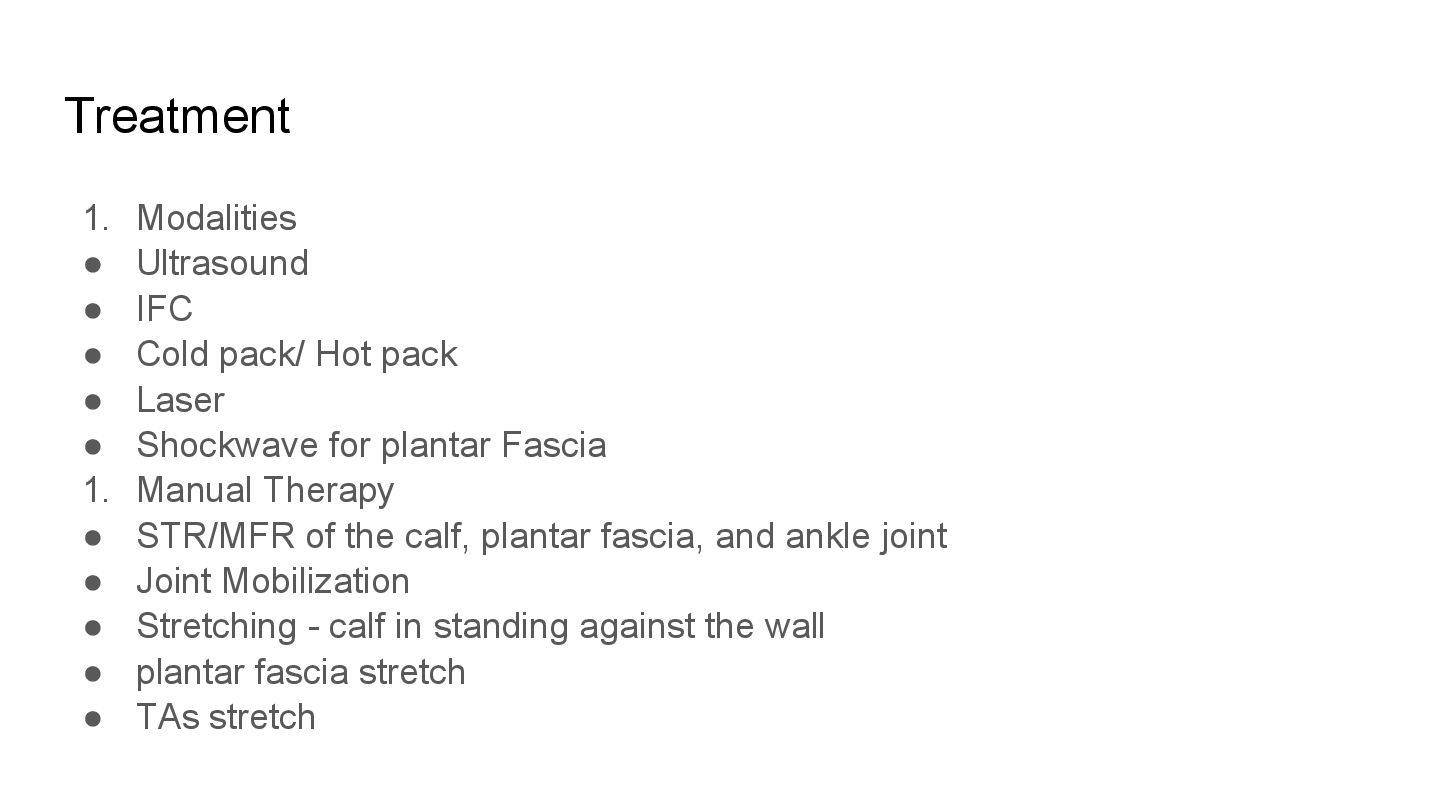
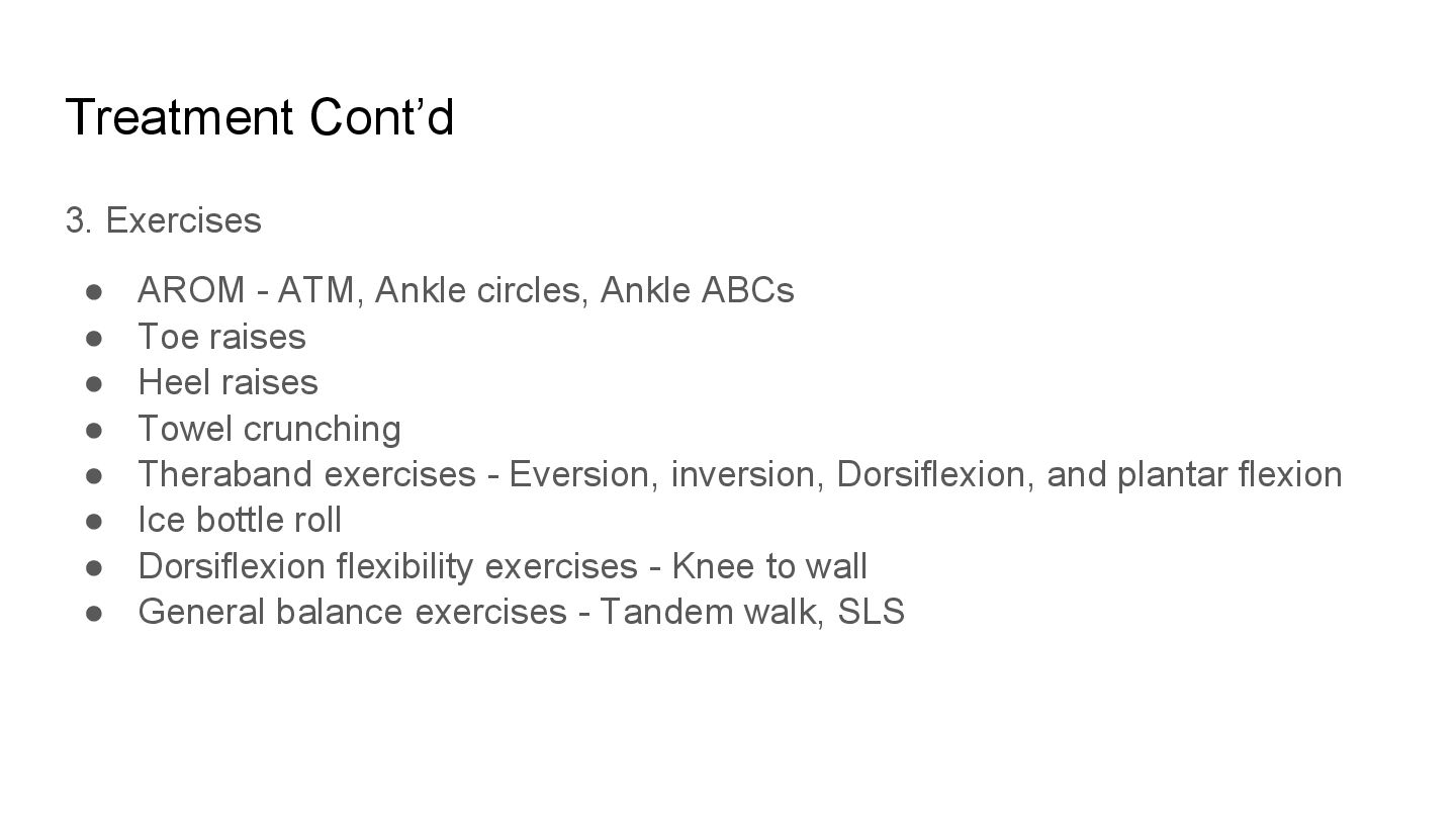
Hot pack • Laser • Shockwave for plantar Fascia 1. Manual Therapy • STR/MFR of the calf, plantar fascia, and ankle joint • Joint Mobilization • Stretching - calf in standing against the wall • plantar fascia stretch • TAs stretch


 emna__ayadi
 davetheseo
 cassininazir
 samanthasiow
 jcasabona
 garrettdimon
 mlcsv
 szymonslowik
 jacobian
 sabderemane
 uxyall
 palkan