Beta, and Gamma Oscillations N. H.L. Lam1, J.-M. Schoffelen2, J. Udden1, A. Hulten1, P. Hagoort2 PAPER INTRODUCTION Introduced by Hayato Maki (NAIST, AHC-LAB) 2017/01/26 1 Aalto University, the department of biomedical engineering, Finland 2 Donders Center for Cognitive Neuroimaging, Netherlands
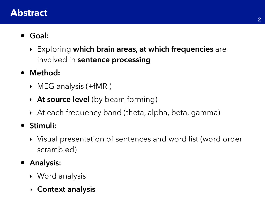
are involved in sentence processing • Method: ‣ MEG analysis (+fMRI) ‣ At source level (by beam forming) ‣ At each frequency band (theta, alpha, beta, gamma) • Stimuli: ‣ Visual presentation of sentences and word list (word order scrambled) • Analysis: ‣ Word analysis ‣ Context analysis Abstract 2
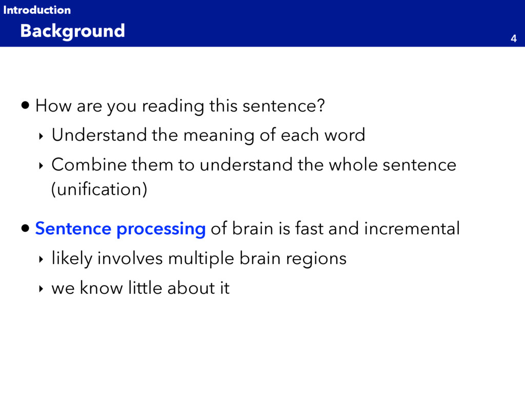
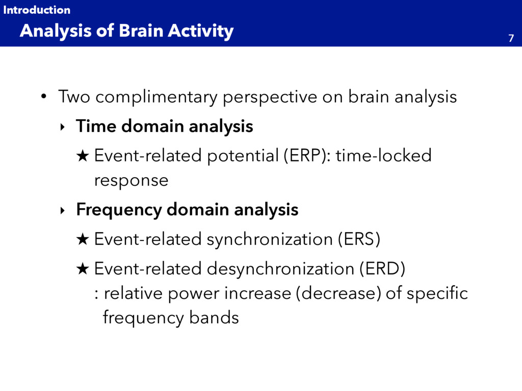
meaning of each word ‣ Combine them to understand the whole sentence (unification) • Sentence processing of brain is fast and incremental ‣ likely involves multiple brain regions ‣ we know little about it Background 4 Introduction
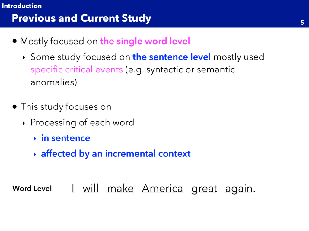
study focused on the sentence level mostly used specific critical events (e.g. syntactic or semantic anomalies) • This study focuses on ‣ Processing of each word ‣ in sentence ‣ affected by an incremental context Previous and Current Study 5 Introduction I will make America great again. Word Level
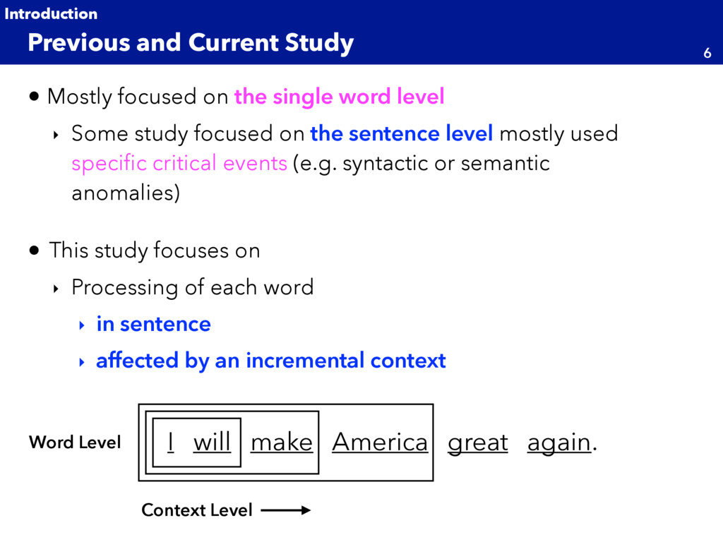
study focused on the sentence level mostly used specific critical events (e.g. syntactic or semantic anomalies) • This study focuses on ‣ Processing of each word ‣ in sentence ‣ affected by an incremental context Previous and Current Study 6 Introduction I will make America great again. Word Level Context Level
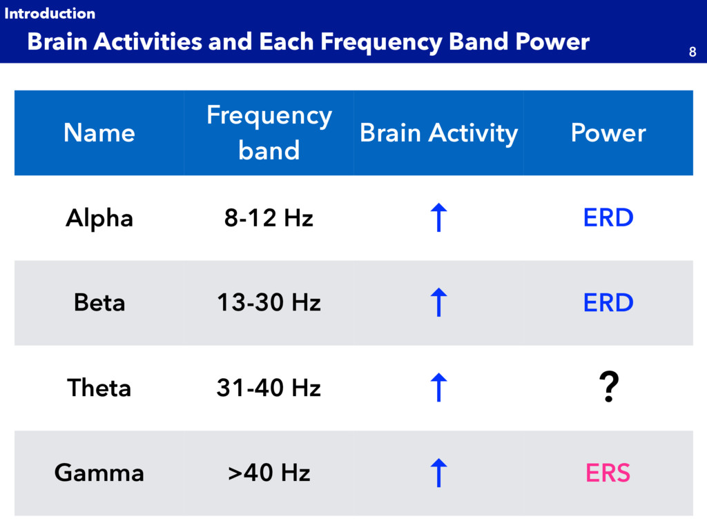
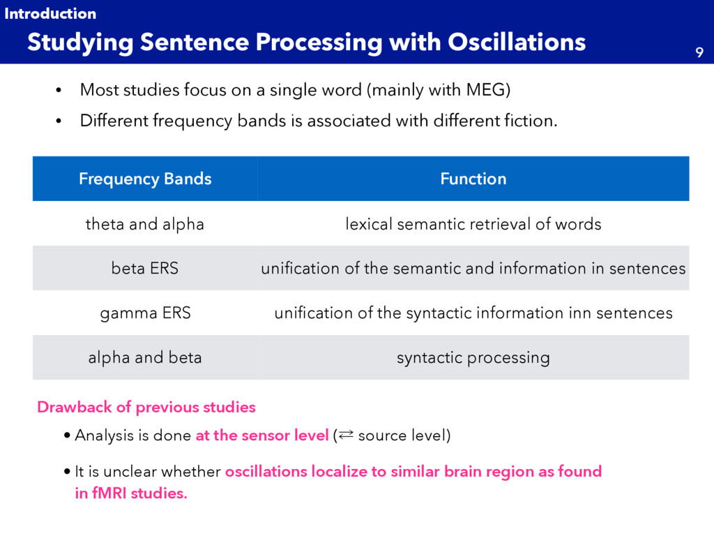
MEG) • Different frequency bands is associated with different fiction. Studying Sentence Processing with Oscillations 9 Introduction Frequency Bands Function theta and alpha lexical semantic retrieval of words beta ERS unification of the semantic and information in sentences gamma ERS unification of the syntactic information inn sentences alpha and beta syntactic processing Drawback of previous studies • Analysis is done at the sensor level (⁶ source level) • It is unclear whether oscillations localize to similar brain region as found in fMRI studies.
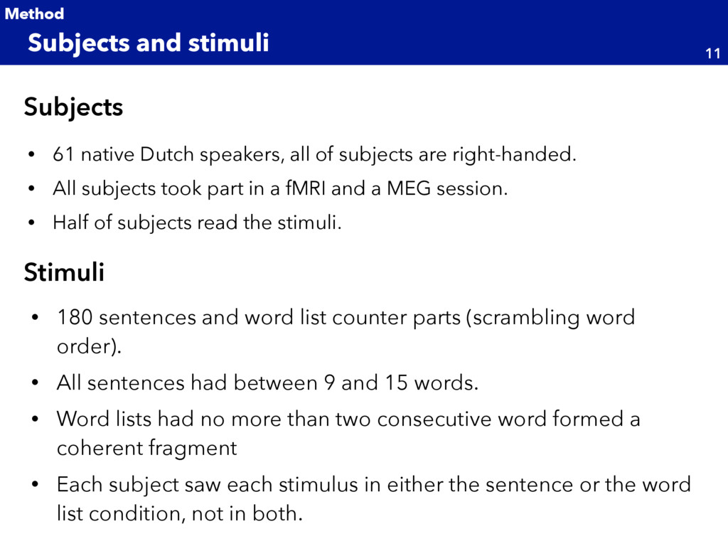
• All subjects took part in a fMRI and a MEG session. • Half of subjects read the stimuli. Subjects and stimuli 11 Method • 180 sentences and word list counter parts (scrambling word order). • All sentences had between 9 and 15 words. • Word lists had no more than two consecutive word formed a coherent fragment • Each subject saw each stimulus in either the sentence or the word list condition, not in both. Subjects Stimuli
announcement (1500ms) Block type announcement (1500ms) • Sentence block or word list block • Each block has 5 sentence (or word list) • 24 block in total ˒ Mini Block Design ˒ In each block Fixation cross (1200-2200ms) Word Presentation (300-1344ms) + This Blank Screen (300ms) ɾɾɾ Yes/No question (after 10% of trials) comprehension Q (Sentence) word content Q (Sent. and WrdL.st)
frequency and down sampled to 300 Hz • 300 Hz low pass applied • EMG and ECG also recorded Data Acquisition 13 Method • ECG artifacts were estimated using denoising source separation (DSS) [Sarela and Valpola, 2005]. • Other artifacts were estimated using field trip toolbox. • Data segments including an artifact were excluded from analysis. • Power line inference of 50 Hz and its harmonics were estimated and subtracted [Schoffelen, 2005]. MEG Preprocess
frequency and down sampled to 300 Hz • 300 Hz low pass applied • EMG and ECG also recorded Data Acquisition 14 Method • ECG artifacts were estimated using denoising source separation (DSS) [Sarela and Valpola, 2005]. • Other artifacts were estimated using field trip toolbox. • Data segments including an artifact were excluded from analysis. • Power line inference of 50 Hz and its harmonics were estimated and subtracted [Schoffelen, 2005]. MEG Preprocess
Localization of oscillatory brain activity Source Localization 15 Method • For source localization, anatomical MRI and MEG were co- registered. Frequency-domain beam forming fMRI data
Localization of oscillatory brain activity Statical Test 16 Method • For source localization, anatomical MRI and MEG were co- registered. Frequency-domain beam forming fMRI data
• 3 time window (centered at 250ms, 350ms, 450ms after word onset) • For each of 3 time window and 5 frequency bands, a separate statical test was done. Word Analysis and Context Analysis 17 Method Word Analysis Context Analysis • In this analysis, we’re interested in the change in neural activity as the sentence unfold. • Comparison of neural responses to late v.s. early words in sentence or word list. • Early words are 2nd, 3rd, and 4th words in sentence (world list). • Late words are 2nd, 3rd, and 4th words from the last word in sentence (world list). (to be continued)
• 3 time window (centered at 250ms, 350ms, 450ms after word onset) • For each of 3 time window and 5 frequency bands, a separate statical test was done. Context Analysis 18 Method Word Analysis Context Analysis • Interested in the change in neural activity as the sentence unfold. • Comparison of neural responses to late v.s. early words in sentence or word list. • Early words are 2nd, 3rd, and 4th words in sentence (world list). • Late words are 2nd, 3rd, and 4th words from the last word in sentence (world list). (to be continued)
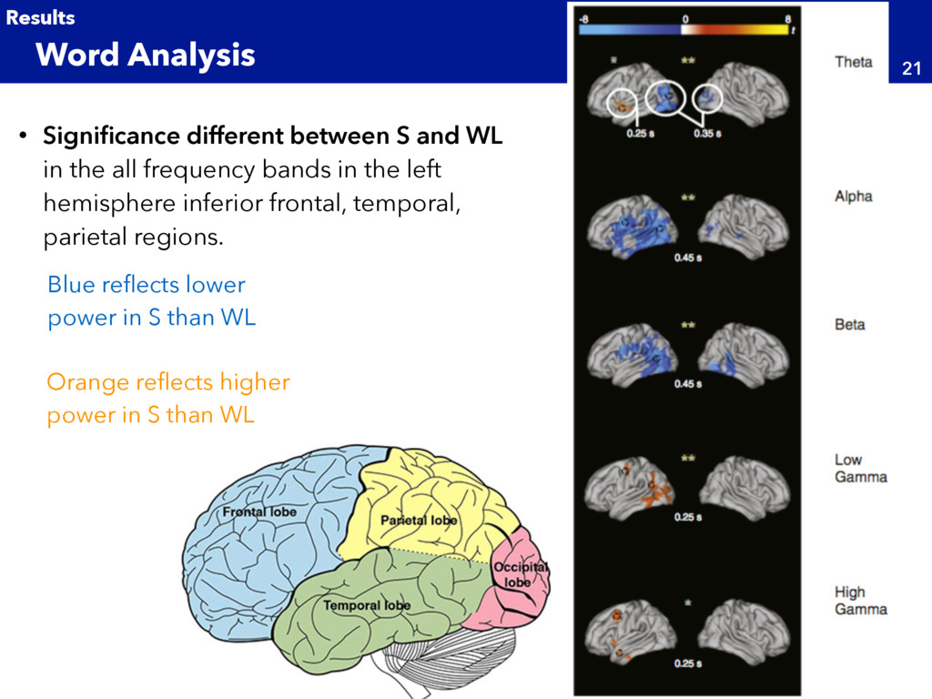
frequency bands in the left hemisphere inferior frontal, temporal, parietal regions. Word Analysis 21 Results Blue reflects lower power in S than WL Orange reflects higher power in S than WL
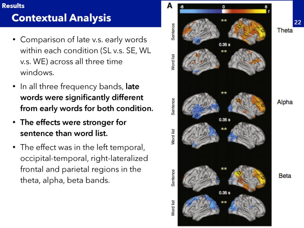
(SL v.s. SE, WL v.s. WE) across all three time windows. • In all three frequency bands, late words were significantly different from early words for both condition. • The effects were stronger for sentence than word list. • The effect was in the left temporal, occipital-temporal, right-lateralized frontal and parietal regions in the theta, alpha, beta bands. Contextual Analysis 22 Results
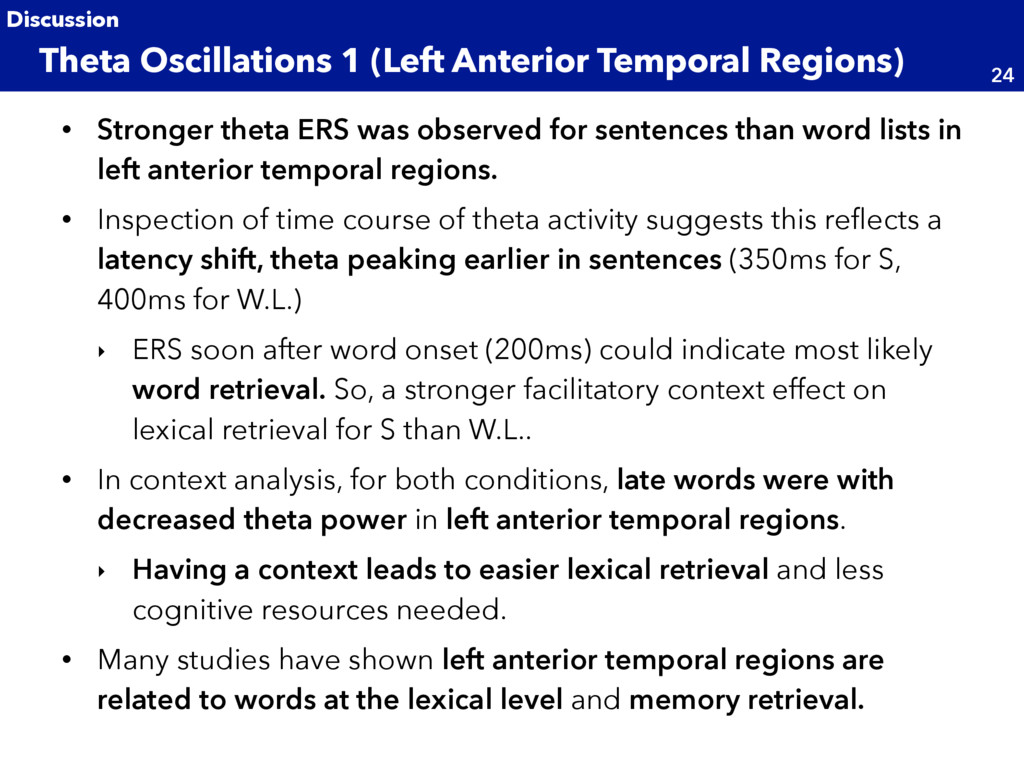
lists in left anterior temporal regions. • Inspection of time course of theta activity suggests this reflects a latency shift, theta peaking earlier in sentences (350ms for S, 400ms for W.L.) ‣ ERS soon after word onset (200ms) could indicate most likely word retrieval. So, a stronger facilitatory context effect on lexical retrieval for S than W.L.. • In context analysis, for both conditions, late words were with decreased theta power in left anterior temporal regions. ‣ Having a context leads to easier lexical retrieval and less cognitive resources needed. • Many studies have shown left anterior temporal regions are related to words at the lexical level and memory retrieval. Theta Oscillations 1 (Left Anterior Temporal Regions) 24 Discussion
are observed in context analysis. • Theta power was significantly higher for lated words than early ones in sentences. • Late words are likely to increase task demand in memory. • Previous studies have showed increase of theta ERS in frontal and parietal sensors has been observed with an increase in working memory load. Theta Oscillations 2 (Bilateral Frontal and Right Parietal Regions) 25 Discussion
in the alpha and beta bands. • Stronger ERD in left-lateralized temporal, parietal, and frontal areas for words in S than W.L.. • ERD in alpha/beta bands is typically interpreted to reflect more activation of underlying population. • This effect could reflect unification of the incoming word in sentences. ‣ A recent study showed ERD between 8-30 Hz in bilateral occipital and parietal regions and left posterior temporal regions, following a semantic or syntactic violation. Alpha and Beta oscillations (Word Analysis) 26 Discussion
late words than early word in left frontal, temporal, and bilateral occipital regions. • This effect could reflect a context facilitatory effect (MUC model) Alpha and Beta oscillations (Context Analysis) 27 Discussion
list was observed at low and high gamma bands. • Low gamma effect in left occipital, parietal, motor, temporal regions. • High gamma effect in left frontal and temporal regions Gamma oscillations 28 Discussion
the left hemisphere. • Low-level (orthographic and word sound analysis) and non-language-specific components activated bilateral fronts-parietal regions. Left and Right Hemisphere 29 Discussion
response to sentence processing at each frequency band (alpha, beta, theta, gamma) • Sentence processing is related to wide area of brain and all frequency bands are involved. • Especially, left temporal and frontal regions are important. Conclusion 30 Discussion
![• Dynamic imaging of coherent sources (DICS) [Gross, 2001] •](https://files.speakerdeck.com/presentations/598d1ad00d5e4a5b9d5aab147ca2c5bf/slide_14.jpg)
![• Dynamic imaging of coherent sources (DICS) [Gross, 2001] •](https://files.speakerdeck.com/presentations/598d1ad00d5e4a5b9d5aab147ca2c5bf/slide_15.jpg)