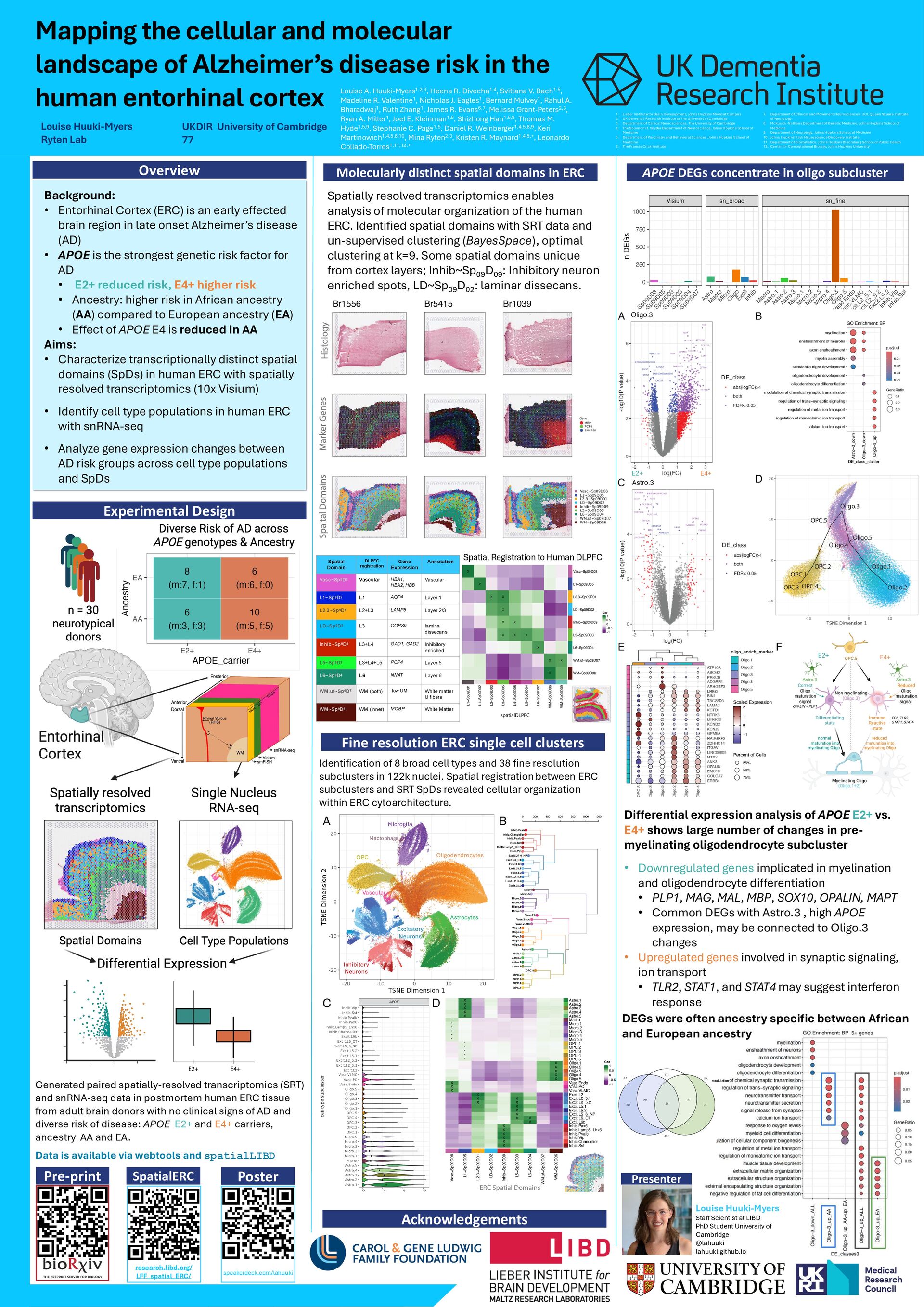
in the human entorhinal cortex Louise Huuki-Myers Ryten Lab UKDIR University of Cambridge 77 Fine resolution ERC single cell clusters APOE DEGs concentrate in oligo subcluster Identification of 8 broad cell types and 38 fine resolution subclusters in 122k nuclei. Spatial registration between ERC subclusters and SRT SpDs revealed cellular organization within ERC cytoarchitecture. Differential expression analysis of APOE E2+ vs. E4+ shows large number of changes in pre- myelinating oligodendrocyte subcluster • Downregulated genes implicated in myelination and oligodendrocyte differentiation • PLP1, MAG, MAL, MBP, SOX10, OPALIN, MAPT • Common DEGs with Astro.3 , high APOE expression, may be connected to Oligo.3 changes • Upregulated genes involved in synaptic signaling, ion transport • TLR2, STAT1, and STAT4 may suggest interferon response modulation Spatially resolved transcriptomics enables analysis of molecular organization of the human ERC. Identified spatial domains with SRT data and un-supervised clustering (BayesSpace), optimal clustering at k=9. Some spatial domains unique from cortex layers; Inhib~Sp 09 D 09 : Inhibitory neuron enriched spots, LD~Sp 09 D 02 : laminar dissecans. Background: • Entorhinal Cortex (ERC) is an early effected brain region in late onset Alzheimer’s disease (AD) • APOE is the strongest genetic risk factor for AD • E2+ reduced risk, E4+ higher risk • Ancestry: higher risk in African ancestry (AA) compared to European ancestry (EA) • Effect of APOE E4 is reduced in AA Aims: • Characterize transcriptionally distinct spatial domains (SpDs) in human ERC with spatially resolved transcriptomics (10x Visium) • Identify cell type populations in human ERC with snRNA-seq • Analyze gene expression changes between AD risk groups across cell type populations and SpDs Experimental Design Overview Spatial Domain DLPFC registration Gene Expression Annotation Vasc~Sp9D8 Vascular HBA1, HBA2, HBB Vascular L1~Sp9D5 L1 AQP4 Layer 1 L2.3~Sp9D1 L2+L3 LAMP5 Layer 2/3 LD~Sp9D2 L3 COPS9 lamina dissecans Inhib~Sp9D9 L3+L4 GAD1, GAD2 Inhibitory enriched L5~Sp9D3 L3+L4+L5 PCP4 Layer 5 L6~Sp9D4 L6 NNAT Layer 6 WM.uf~Sp9D7 WM (both) low UMI White matter U fibers WM~Sp9D6 WM (inner) MOBP White Matter Molecularly distinct spatial domains in ERC A qr code with black and white text AI-generated content may be incorrect. Pre-print Acknowledgements Louise A. Huuki-Myers1,2,3, Heena R. Divecha1,4, Svitlana V. Bach1,5, Madeline R. Valentine1, Nicholas J. Eagles1, Bernard Mulvey1, Rahul A. Bharadwaj1, Ruth Zhang1, James R. Evans6,7, Melissa Grant-Peters2,3, Ryan A. Miller1, Joel E. Kleinman1,5, Shizhong Han1,5,8, Thomas M. Hyde1,5,9, Stephanie C. Page1,5, Daniel R. Weinberger1,4,5,8,9, Keri Martinowich1,4,5,8,10, Mina Ryten2,3, Kristen R. Maynard1,4,5,+, Leonardo Collado-Torres1,11,12,+ 1. Lieber Institute for Brain Development, Johns Hopkins Medical Campus 2. UK Dementia Research Institute at The University of Cambridge 3. Department of Clinical Neurosciences, The University of Cambridge 4. The Solomon H. Snyder Department of Neuroscience, Johns Hopkins School of Medicine 5. Department of Psychiatry and Behavioral Sciences, Johns Hopkins School of Medicine 6. The Francis Crick Institute 7. Department of Clinical and Movement Neurosciences, UCL Queen Square Institute of Neurology 8. McKusick-Nathans Department of Genetic Medicine, Johns Hopkins School of Medicine 9. Department of Neurology, Johns Hopkins School of Medicine 10. Johns Hopkins Kavli Neuroscience Discovery Institute 11. Department of Biostatistics, Johns Hopkins Bloomberg School of Public Health 12. Center for Computational Biology, Johns Hopkins University DEGs were often ancestry specific between African and European ancestry Spatial Registration to Human DLPFC SpatialERC research.libd.org/ LFF_spatial_ERC/ Poster speakerdeck.com/lahuuki Generated paired spatially-resolved transcriptomics (SRT) and snRNA-seq data in postmortem human ERC tissue from adult brain donors with no clinical signs of AD and diverse risk of disease: APOE E2+ and E4+ carriers, ancestry AA and EA. Data is available via webtools and spatialLIBD Oligodendrocytes Microglia OPC Vascular Astrocytes Macrophage Excitatory Neurons Inhibitory Neurons E4+ E2+ Presenter Louise Huuki-Myers Staff Scientist at LIBD PhD Student University of Cambridge @lahuuki lahuuki.github.io