Upgrade to Pro — share decks privately, control downloads, hide ads and more …

Breast Cancer

Robert Warner MD
October 12, 2013
95

Breast Cancer

Robert Warner MD

October 12, 2013
Tweet

Transcript

  1. House Calls Radio Show Sunday 9:30 am The Dean &

    Don Show Tuesday 9:35 am KMA 960 AM kma960.com HeartlandOncology.com On the Radio
  2. What is cancer? • Unregulated cell growth with the ability

    to invade nearby areas and spread to distant areas.
  3. Breast Cancer • 23% of world wide cancers • Most

    frequently diagnosed cancer in women globally • 500,000 deaths world wide annually • Most frequent cause of world cancer deaths in women globally • 2nd most common cause of female cancer deaths in the United States
  4. Breast Cancer 1% 1% 2% 2% 7% 8% 78% Infiltrating

    Ductal Invasive Lobular Ductal/Lobular Mucinous Tubular Medullary Papillary
  5. TX Primary tumor cannot be assessed T0 No evidence of

    primary tumor Tis Carcinoma in situ Tis (DCIS) Ductal carcinoma in situ Tis (LCIS) Lobular carcinoma in situ Tis (Paget's) Paget's disease (Paget disease) of the nipple NOT associated with invasive carcinoma and/or carcinoma in situ (DCIS and/or LCIS) in the underlying breast parenchyma. Carcinomas in the breast parenchyma associated with Paget's disease are categorized based on the size and characteristics of the parenchymal disease, although the presence of Paget's disease should still be noted. T1 Tumor ≤20 mm in greatest dimension T1mi Tumor ≤1 mm in greatest dimension T1a Tumor >1 mm but ≤5 mm in greatest dimension T1b Tumor >5 mm but ≤10 mm in greatest dimension T1c Tumor >10 mm but ≤20 mm in greatest dimension T2 Tumor >20 mm but ≤50 mm in greatest dimension T3 Tumor >50 mm in greatest dimension T4◊ Tumor of any size with direct extension to the chest wall and/or to the skin (ulceration or skin nodules) T4a Extension to the chest wall, not including only pectoralis muscle adherence/invasion T4b Ulceration and/or ipsilateral satellite nodules and/or edema (including peau d'orange) of the skin, which do not meet the criteria for inflammatory carcinoma T4c Both T4a and T4b T4d Inflammatory carcinoma§ Posttreatment ypT.¥ The use of neoadjuvant therapy does not change the clinical (pretreatment) stage. Clinical (pretreatment) T will be defined by clinical and radiographic findings, while y pathologic (posttreatment) T will be determined by pathologic size and extension. The ypT will be measured as the largest single focus of invasive tumor, with the modifier "m" indicating multiple foci. The measurement of the largest tumor focus should not include areas of fibrosis within the tumor bed Primary tumor (T)*•Δ
  6. NX Regional lymph nodes cannot be assessed (eg, previously removed)

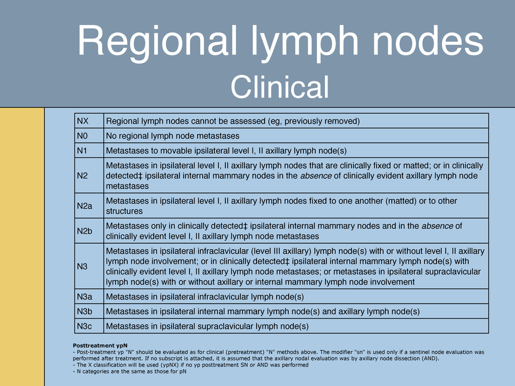
    N0 No regional lymph node metastases N1 Metastases to movable ipsilateral level I, II axillary lymph node(s) N2 Metastases in ipsilateral level I, II axillary lymph nodes that are clinically fixed or matted; or in clinically detected‡ ipsilateral internal mammary nodes in the absence of clinically evident axillary lymph node metastases N2a Metastases in ipsilateral level I, II axillary lymph nodes fixed to one another (matted) or to other structures N2b Metastases only in clinically detected‡ ipsilateral internal mammary nodes and in the absence of clinically evident level I, II axillary lymph node metastases N3 Metastases in ipsilateral infraclavicular (level III axillary) lymph node(s) with or without level I, II axillary lymph node involvement; or in clinically detected‡ ipsilateral internal mammary lymph node(s) with clinically evident level I, II axillary lymph node metastases; or metastases in ipsilateral supraclavicular lymph node(s) with or without axillary or internal mammary lymph node involvement N3a Metastases in ipsilateral infraclavicular lymph node(s) N3b Metastases in ipsilateral internal mammary lymph node(s) and axillary lymph node(s) N3c Metastases in ipsilateral supraclavicular lymph node(s) Posttreatment ypN - Post-treatment yp "N" should be evaluated as for clinical (pretreatment) "N" methods above. The modifier "sn" is used only if a sentinel node evaluation was performed after treatment. If no subscript is attached, it is assumed that the axillary nodal evaluation was by axillary node dissection (AND). - The X classification will be used (ypNX) if no yp posttreatment SN or AND was performed - N categories are the same as those for pN Regional lymph nodes Clinical
  7. pNX Regional lymph nodes cannot be assessed (eg, previously removed,

    or not removed for pathologic study) pN0 No regional lymph node metastasis identified histologically pN0(i-) No regional lymph node metastases histologically, negative immunohistochemistry (IHC) pN0(i+) Malignant cells in regional lymph node(s) no greater than 0.2 mm (detected by H&E or IHC including isolated tumor cell clusters (ITC)) pN0(mol-) No regional lymph node metastases histologically, negative molecular findings (RT-PCR)•• pN0(mol+) Positive molecular findings (RT-PCR)••, but no regional lymph node metastases detected by histology or IHC pN1 Micrometastases; or metastases in 1-3 axillary lymph nodes; and/or in internal mammary nodes with metastases detected by sentinel lymph node biopsy but not clinically detected ΔΔ pN1mi Micrometastases (greater than 0.2 mm and/or more than 200 cells, but none greater than 2.0 mm) pN1a Metastases in 1-3 axillary lymph nodes, at least one metastasis greater than 2.0 mm pN1b Metastases in internal mammary nodes with micrometastases or macrometastases detected by sentinel lymph node biopsy but not clinically detected ΔΔ pN1c Metastases in 1-3 axillary lymph nodes and in internal mammary lymph nodes with micrometastases or macrometastases detected by sentinel lymph node biopsy but not clinically detected pN2 Metastases in 4-9 axillary lymph nodes; or in clinically detected◊◊ internal mammary lymph nodes in the absence of axillary lymph node metastases pN2a Metastases in 4-9 axillary lymph nodes (at least one tumor deposit greater than 2.0 mm) pN2b Metastases in clinically detected◊◊ internal mammary lymph nodes in the absence of axillary lymph node metastases pN3 Metastases in ten or more axillary lymph nodes; or in infraclavicular (level III axillary) lymph nodes; or in clinically detected◊◊ ipsilateral internal mammary lymph nodes in the presence of one or more positive level I, II axillary lymph nodes; or in more than three axillary lymph nodes and in internal mammary lymph nodes with micrometastases or macrometastases detected by sentinel lymph node biopsy but not clinically detected ΔΔ; or in ipsilateral supraclavicular lymph nodes pN3a Metastases in ten or more axillary lymph nodes (at least one tumor deposit greater than 2.0 mm); or metastases to the infraclavicular (level III axillary lymph) nodes pN3b Metastases in clinically detected◊◊ ipsilateral internal mammary lymph nodes in the presence of one or more positive axillary lymph nodes; or in more than three axillary lymph nodes and in internal mammary lymph nodes with micrometastases or macrometastases detected by sentinel lymph node biopsy but not clinically detected ΔΔ pN3c Metastases in ipsilateral supraclavicular lymph nodes Regional lymph nodes Pathologic
  8. M0 No clinical or radiographic evidence of distant metastases cM0(i+)

    No clinical or radiographic evidence of distant metastases, but deposits of molecularly or microscopically detected tumor cells in circulating blood, bone marrow, or other nonregional nodal tissue that are no larger than 0.2 mm in a patient without symptoms or signs of metastases M1 Distant detectable metastases as determined by classic clinical and radiographic means and/or histologically proven larger than 0.2 mm Distant metastasis (M) Posttreatment yp M classification. The M category for patients treated with neoadjuvant therapy is the category assigned in the clinical stage, prior to initiation of neoadjuvant therapy. Identification of distant metastases after the start of therapy in cases where pretherapy evaluation showed no metastases is considered progression of disease. If a patient was designated to have detectable distant metastases (M1) before chemotherapy, the patient will be designated as M1 throughout.
  9. 0 Tis N0 M0 IA T1 N0 M0 IB T0

    N1mi M0 T1 N1mi M0 IIA T0 N1 M0 T1 N1 M0 T2 N0 M0 IIB T2 N1 M0 T3 N0 M0 IIIA T0 N2 M0 T1 N2 M0 T2 N2 M0 T3 N1 M0 T3 N2 M0 IIIB T4 N0 M0 T4 N1 M0 T4 N2 M0 IIIC Any T N3 M0 IV Any T Any N M1 Anatomic Stage/ Prognostic Groups
  10. Diagnosis • Introduced in 1965 • Women who begin annual

    screening mammography at age 35 and continue until age 75 have a projected benefit of reduced mortality exceeding the radiation risk by a factor of more than 25 • Digital mammography is associated with a lower radiation dose than film screen mammography for the same image quality Mammography
  11. Diagnosis Mammography Assessment category Recommendation Probability of malignancy 0: Incomplete

    Need for further evaluation Not applicable 1: Normal Normal interval follow-up 0 percent 2: Benign Normal interval follow-up 0 percent 3: Probably benign A short interval follow-up is recommended <2 percent 4: Suspicious abnormality A biopsy should be considered ≥2 to <95 percent 5: Highly suggestive of malignancy Biopsy or surgery should be performed ≥95 percent 6: Biopsy-proven carcinoma Appropriate action should be taken Source: Breast Imaging Reporting and Data System (BI-RADS) Atlas. 4th Edition. American College of Radiology, Reston, VA, 2003
  12. • 90% sensitive ◦ Although suboptimal in younger women and

    women with dense breasts • Mammography may increase the risk of cancer development in some genetic syndromes Diagnosis Mammography
  13. • Used to assess a palpable mass which is mammogram

    negative, or to further characterize a positive mammogram • Can delineate cysts and fibroadenomas from malignancies, which mammograms and physical examination cannot Diagnosis Breast Ultrasound
  14. • 88%-100% Sensitivity for breast cancers • Only 72% specific

    in differentiating malignant from benign lesions • MRI is more sensitive than mammography, ultra sound or physical exam Diagnosis Breast MRI
  15. • Fine needle aspiration biopsy ◦ If palpable • Stereotactic

    mammographic guided biopsy ◦ If positive on mammogram • Core needle biopsy; often using ultra sound • MRI guided breast biopsy ◦ If non palpable and not visualized on mammogram Diagnosis Tissue Biopsy
  16. • Early stage-clinical stage I, IIA, IIB, and IIIA with

    no involved lymph nodes • Locally advanced-all other stage III • Metastatic disease Patient Stratification Staging
  17. • CT, chest • CT, abdomen • CT, pelvis •

    Bone Scan • MRI, bone • MRI, brain • PET scan Localizing symptoms Abnormal laboratory findings Abnormal physical exam Locally advanced disease High grade disease Patient Stratification Staging
  18. • Prognostic Molecular Profiles: • Oncotype DX; 21 gene recurrence

    score • Mammaprint; 70 gene recurrence score • Used for prognosis and treatment options ER+ = Good Prognosis PR+ = Good Prognosis HER2/neu = Poor Prognosis Patient Stratification Receptor Status
  19. • Lumpectomy with Primary Radiation Therapy • Contraindicated if adequate

    cosmesis cannot be obtained due to size or location of malignancy • Women over 70 with less than a 2 cm tumor and no involved lymph nodes do not need radiation therapy • People with hereditary breast ovarian cancer syndrome may have an increased risk of developing malignancy with exposure to radiation • Sentinel lymph node biopsy; to determine stage regardless of mastectomy or lumpectomy Treatment Breast Conserving Therapy
  20. • Equally effective compared to lumpectomy with radiation therapy •

    Replaced the radical mastectomy (Halsted procedure) • Primary radiation therapy not required Treatment Modified Radical Mastectomy
  21. • IV or po cytotoxic agent ("chemotherapy") or hormonal manipulation

    • Improve the surgical outcome-clinical Stage III • Allow breast conserving therapy • Allows for early evaluation of effectiveness of systemic therapy • Overall survival may be improved Treatment Neoadjuvant Therapy
  22. • Dose dense adriamycin+cytoxan, taxol • Carboplatin+taxol • Herceptin if

    Her2/neu status is positive • Hormonal manipulation if ER+/PR+ • Avastin? Treatment Neoadjuvant Therapy
  23. • As primary treatment following lumpectomy: ◦ Whole breast RT

    is used, typically 4600-5000 rads, followed by a boast to the lumpectomy cavity ◦ In select cases (ER+, node -, low grade, older patient) 4250 rads may be adequate ◦ If node + disease, regional RT is added to whole breast RT ◦ BRCA mutation have no evidence of greater radiation sensitivity ◦ External beam RT is also used to palliate certain metastatic areas, eg bone or brain Treatment Radiation Therapy
  24. • Used as prevention relative to risk factors • Stage

    • Grade • Hormonal Receptor Status • HER2/neu Status Treatment Adjuvant Treatment
  25. • Dose dense adriamycin+cytoxan, taxol-6 month duration • Carboplatin+taxol-6 month

    duration • Herceptin-1 year duration • Hormonal manipulation • Tamoxifen, premenopausal-5 year duration (possibly 10 years) • Arimidex, postmenopausal-5 year duration Treatment Adjuvant Treatment
  26. • Hormonal Receptor Positive • Arimidex • Aromasen • Femara

    • Faslodex • Tamoxifen • Reloxifen • Megace • Halotestin • DES • Aminoglutethamide Treatment Metastatic Disease
  27. • Her2/neu Receptor Positive • Herceptin • Kadcyla • Tykerb

    • Perjeta • Vascular endothelial growth factor receptor inhibitor • Avastin Treatment Metastatic Disease
  28. Supportive Agents • Bisphosphonates, Bone Mets • Anti Nausea Meds

    ◦ Aloxi ◦ Zofran ◦ Kytril ◦ Anzemet ◦ Emend ◦ Sancuso • White Cell Support ◦ Neupogen ◦ Neulasta • Red Blood Cell Support ◦ Procrit ◦ Aranesp • Venous Access Devices ◦ IV fluids
  29. Genetics • Primary cause in 5-10% of all cases •

    Hereditary breast-ovarian cancer syndrome; 5% of breast cancer cases • Mutation of 2 tumor supressor genes • BReast CAncer type 1 (BRCA 1): 17q • BReast CAncer type 2 (BRCA 2): 13q
  30. Percent of population Percent of all breast cancer cases Average

    risk of breast cancer to age 70 Family history of breast cancer Positive BRCA1 or BRCA2 mutation Rest of population ~10% 15 - 20% 10 - 13% ~0.1% 5 - 6% 50 - 85% ~90% 80 - 85% 7% Genetics HBOC syndrome
  31. Family history Positive Mutation Rest of Population Percent of all

    breast cancer cases Average risk of breast cancer to age 70 Genetics HBOC syndrome
  32. • Positive test does not mean that the patient will

    develop breast or ovarian cancer • Negative test does not mean that the patient cannot develop breast or ovarian cancer Genetics HBOC syndrome
  33. • Non-Ashkenazi Jewish women ◦ Two first-degree relatives with breast

    cancer, one of whom was diagnosed at age 50 or younger ◦ A combination of three or more first or second-degree relatives with breast cancer regardless of age at diagnosis ◦ A combination of both breast and ovarian cancer among first and second- degree relatives ◦ A first-degree relative with bilateral breast cancer ◦ A combination of two or more first or second degree relatives with ovarian cancer, regardless of age at diagnosis ◦ A first or second-degree relative with both breast and ovarian cancer at any age ◦ History of breast cancer in a male relative • Women of Ashkenazi Jewish descent ◦ Any first-degree relative (or two second degree relatives on the same side of the family) with breast or ovarian cancer Genetics BRCA Mutation Testing
  34. Breast Cancer Risk Factors • Hereditary • Smoking • High

    Fat Diet • Alcohol Intake • Obesity • Radiation • Shift Work • Organic Solvents • Pesticides
  35. House Calls Radio Show Sunday 9:30 am The Dean &

    Don Show Tuesday 9:35 am KMA 960 AM kma960.com HeartlandOncology.com On the Radio