Upgrade to Pro — share decks privately, control downloads, hide ads and more …

低体温療法とaEEG脳波の相関を語る

imi
April 07, 2015
190

 低体温療法とaEEG脳波の相関を語る

imi

April 07, 2015
Tweet

Transcript

  1. Continuous and simplified EEG (aEEG) to monitor brain recovery after

    Cardiac Arrest Hans Friberg, Associate professor Center for Resuscitation Science Lund University Sweden CENTER FOR RESUSCITATION SCIENCE IN THE ØRESUND REGION The 42nd Annual Meeting of The Japanese Society of Intensive Care Medicine
  2. Lund University / Faculty / Department / Unit / Document

    / Date Natural course of brain recovery following cardiac arrest COMA Vegetative state Cardiac arrest Brain stem recovery Recovery of cortical function Brain Dead hours – days (– weeks)
  3. Lund University / Faculty / Department / Unit / Document

    / Date • 86/162 (53%) died in hospital • Most (>70%) die from brain injury Cause of death after cardiac arrest Dragancea I et al. Resuscitation 2012
  4. Lund University / Faculty / Department / Unit / Document

    / Date Seizures are common: A. Clinical seizures B. Electrographic seizures ± A How common? Brain injury after cardiac arrest
  5. Lund University / Faculty / Department / Unit / Document

    / Date Nielsen et al. CCM 2011 Jan;39(1):57-64 1000 patients – clinical or electrographic seizures in 24% • Good outcome in 17% at 6 months Seizures after cardiac arrest – The Hypothermia Registry –
  6. Lund University / Faculty / Department / Unit / Document

    / Date Seder et al. CCM 2015, e-pub ahead of print 2500 patients – myoclonic seizures in 18% • Epileptiform activity in 55% • Good outcome in 9% Seizures after cardiac arrest – International Cardiac Arrest Registry –
  7. Lund University / Faculty / Department / Unit / Document

    / Date Rundgren et al. CCM 2010;38:1838 Electrographic seizures in 26/95 patients – 27% • 2/26 patients (8%) good outcome Seizures after cardiac arrest
  8. Lund University / Faculty / Department / Unit / Document

    / Date Seizures in the TTM-trial (Nielsen et al. 2013) Seizure type TTM33 (n=473) TTM36 (n=466) All (n=939) No Seizure 326 (69%) 345 (74%) 671 (71%) Any seizure 147 (31%) 121 (26%) 268 (29%) Tonic-Clonic 37 (8%) 34 (7%) 71 (8%) Myoclonic 132 (28%) 108 (23%) 240 (26%) ”Any seizure” • 29/268 patients (11%) good outcome Nielsen et al. N Engl J Med 2013
  9. Lund University / Faculty / Department / Unit / Document

    / Date Seizures are common: A. Clinical seizures B. Electrographic seizures ± A How do we detect? Seizures after cardiac arrest
  10. Lund University / Faculty / Department / Unit / Document

    / Date • Electrophysiologic monitoring – EEG – cEEG – aEEG – SSEP Neurophysiology
  11. Lund University / Faculty / Department / Unit / Document

    / Date Electrode placement routine multichannel EEG
  12. Lund University / Faculty / Department / Unit / Document

    / Date Normal adult EEG eyes shut eyes open
  13. Lund University / Faculty / Department / Unit / Document

    / Date EEG in the ICU Claassen J et al. ICM 2013
  14. Lund University / Faculty / Department / Unit / Document

    / Date EEG after cardiac arrest • Recommends EEG during hypothermia and within 24h after rewarming to rule out non-convulsive seizures • Suggests EEG to assist with prognostication, particularly in patients treated with hypothermia • Suggests continuous EEG to assist with prognostication of coma Further data are necessary to understand its full potential Claassen J et al. ICM 2013
  15. Lund University / Faculty / Department / Unit / Document

    / Date EEG after cardiac arrest – limitations • Different grading systems, definitions & terminology • EEG can be affected by sedation • Inter-rater variability • Cost – continuous multichannel EEG • Lack of large prospective studies on the prevalence and predictive value of EEG-changes
  16. Lund University / Faculty / Department / Unit / Document

    / Date Objective: Standardize terminology of EEG-patterns in the critically ill Goal: Aid communication and facilitate future research Hirsch LJ et al. J Clin Neurophysiol 2012 Critical Care EEG terminology – ACNS
  17. Lund University / Faculty / Department / Unit / Document

    / Date EEG interpretation – cardiac arrest • Background activity – Flat (very low voltage) – Suppression-burst – Continuous • Epileptiform activity – Sporadic – Frequent – Electrographic status ep • Reactivity – yes/no – Sound – Pain • Routine EEG or continuous EEG-monitoring?
  18. Lund University / Faculty / Department / Unit / Document

    / Date • Traditional multichannel montage • Simplified montage, few channels • Trend curves with compressed time scale • Simplified montage and trend curves – aEEG Continuous EEG – cEEG Friedman et al. Anesthesia and Analgesia 2009
  19. Lund University / Faculty / Department / Unit / Document

    / Date aEEG amplitude integrated electroencephalography Established method in neonatal ICUs! Preferred method in adult ICUs! • simplified EEG – few channels • trend analysis – to facilitate!
  20. Lund University / Faculty / Department / Unit / Document

    / Date • Simplified montage – 2 channels • Initiated by ICU nurses on adm. Trend curves aEEG = amplitude integrated EEG 4-6 hours per screen Original-EEG 10 sec per screen
  21. Lund University / Faculty / Department / Unit / Document

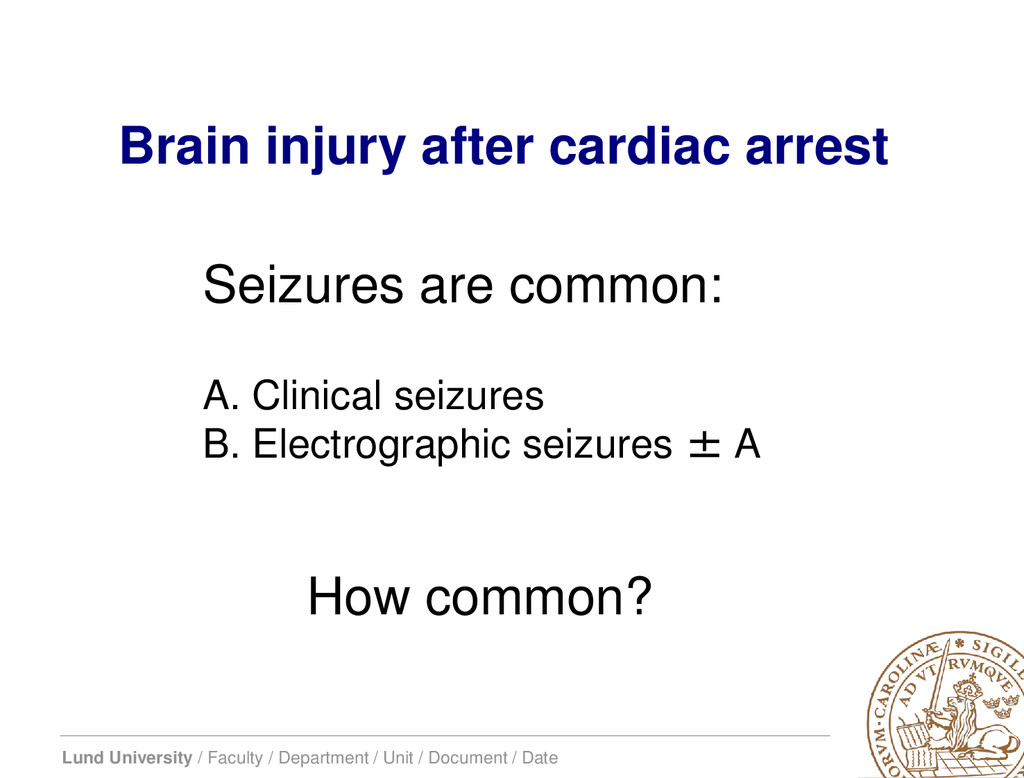
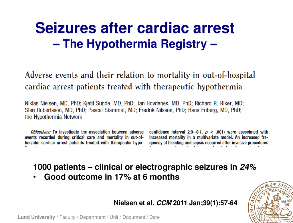
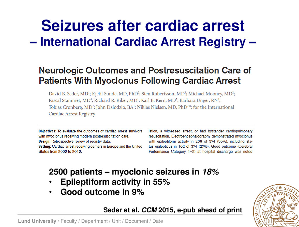
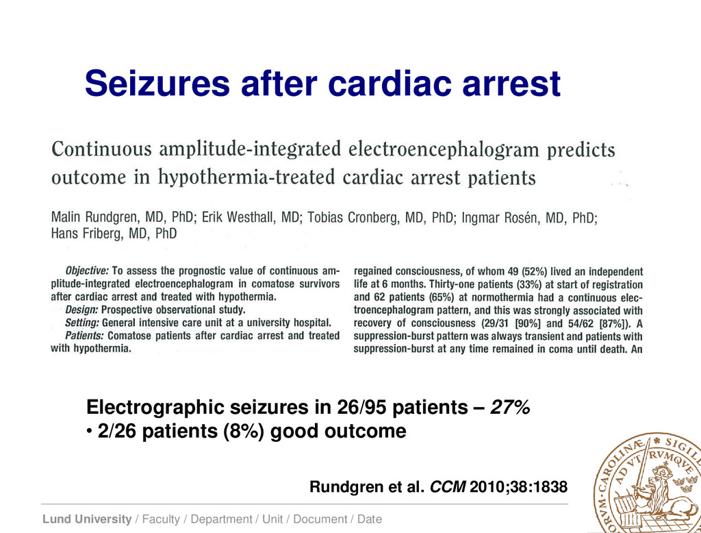
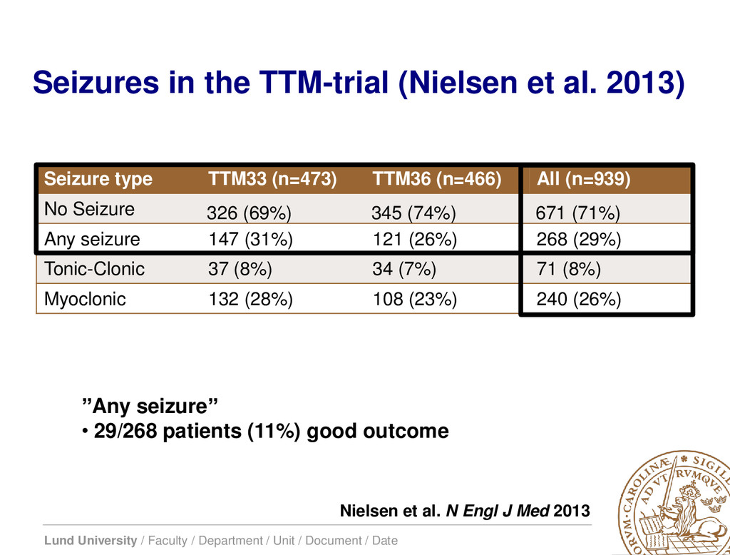
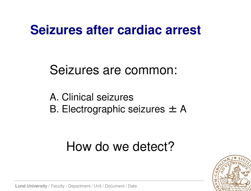
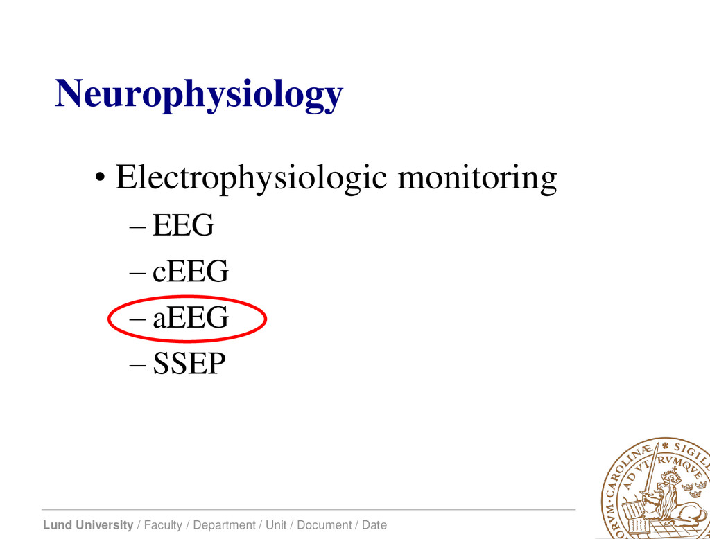
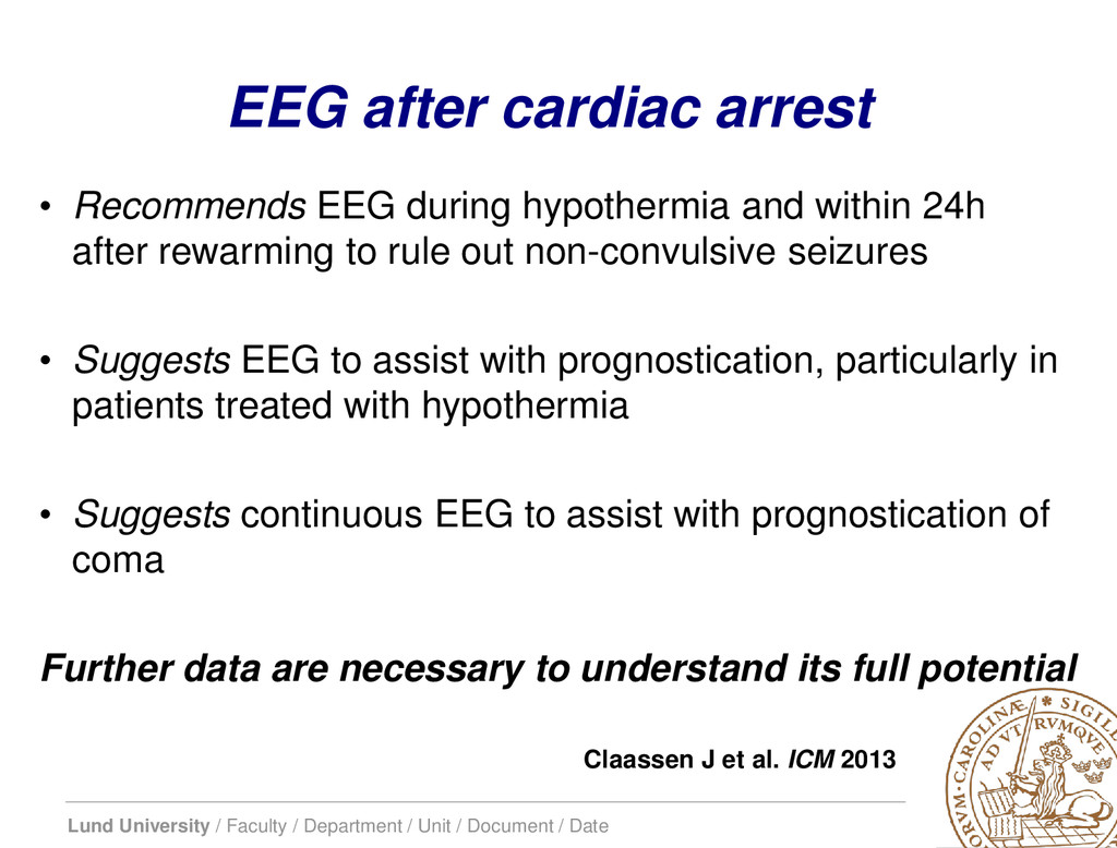
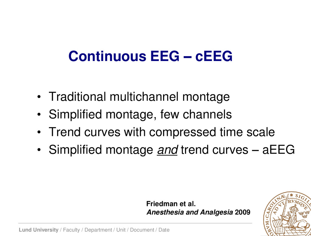
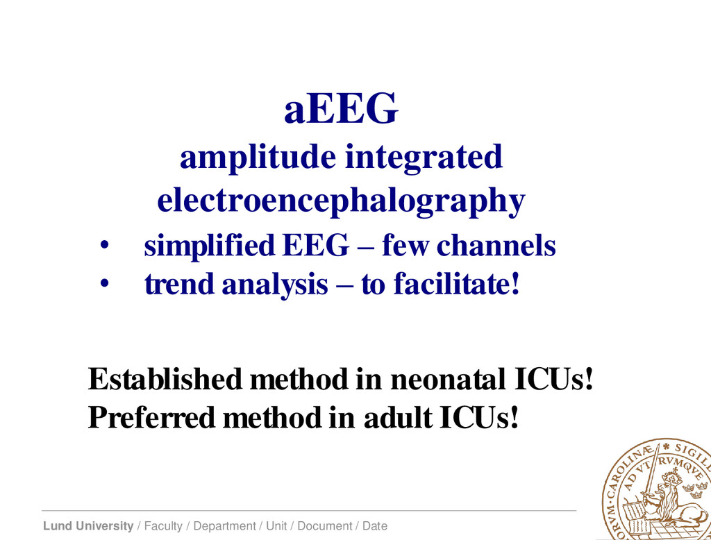
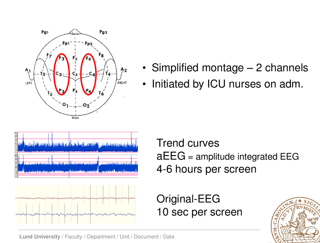
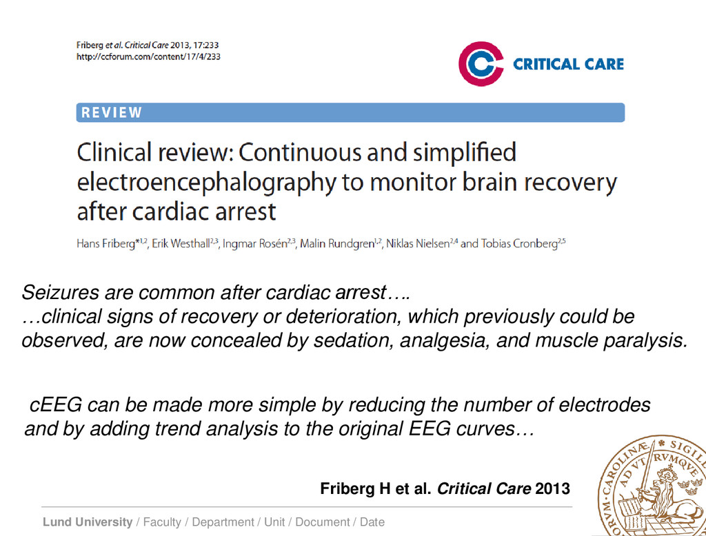
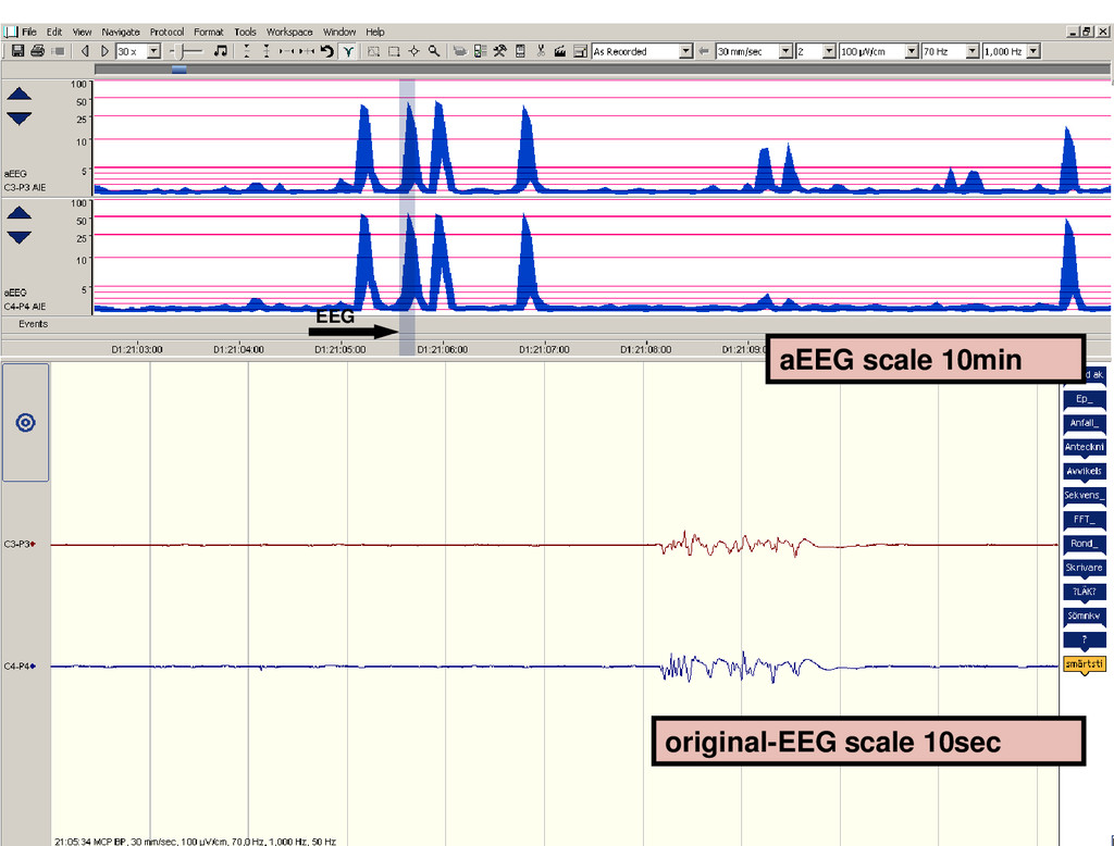
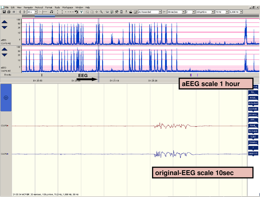
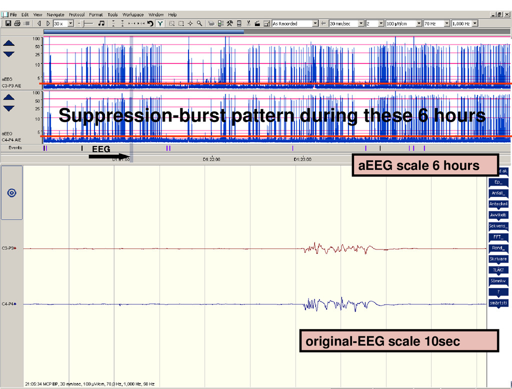
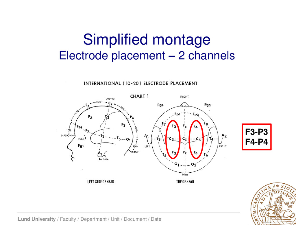
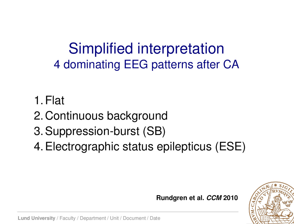
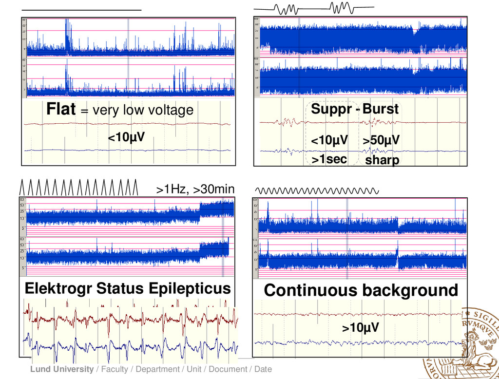
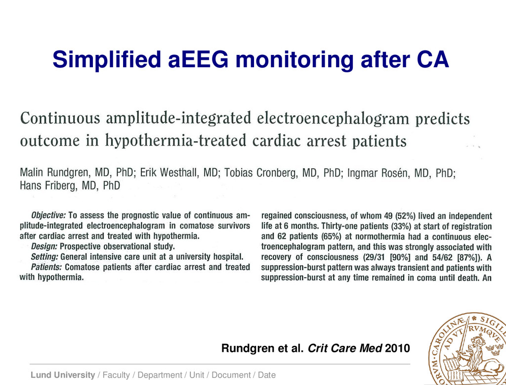
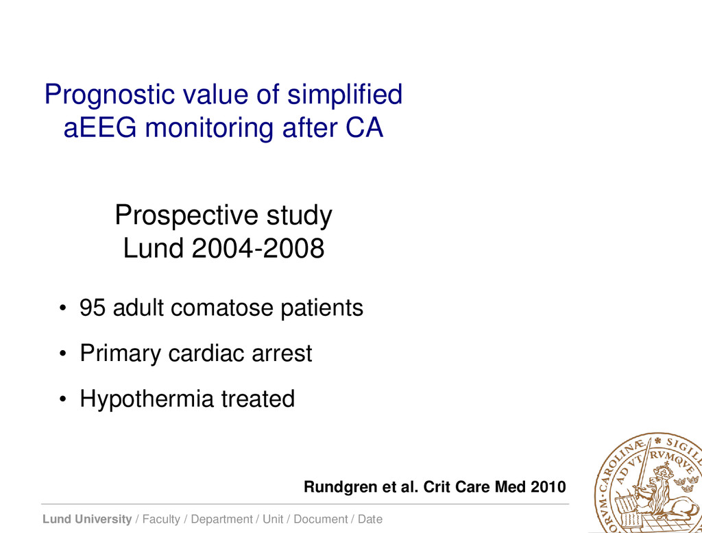
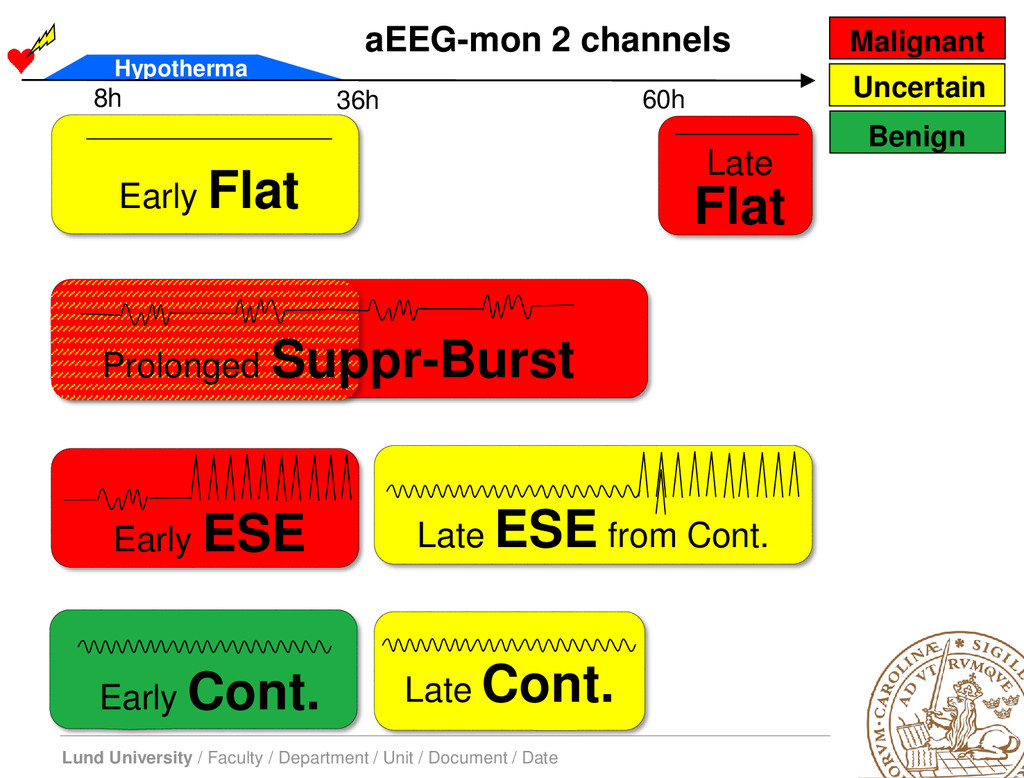
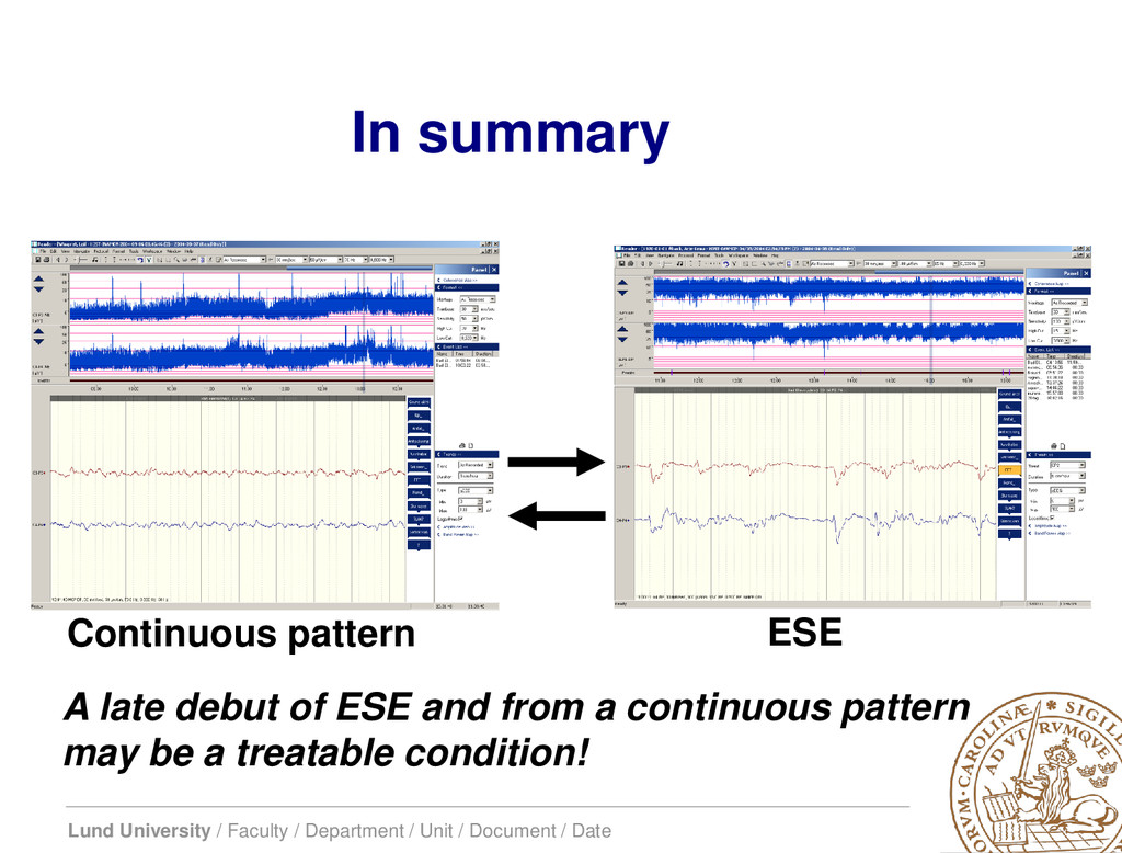
    / Date Friberg H et al. Critical Care 2013 Seizures are common after cardiac arrest…. …clinical signs of recovery or deterioration, which previously could be observed, are now concealed by sedation, analgesia, and muscle paralysis. cEEG can be made more simple by reducing the number of electrodes and by adding trend analysis to the original EEG curves…
  22. Lund University / Faculty / Department / Unit / Document

    / Date • To reach general use, cEEG needs to be – Simple • Reduce the number of leads • Add trend analysis to the original EEG curves – Possible to apply bedside, preferably by ICU-staff – Cost-effective Continuous EEG (cEEG) after cardiac arrest
  23. Lund University / Faculty / Department / Unit / Document

    / Date • Diagram of signal pathways – simplified EEG/aEEG • EEG signal modification: – Asymmetric filtering – 2-15 Hz – Semilogaritmic scale • Compressed timescale – to show trends • Easier interpretation
  24. Lund University / Faculty / Department / Unit / Document

    / Date Amplification (dB) -40 -35 -30 -25 -20 -15 -10 -5 0 5 0 5 10 15 20 25 30 Hz dB Amplification ≈ 2–15 Hz aEEG – assymetric filter Movements Breathing Muscle activity
  25. Lund University / Faculty / Department / Unit / Document

    / Date 5 10 25 50 100 0 Trend curve (aEEG), ≈ 4 hours/screen Original-EEG, scale, ≈ 10 sec/screen
  26. Lund University / Faculty / Department / Unit / Document

    / Date EEG max ampl aEEG max ≈ 20-35 µV EEG min ampl aEEG min ≈ 2-4 µV EEG aEEG Original EEG
  27. Lund University / Faculty / Department / Unit / Document

    / Date aEEG scale 1 minute original-EEG scale 1 minute EEG
  28. Lund University / Faculty / Department / Unit / Document

    / Date aEEG scale 10min original-EEG scale 10sec EEG
  29. Lund University / Faculty / Department / Unit / Document

    / Date aEEG scale 1 hour original-EEG scale 10sec EEG
  30. Lund University / Faculty / Department / Unit / Document

    / Date aEEG scale 6 hours original-EEG scale 10sec EEG Suppression-burst pattern during these 6 hours
  31. Lund University / Faculty / Department / Unit / Document

    / Date • Simplified montage – Reduced montage, two leads – Amplitude-integrated trend curves (aEEG) • Simplified interpretation – 4 defined dominating patterns Continuous EEG after cardiac arrest – aEEG Rundgren et al. ICM 2006 Rundgren et al. CCM 2010
  32. Lund University / Faculty / Department / Unit / Document

    / Date F3-P3 F4-P4 Simplified montage Electrode placement – 2 channels
  33. Lund University / Faculty / Department / Unit / Document

    / Date Simplified interpretation 4 dominating EEG patterns after CA 1.Flat 2.Continuous background 3.Suppression-burst (SB) 4.Electrographic status epilepticus (ESE) Rundgren et al. CCM 2010
  34. Lund University / Faculty / Department / Unit / Document

    / Date Flat = very low voltage Elektrogr Status Epilepticus >1Hz, >30min Continuous background <10µV >10µV Suppr - Burst <10µV >50µV >1sec sharp
  35. Lund University / Faculty / Department / Unit / Document

    / Date Rundgren et al. Crit Care Med 2010 Simplified aEEG monitoring after CA
  36. Lund University / Faculty / Department / Unit / Document

    / Date Prognostic value of simplified aEEG monitoring after CA • 95 adult comatose patients • Primary cardiac arrest • Hypothermia treated Rundgren et al. Crit Care Med 2010 Prospective study Lund 2004-2008
  37. Lund University / Faculty / Department / Unit / Document

    / Date aEEG background pattern and outcome Rundgren et al. Crit Care Med 2010
  38. Lund University / Faculty / Department / Unit / Document

    / Date › 26/95 patients (27%) developed ESE › 16 from suppression- burst pattern › 10 from a continuous pattern › 2 patients regained consciousness Electrographic status epilepticus 0 1 2 3 4 5 NT-21- 25h NT-16- 20h NT-11- 15h NT-6- 10h NT-NT- 5h NT- NT+5h NT+6- +10h NT+11- +15h NT+16- +20h ESE from contin ESE from SB -25 -20 -15 -10 -5 NT +5 +10 +15 +20 Rundgren, Westhall et al. CCM 2010
  39. Lund University / Faculty / Department / Unit / Document

    / Date • 57/95 patients recovered consciousness • An initial flat pattern is common – no correlation to outcome • Recovery of a continuous background pattern is a good early predictor of recovery of consciousness – PPV 0.87 • All other patterns than a continuous background at normo- thermia are strong predictors of poor outcome – NPV 0.91 • Electrographic SE develops from two different background patterns, which may have therapeutic implications Rundgren et al. CCM 2010 Simplified aEEG monitoring after CA
  40. Lund University / Faculty / Department / Unit / Document

    / Date Late Cont. Prolonged Suppr-Burst Early Cont. Early Flat Late Flat Early ESE Late ESE from Cont. 8h 36h 60h Hypotherma aEEG-mon 2 channels Uncertain Malignant Benign
  41. Lund University / Faculty / Department / Unit / Document

    / Date The rationale: • Simple to apply and simple to use • A trend monitor of brain function – Aids in prognostication – Seizure detection • Guide anticonvulsive treatment • Early and continuous prognostic information – good as well as poor outcome Simplified aEEG monitoring after CA
  42. Lund University / Faculty / Department / Unit / Document

    / Date A patient with late debut of ESE, 3 weeks in the ICU and good outcome Continuous background Flat Electrographic status epilepticus Hours after cardiac arrest: 6 24 72 48 12 18 30 36 42 54 60 66 EEG pattern: No registration No registration ** Normothermia 1 sec 100μV Westhall et al. Ther Hypothermia Temp Manag 2013
  43. Lund University / Faculty / Department / Unit / Document

    / Date 1 hour aEEG: 1 sec Original-EEG: 100μV Continuous background (A) develops into electrographic status epilepticus (B) A. B.
  44. Lund University / Faculty / Department / Unit / Document

    / Date In summary Continuous pattern ESE A late debut of ESE and from a continuous pattern may be a treatable condition!
  45. Lund University / Faculty / Department / Unit / Document

    / Date Electrographic seizures Our treatment strategy: I. Continuous evaluation (simplified aEEG) II. Suppression of electrographic seizures – 24 h III. In selected cases – prolonged treatment 1-2 w I. ESE from a continuous pattern II. Preserved N20 potentials III. Low Neuron Specific Enolase (NSE) IV. Patience…
  46. Lund University / Faculty / Department / Unit / Document

    / Date Continuous EEG after cardiac arrest – aEEG • Simplified montage – Reduced montage, two leads – Amplitude-integrated trend curves (aEEG) • Simplified interpretation – 4 defined dominating patterns Rundgren et al. ICM 2006 Rundgren et al. CCM 2010 Conclusions I
  47. Lund University / Faculty / Department / Unit / Document

    / Date • aEEG is feasible to perform and a valuable tool after cardiac arrest – prognosis, seizure detection • Needs to be validated against gold standard • Collaboration with neurophysiologists and education of staff are keys to sucess Conclusions II
  48. Lund University / Faculty / Department / Unit / Document

    / Date • Heart damage – Clinical findings – TnT – ECG • Brain damage – Clinical findings – NSE (or other) – EEG Final thought on assessment of tissue damage… Cronberg T et al. Neurology 2011 Rossetti et al. Neurology 2012