Upgrade to Pro — share decks privately, control downloads, hide ads and more …

Basic Retinal Anatomy

Avatar for Optovue Academy Optovue Academy
June 07, 2015
200

Basic Retinal Anatomy

Avatar for Optovue Academy

Optovue Academy

June 07, 2015

Transcript

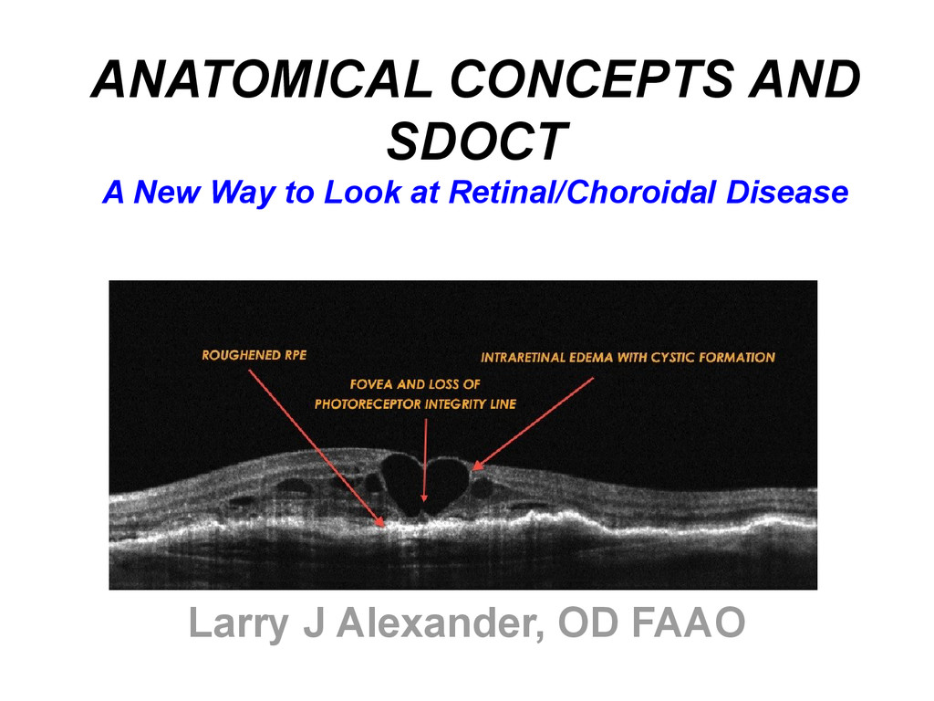
  1. ANATOMICAL CONCEPTS AND SDOCT A New Way to Look at

    Retinal/Choroidal Disease Larry J Alexander, OD FAAO
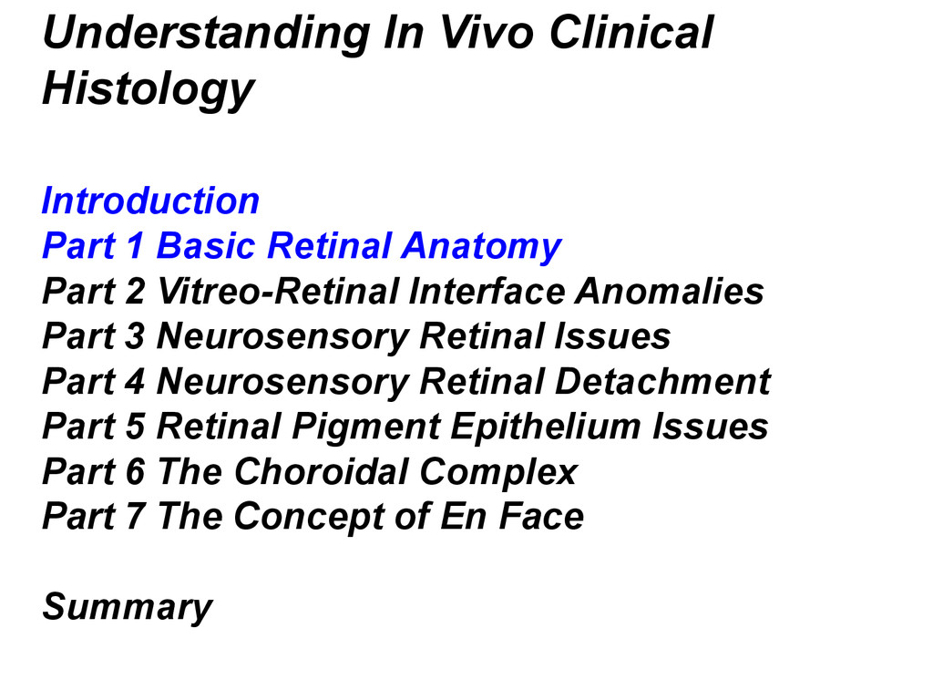
  2. Understanding In Vivo Clinical Histology Introduction Part 1 Basic Retinal

    Anatomy Part 2 Vitreo-Retinal Interface Anomalies Part 3 Neurosensory Retinal Issues Part 4 Neurosensory Retinal Detachment Part 5 Retinal Pigment Epithelium Issues Part 6 The Choroidal Complex Part 7 The Concept of En Face Summary
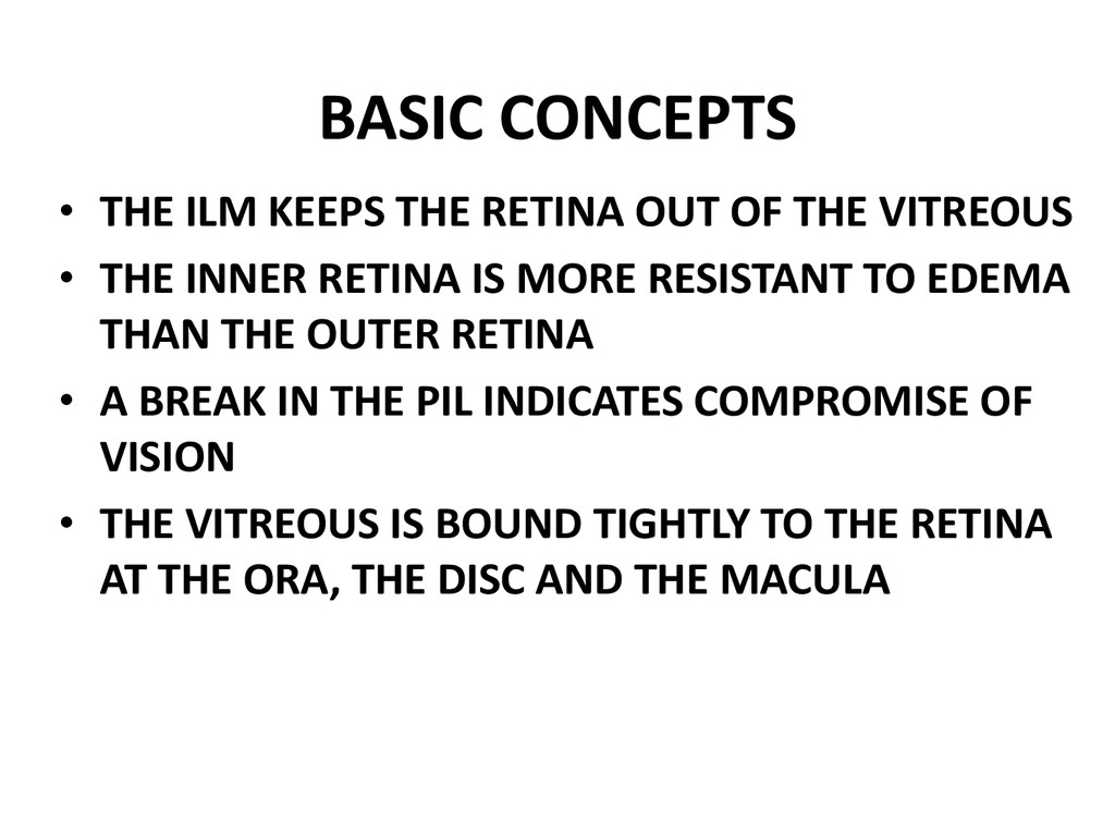
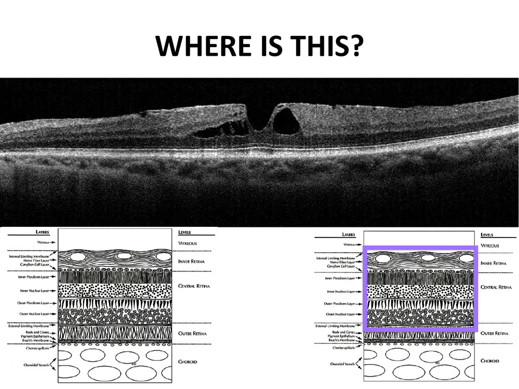
  3. BASIC CONCEPTS • THE ILM KEEPS THE RETINA OUT OF

    THE VITREOUS • THE INNER RETINA IS MORE RESISTANT TO EDEMA THAN THE OUTER RETINA • A BREAK IN THE PIL INDICATES COMPROMISE OF VISION • THE VITREOUS IS BOUND TIGHTLY TO THE RETINA AT THE ORA, THE DISC AND THE MACULA
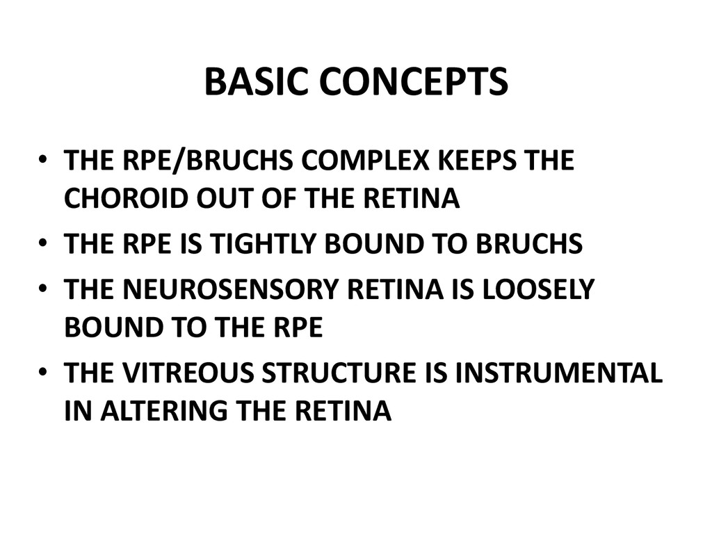
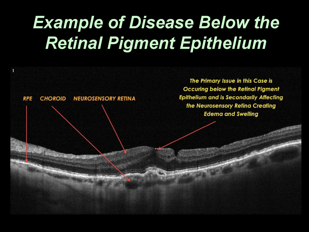
  4. BASIC CONCEPTS • THE RPE/BRUCHS COMPLEX KEEPS THE CHOROID OUT

    OF THE RETINA • THE RPE IS TIGHTLY BOUND TO BRUCHS • THE NEUROSENSORY RETINA IS LOOSELY BOUND TO THE RPE • THE VITREOUS STRUCTURE IS INSTRUMENTAL IN ALTERING THE RETINA
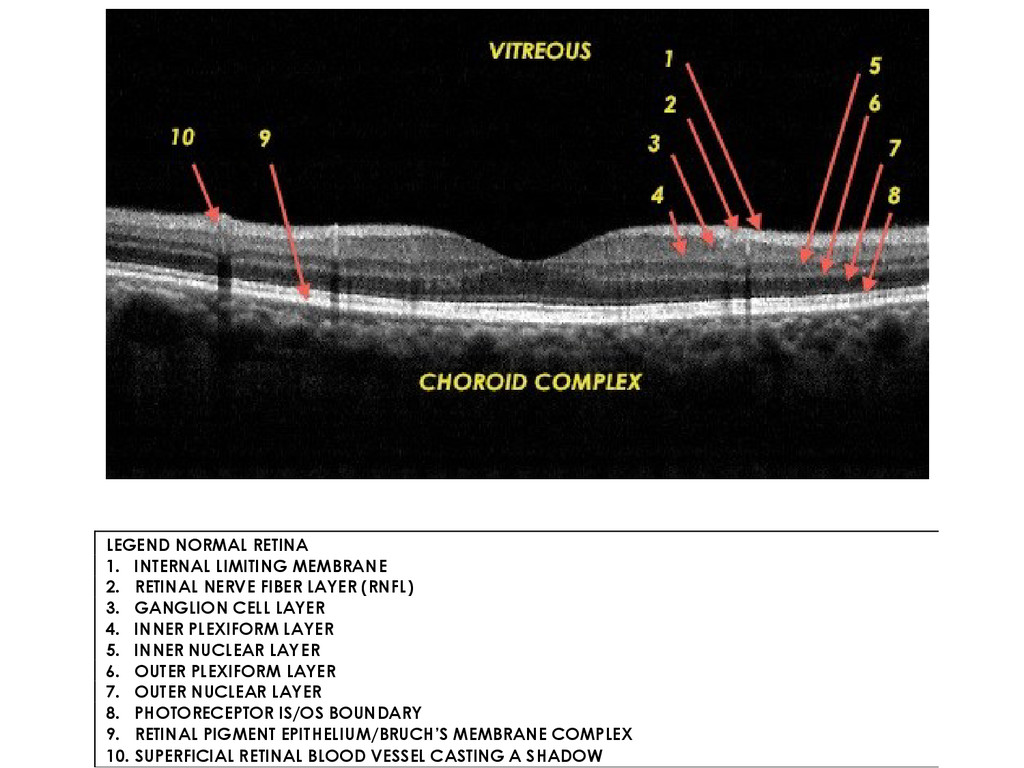
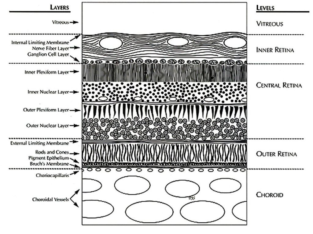
  5. LEGEND NORMAL RETINA 1. INTERNAL LIMITING MEMBRANE 2. RETINAL NERVE

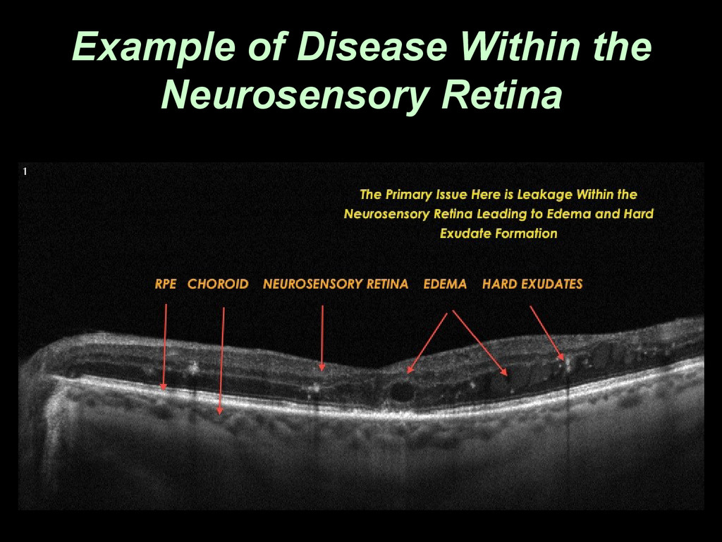
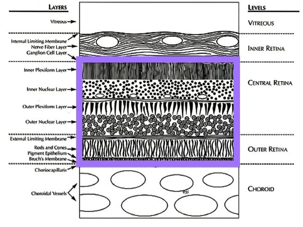
    FIBER LAYER (RNFL) 3. GANGLION CELL LAYER 4. INNER PLEXIFORM LAYER 5. INNER NUCLEAR LAYER 6. OUTER PLEXIFORM LAYER 7. OUTER NUCLEAR LAYER 8. PHOTORECEPTOR IS/OS BOUNDARY 9. RETINAL PIGMENT EPITHELIUM/BRUCH’S MEMBRANE COMPLEX 10. SUPERFICIAL RETINAL BLOOD VESSEL CASTING A SHADOW
  6. BASIC CONCEPTS • IF YOU UNDERSTAND SIMPLE CLINICAL ANATOMY…. •

    YOU WILL UNDERSTAND SDOCT… • BUT IF YOU DO NOT… • YOU WILL NEVER UNDERSTAND SDOCT
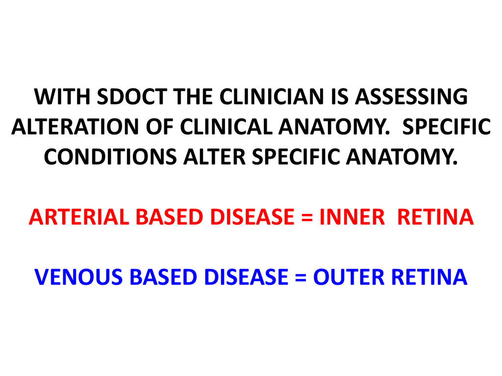
  7. WITH SDOCT THE CLINICIAN IS ASSESSING ALTERATION OF CLINICAL ANATOMY.

    SPECIFIC CONDITIONS ALTER SPECIFIC ANATOMY. ARTERIAL BASED DISEASE = INNER RETINA VENOUS BASED DISEASE = OUTER RETINA
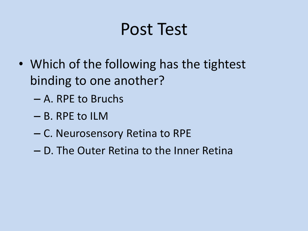
  8. Post Test • Which of the following has the tightest

    binding to one another? – A. RPE to Bruchs – B. RPE to ILM – C. Neurosensory Retina to RPE – D. The Outer Retina to the Inner Retina
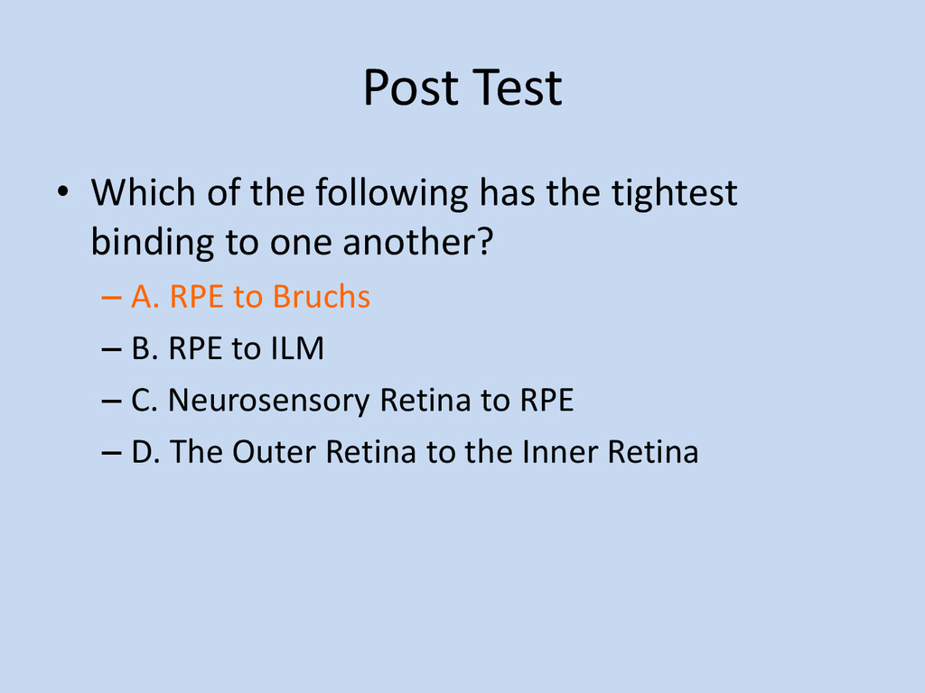
  9. Post Test • Which of the following has the tightest

    binding to one another? – A. RPE to Bruchs – B. RPE to ILM – C. Neurosensory Retina to RPE – D. The Outer Retina to the Inner Retina
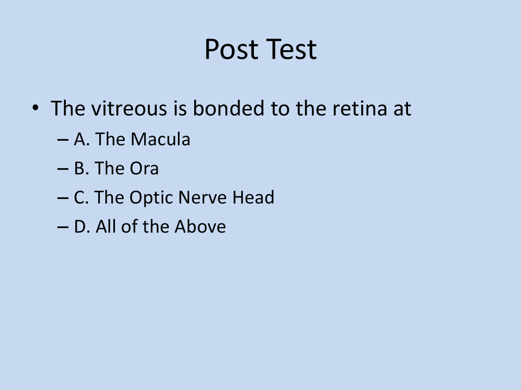
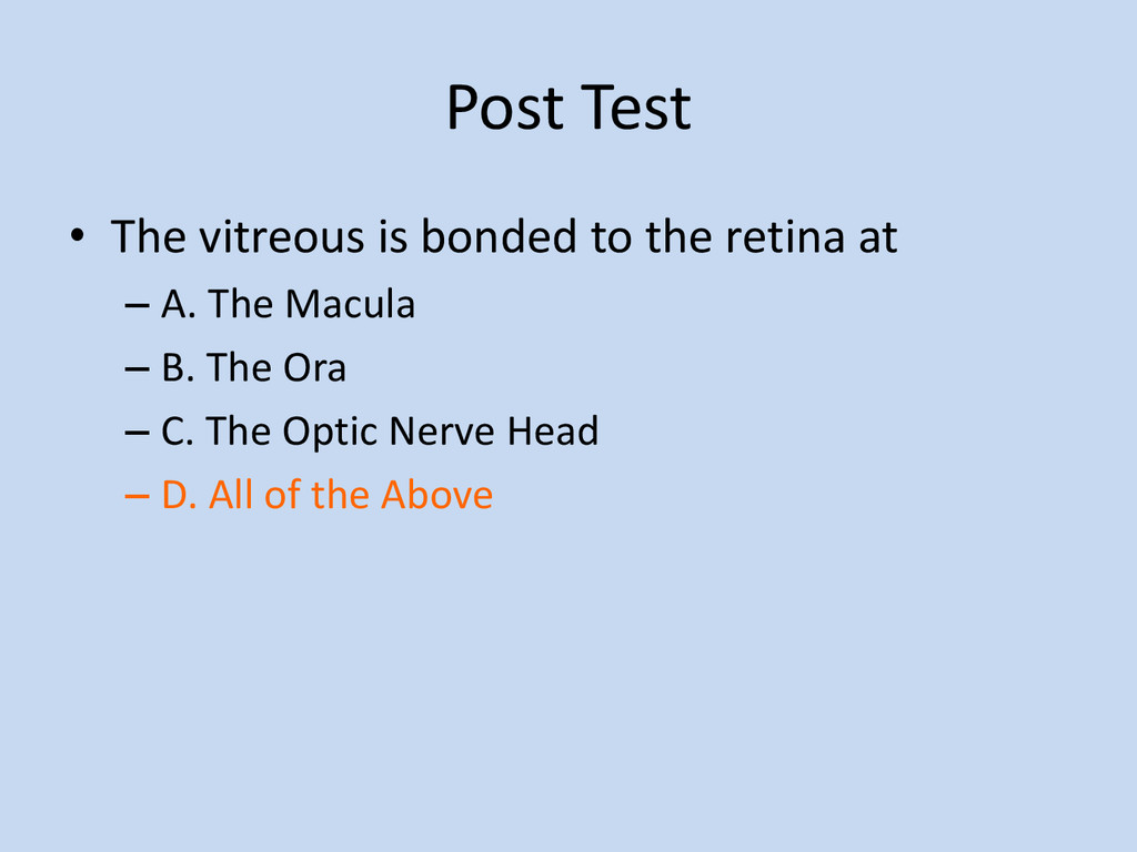
  10. Post Test • The vitreous is bonded to the retina

    at – A. The Macula – B. The Ora – C. The Optic Nerve Head – D. All of the Above
  11. Post Test • The vitreous is bonded to the retina

    at – A. The Macula – B. The Ora – C. The Optic Nerve Head – D. All of the Above
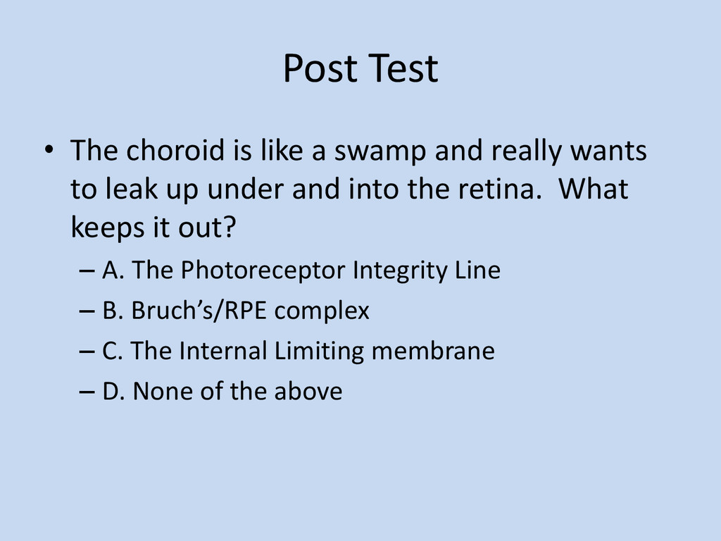
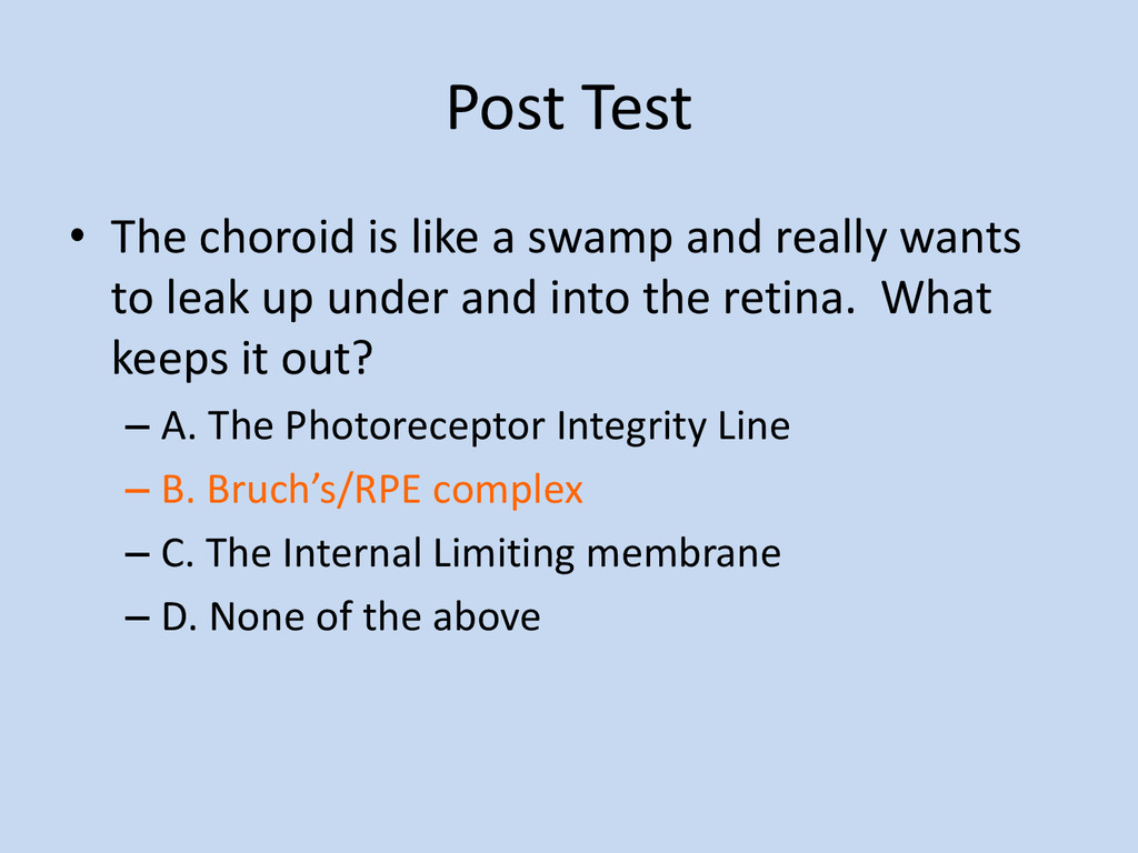
  12. Post Test • The choroid is like a swamp and

    really wants to leak up under and into the retina. What keeps it out? – A. The Photoreceptor Integrity Line – B. Bruch’s/RPE complex – C. The Internal Limiting membrane – D. None of the above
  13. Post Test • The choroid is like a swamp and

    really wants to leak up under and into the retina. What keeps it out? – A. The Photoreceptor Integrity Line – B. Bruch’s/RPE complex – C. The Internal Limiting membrane – D. None of the above
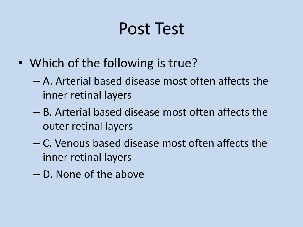
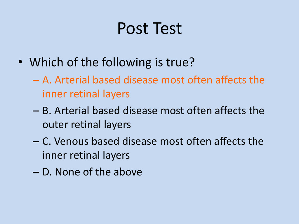
  14. Post Test • Which of the following is true? –

    A. Arterial based disease most often affects the inner retinal layers – B. Arterial based disease most often affects the outer retinal layers – C. Venous based disease most often affects the inner retinal layers – D. None of the above
  15. Post Test • Which of the following is true? –

    A. Arterial based disease most often affects the inner retinal layers – B. Arterial based disease most often affects the outer retinal layers – C. Venous based disease most often affects the inner retinal layers – D. None of the above
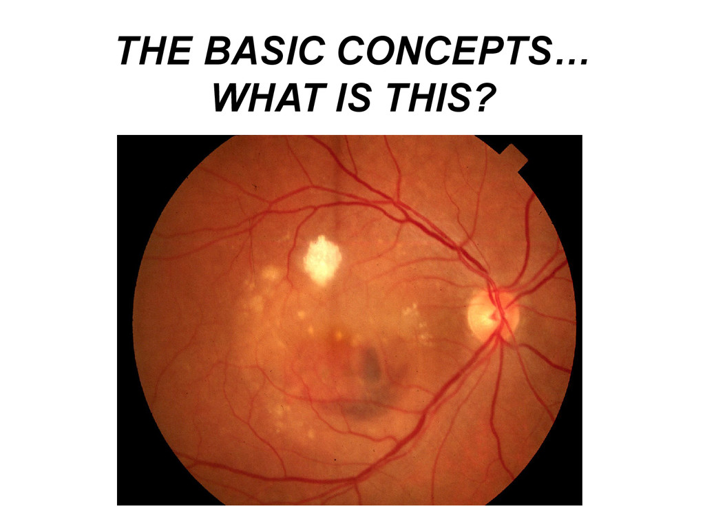
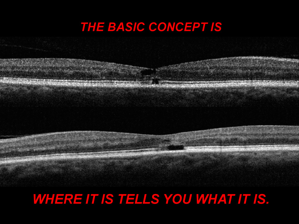
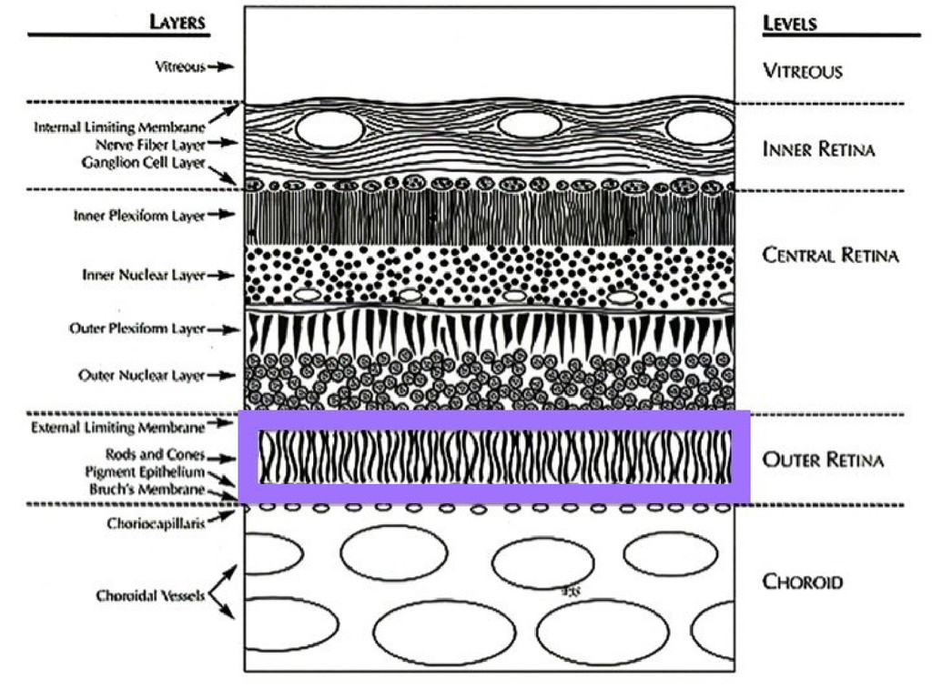
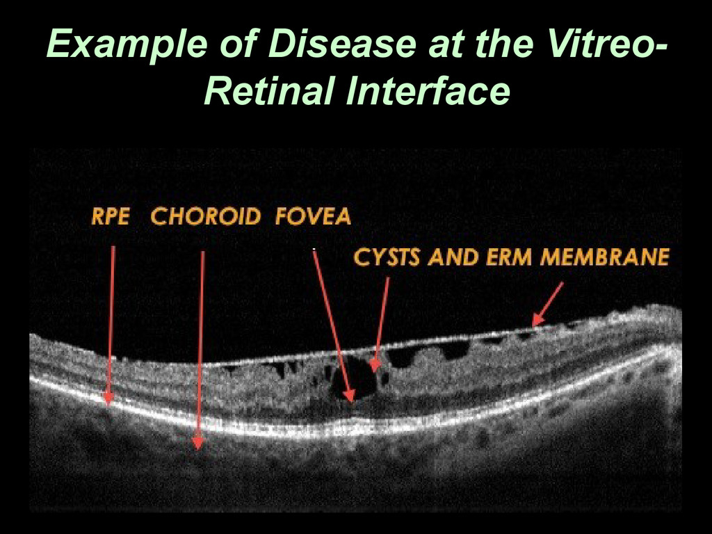
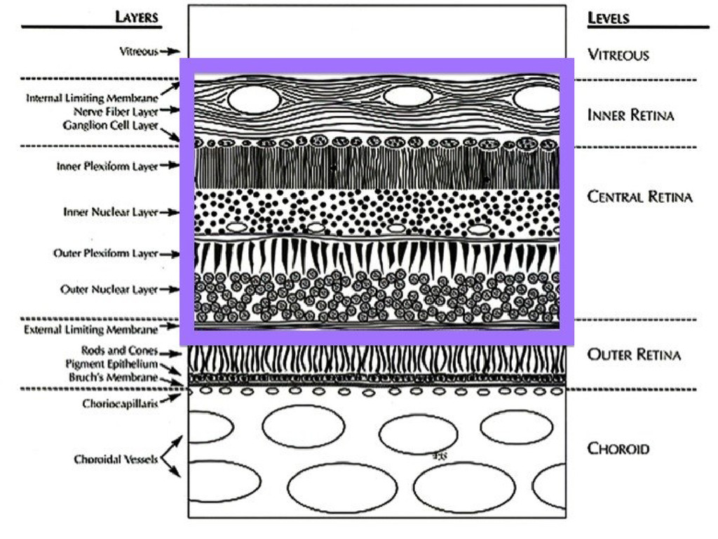
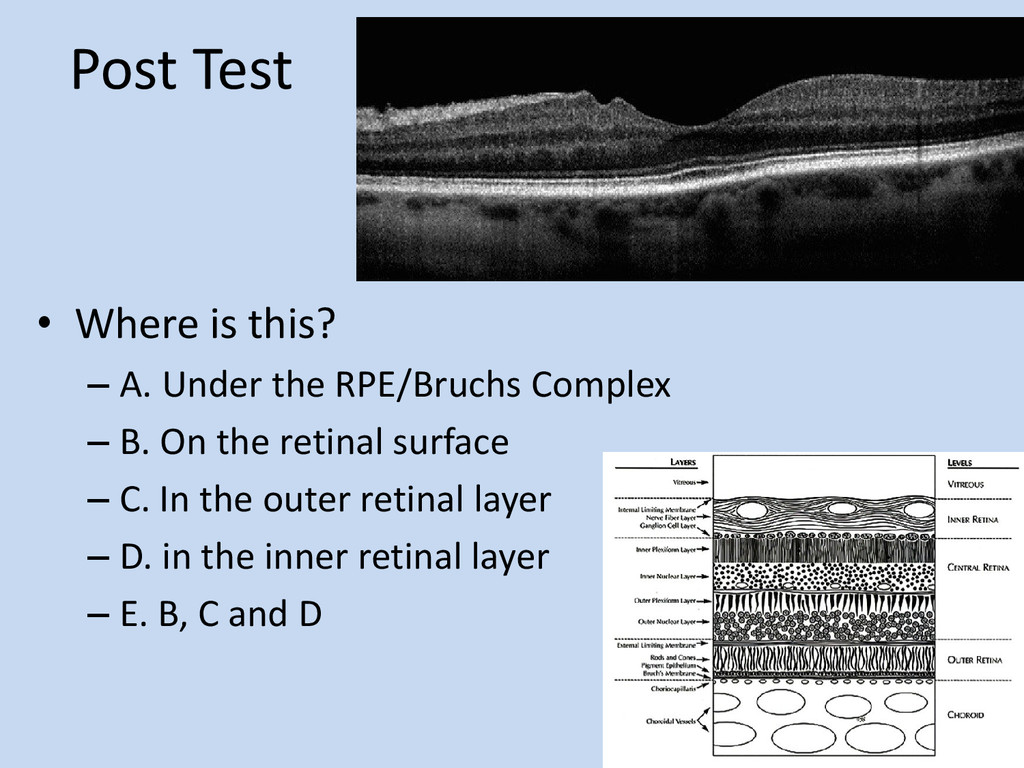
  16. Post Test • Where is this? – A. Under the

    RPE/Bruchs Complex – B. On the retinal surface – C. In the outer retinal layer – D. in the inner retinal layer – E. B, C and D
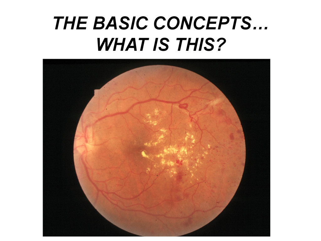
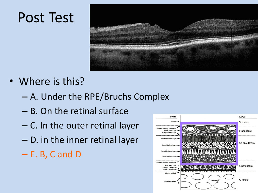
  17. Post Test • Where is this? – A. Under the

    RPE/Bruchs Complex – B. On the retinal surface – C. In the outer retinal layer – D. in the inner retinal layer – E. B, C and D