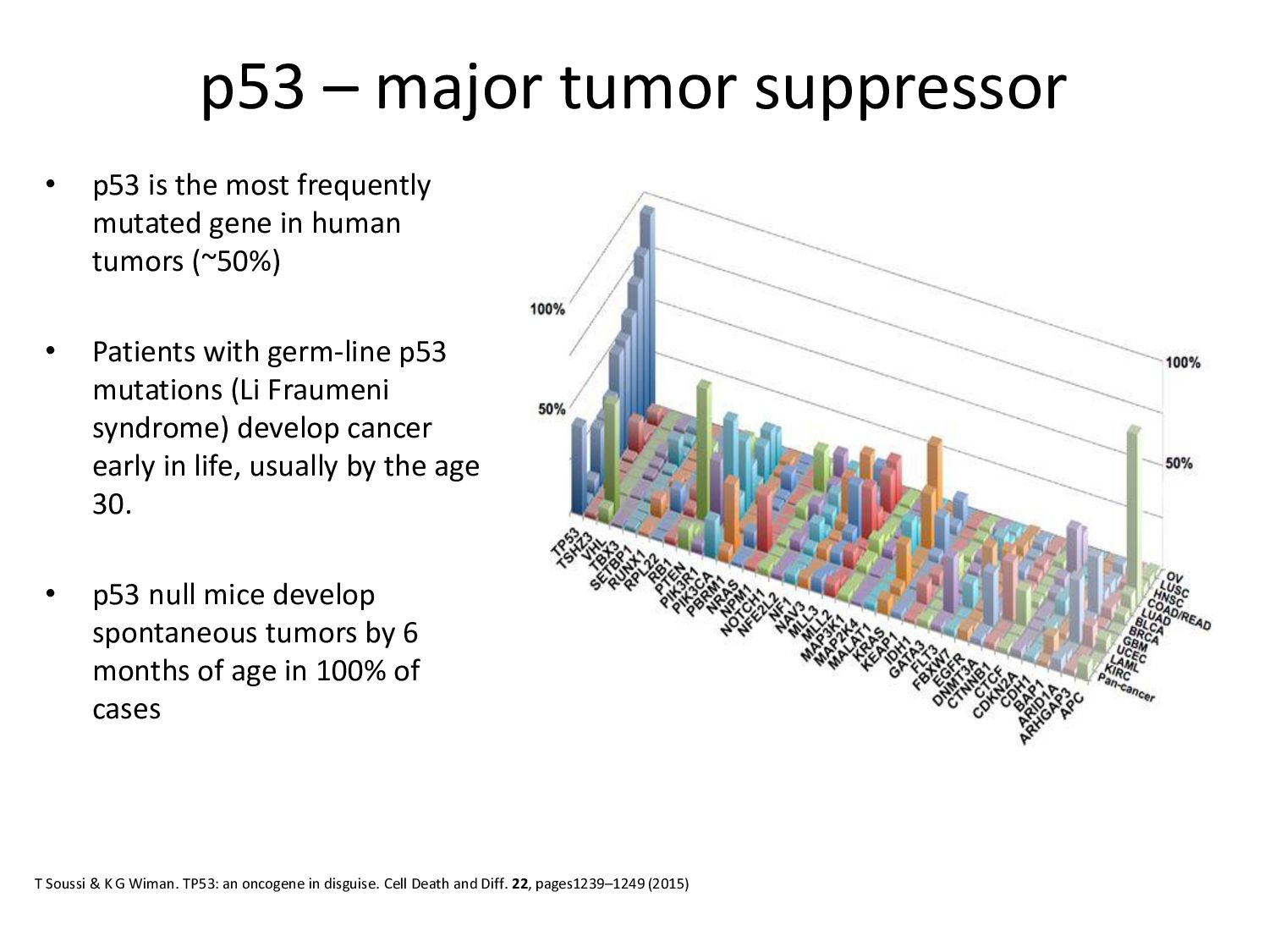
frequently mutated gene in human tumors (~50%) • Patients with germ-line p53 mutations (Li Fraumeni syndrome) develop cancer early in life, usually by the age 30. • p53 null mice develop spontaneous tumors by 6 months of age in 100% of cases T Soussi & K G Wiman. TP53: an oncogene in disguise. Cell Death and Diff. 22, pages1239–1249 (2015)
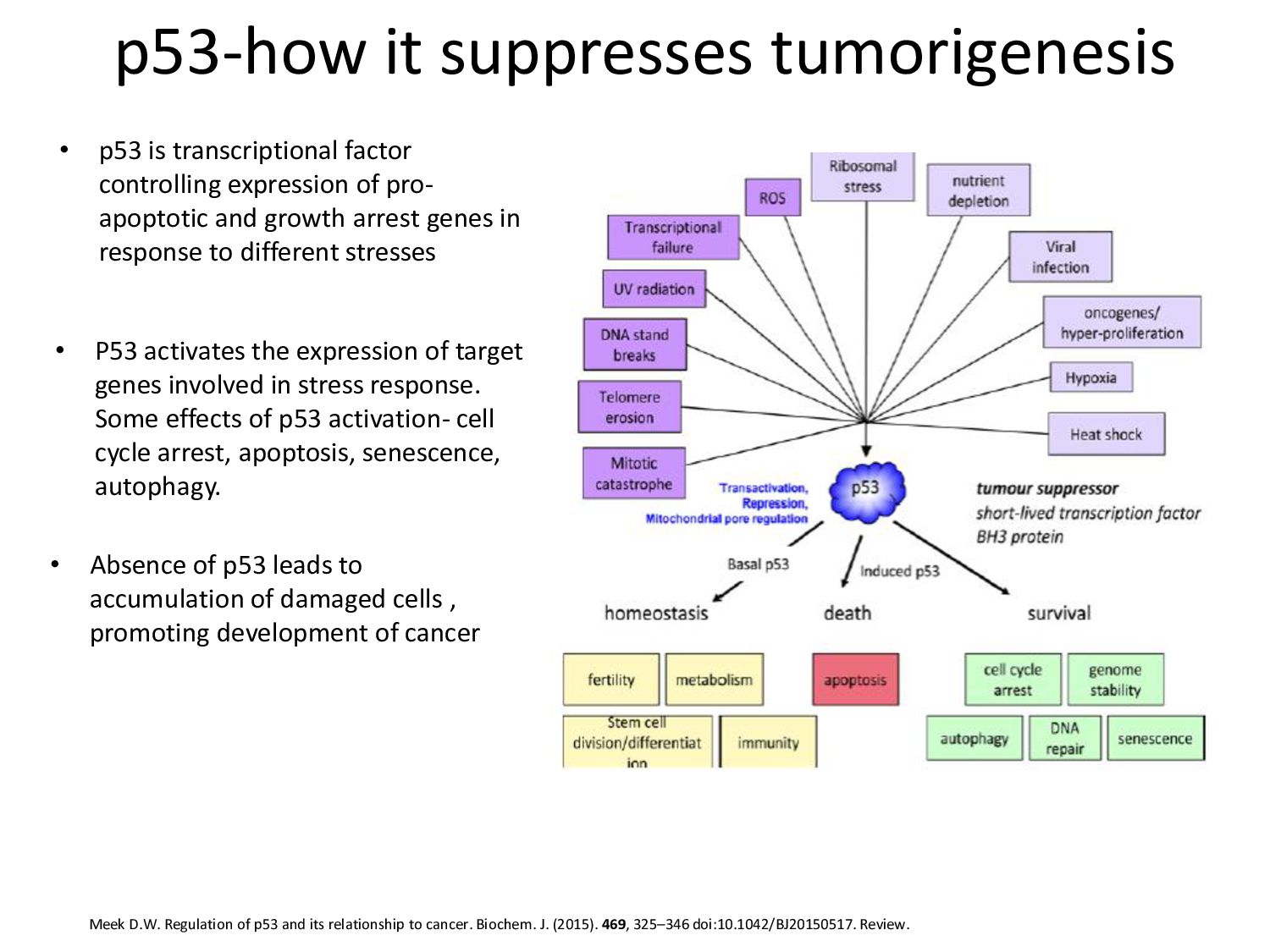
its relationship to cancer. Biochem. J. (2015). 469, 325–346 doi:10.1042/BJ20150517. Review. • P53 activates the expression of target genes involved in stress response. Some effects of p53 activation- cell cycle arrest, apoptosis, senescence, autophagy. • Absence of p53 leads to accumulation of damaged cells , promoting development of cancer • p53 is transcriptional factor controlling expression of pro- apoptotic and growth arrest genes in response to different stresses
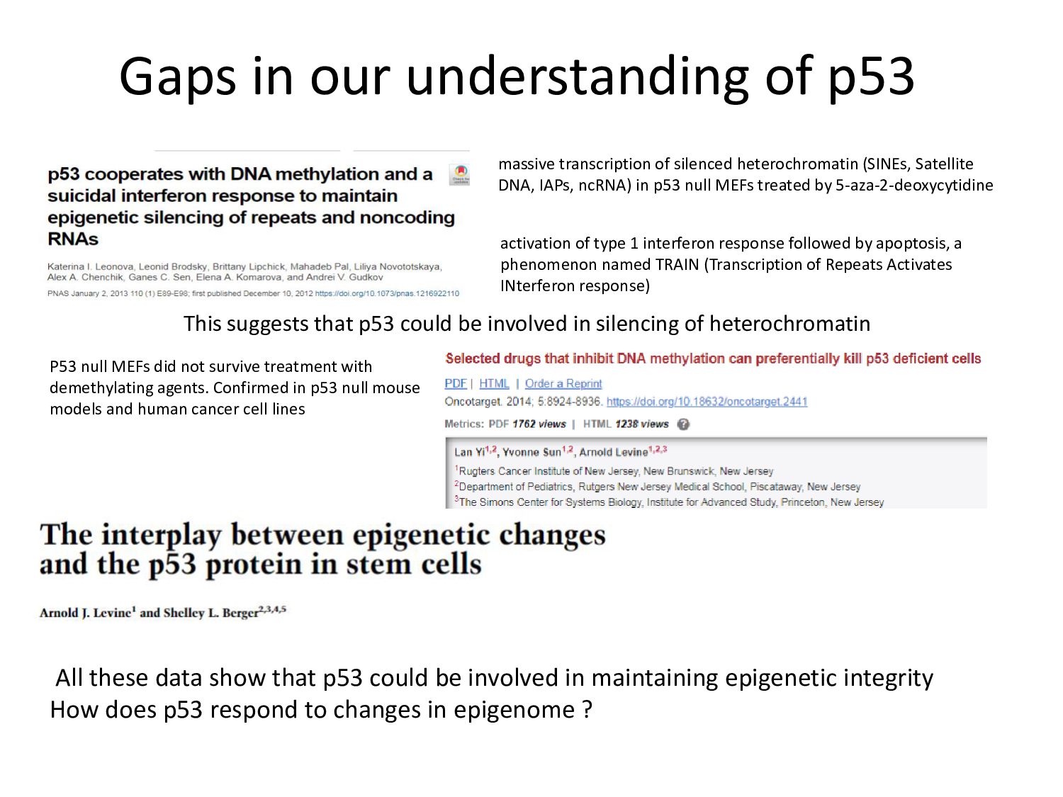
not survive treatment with demethylating agents. Confirmed in p53 null mouse models and human cancer cell lines massive transcription of silenced heterochromatin (SINEs, Satellite DNA, IAPs, ncRNA) in p53 null MEFs treated by 5-aza-2-deoxycytidine activation of type 1 interferon response followed by apoptosis, a phenomenon named TRAIN (Transcription of Repeats Activates INterferon response) All these data show that p53 could be involved in maintaining epigenetic integrity How does p53 respond to changes in epigenome ? This suggests that p53 could be involved in silencing of heterochromatin
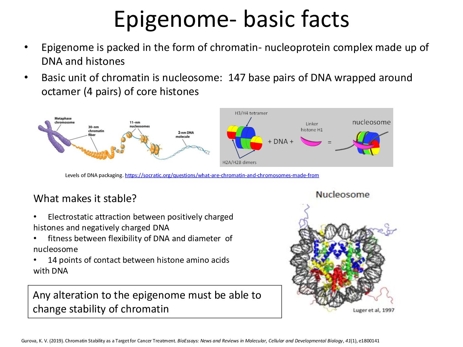
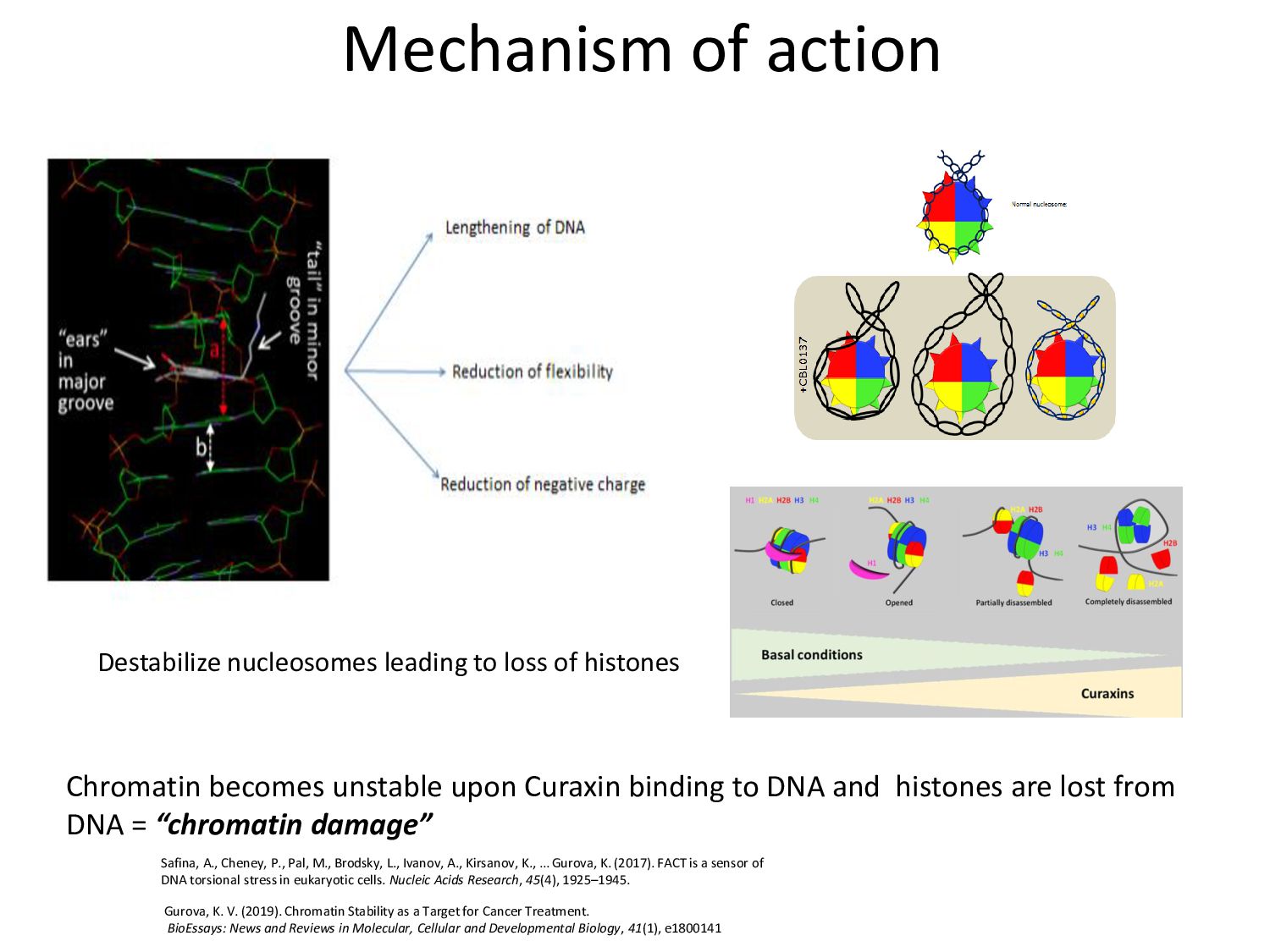
of chromatin- nucleoprotein complex made up of DNA and histones • Basic unit of chromatin is nucleosome: 147 base pairs of DNA wrapped around octamer (4 pairs) of core histones Levels of DNA packaging. https://socratic.org/questions/what-are-chromatin-and-chromosomes-made-from Gurova, K. V. (2019). Chromatin Stability as a Target for Cancer Treatment. BioEssays: News and Reviews in Molecular, Cellular and Developmental Biology, 41(1), e1800141 What makes it stable? • Electrostatic attraction between positively charged histones and negatively charged DNA • fitness between flexibility of DNA and diameter of nucleosome • 14 points of contact between histone amino acids with DNA Any alteration to the epigenome must be able to change stability of chromatin
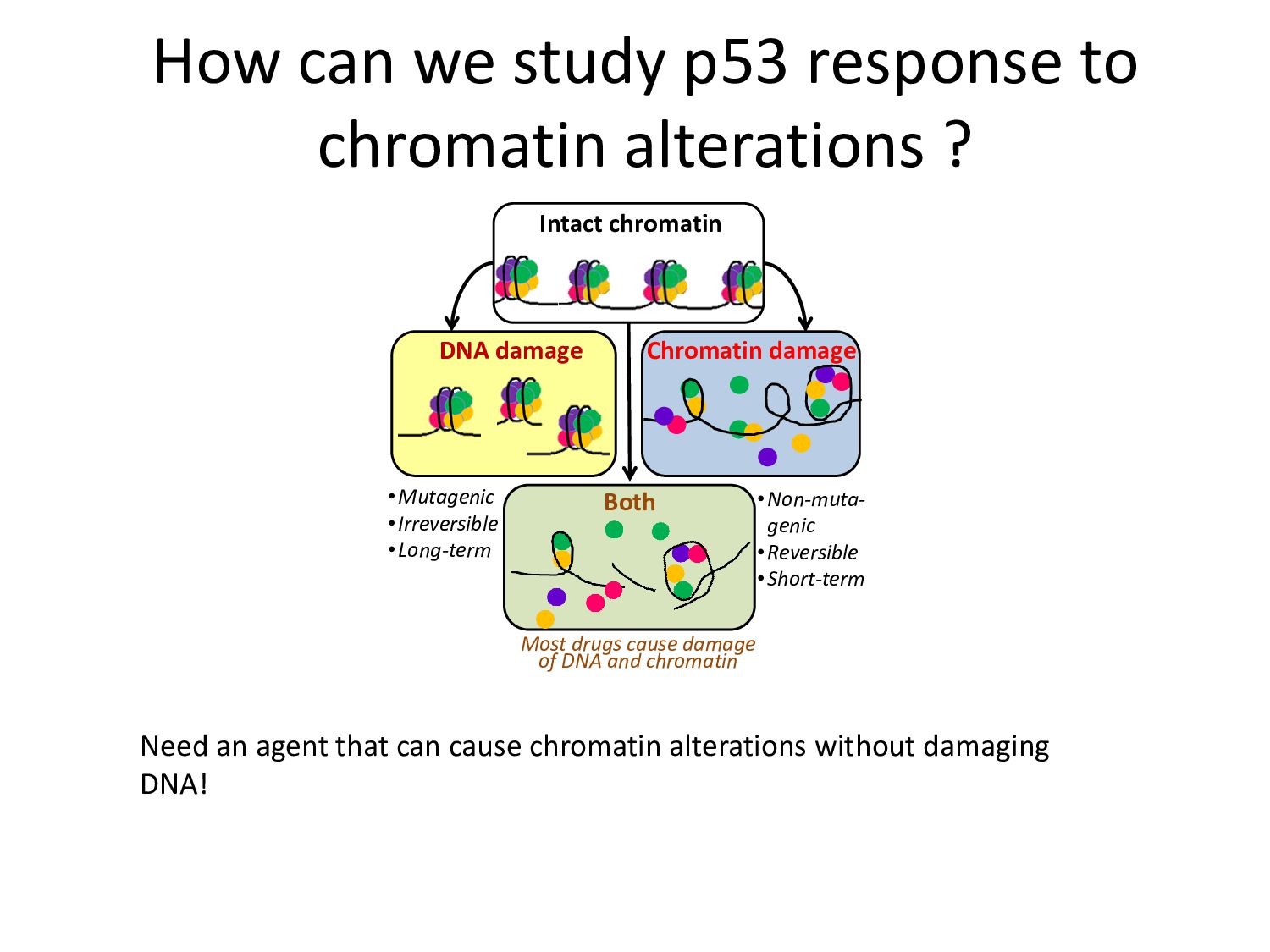
Need an agent that can cause chromatin alterations without damaging DNA! Intact chromatin DNA damage Chromatin damage Both •Mutagenic •Irreversible •Long-term •Non-muta- genic •Reversible •Short-term Most drugs cause damage of DNA and chromatin
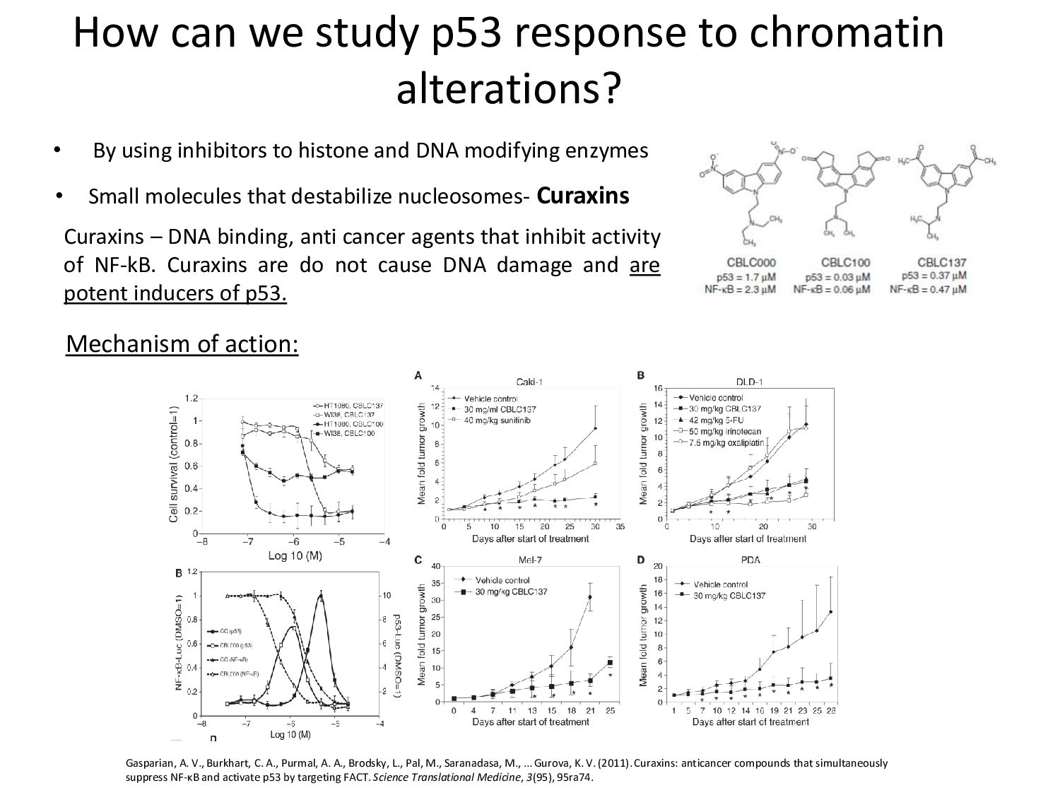
By using inhibitors to histone and DNA modifying enzymes Mechanism of action: Gasparian, A. V., Burkhart, C. A., Purmal, A. A., Brodsky, L., Pal, M., Saranadasa, M., … Gurova, K. V. (2011). Curaxins: anticancer compounds that simultaneously suppress NF-κB and activate p53 by targeting FACT. Science Translational Medicine, 3(95), 95ra74. Curaxins – DNA binding, anti cancer agents that inhibit activity of NF-kB. Curaxins are do not cause DNA damage and are potent inducers of p53. • Small molecules that destabilize nucleosomes- Curaxins
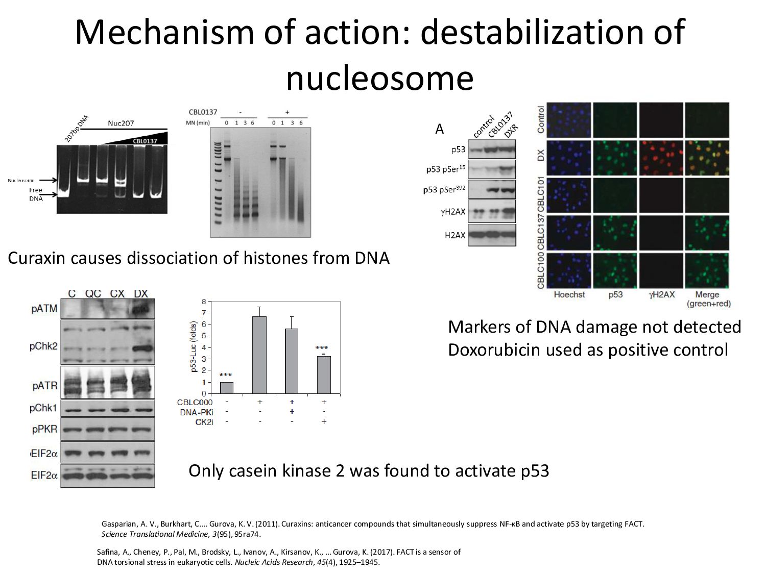
C.… Gurova, K. V. (2011). Curaxins: anticancer compounds that simultaneously suppress NF-κB and activate p53 by targeting FACT. Science Translational Medicine, 3(95), 95ra74. Curaxin causes dissociation of histones from DNA Markers of DNA damage not detected Doxorubicin used as positive control Safina, A., Cheney, P., Pal, M., Brodsky, L., Ivanov, A., Kirsanov, K., … Gurova, K. (2017). FACT is a sensor of DNA torsional stress in eukaryotic cells. Nucleic Acids Research, 45(4), 1925–1945. Fig.3. Comparison of p53 induction by chromatin and DNA damage. A. Immunoblotting of lysates of HT1080 cells treated with CBL0137 or dox (DXR) for 16 hrs. B-C. Comparison of the list of known p53 targets (MSig lists of genes which expression was changed in HT1080 cells 16 hrs afte treatment with CBL0137, doxorubicin or gamma-irradiation (6Gy) asses microarray hybridization. A B Only casein kinase 2 was found to activate p53
DNA and histones are lost from DNA = “chromatin damage” Destabilize nucleosomes leading to loss of histones Gurova, K. V. (2019). Chromatin Stability as a Target for Cancer Treatment. BioEssays: News and Reviews in Molecular, Cellular and Developmental Biology, 41(1), e1800141 Safina, A., Cheney, P., Pal, M., Brodsky, L., Ivanov, A., Kirsanov, K., … Gurova, K. (2017). FACT is a sensor of DNA torsional stress in eukaryotic cells. Nucleic Acids Research, 45(4), 1925–1945.
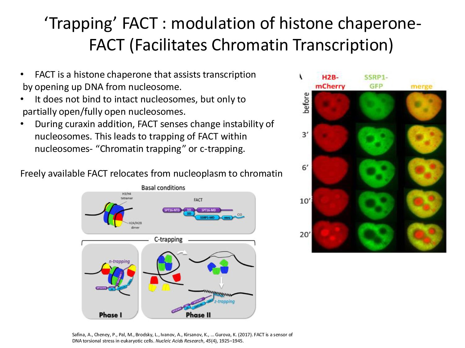
Transcription) • FACT is a histone chaperone that assists transcription by opening up DNA from nucleosome. • It does not bind to intact nucleosomes, but only to partially open/fully open nucleosomes. • During curaxin addition, FACT senses change instability of nucleosomes. This leads to trapping of FACT within nucleosomes- “Chromatin trapping” or c-trapping. Freely available FACT relocates from nucleoplasm to chromatin Safina, A., Cheney, P., Pal, M., Brodsky, L., Ivanov, A., Kirsanov, K., … Gurova, K. (2017). FACT is a sensor of DNA torsional stress in eukaryotic cells. Nucleic Acids Research, 45(4), 1925–1945.
propose two models: Addition of curaxins changes levels of free FACT/bound FACT. • FACT unavailable for phosphorylation by Casein Kinase 2 (CK2). This signals activation of p53 (at Ser392-P) by CK2. • Dissociated histones relocate to nucleolus, releasing MDM2 inhibitors like ARF. Anti-tumor effect of curaxins is mediated through two ways: – Reducing availability of FACT for general transcription – Activation of p53 through FACT- CK2 complex Question: Could p53 response to chromatin damage be different from response to DNA damage ? Fig.2. Potential pathways of p53 activation by chromatin damage. Solid red arrows – established events, dashed – proposed. Standard color code is used for core histones.
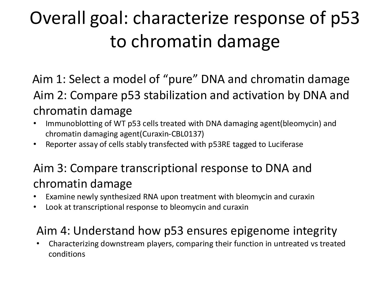
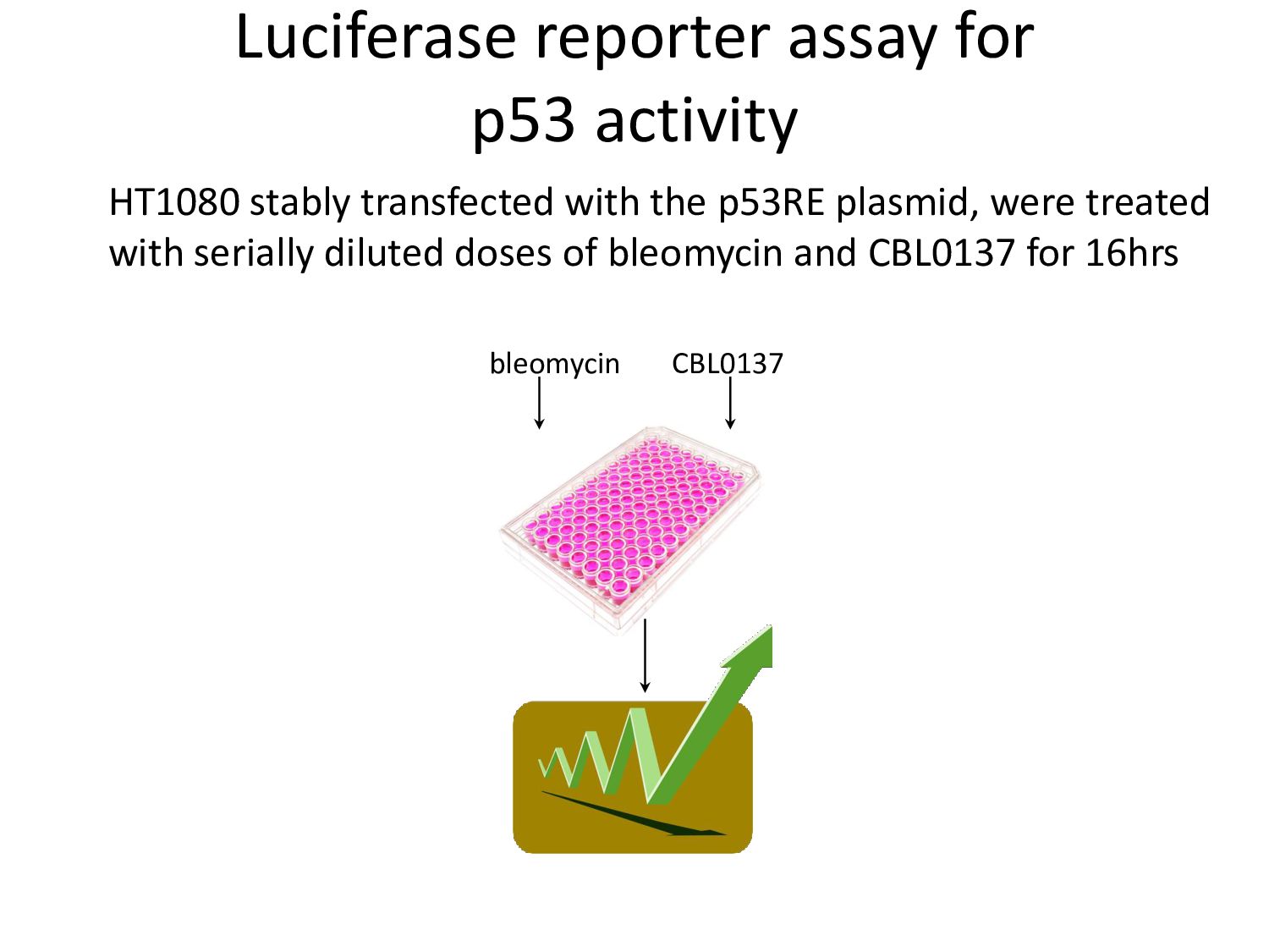
1: Select a model of “pure” DNA and chromatin damage Aim 2: Compare p53 stabilization and activation by DNA and chromatin damage • Immunoblotting of WT p53 cells treated with DNA damaging agent(bleomycin) and chromatin damaging agent(Curaxin-CBL0137) • Reporter assay of cells stably transfected with p53RE tagged to Luciferase Aim 3: Compare transcriptional response to DNA and chromatin damage • Examine newly synthesized RNA upon treatment with bleomycin and curaxin • Look at transcriptional response to bleomycin and curaxin Aim 4: Understand how p53 ensures epigenome integrity • Characterizing downstream players, comparing their function in untreated vs treated conditions
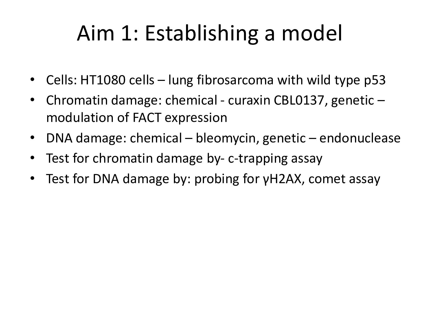
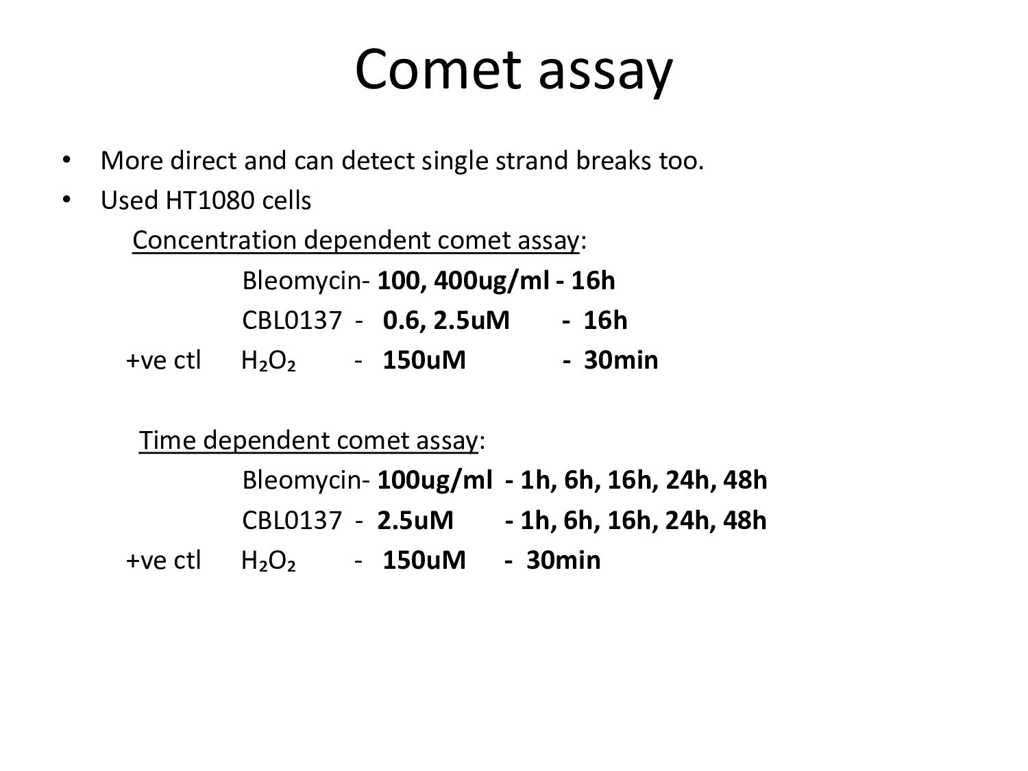
lung fibrosarcoma with wild type p53 • Chromatin damage: chemical - curaxin CBL0137, genetic – modulation of FACT expression • DNA damage: chemical – bleomycin, genetic – endonuclease • Test for chromatin damage by- c-trapping assay • Test for DNA damage by: probing for γH2AX, comet assay
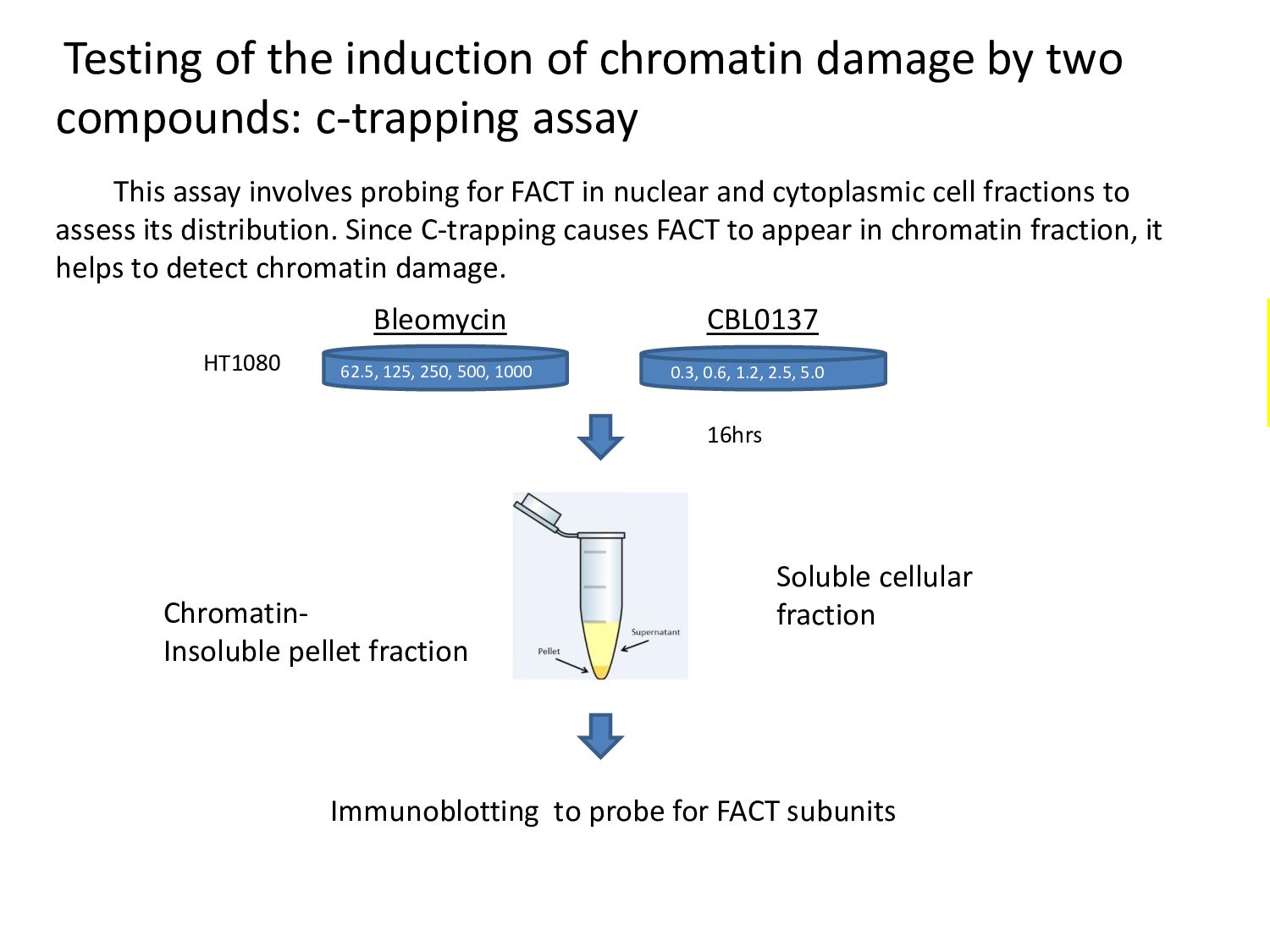
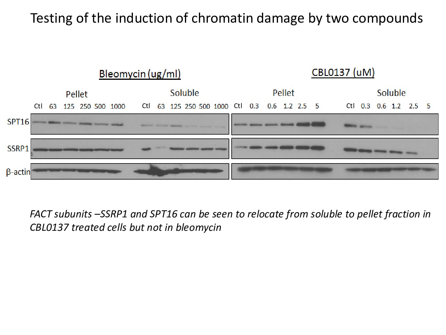
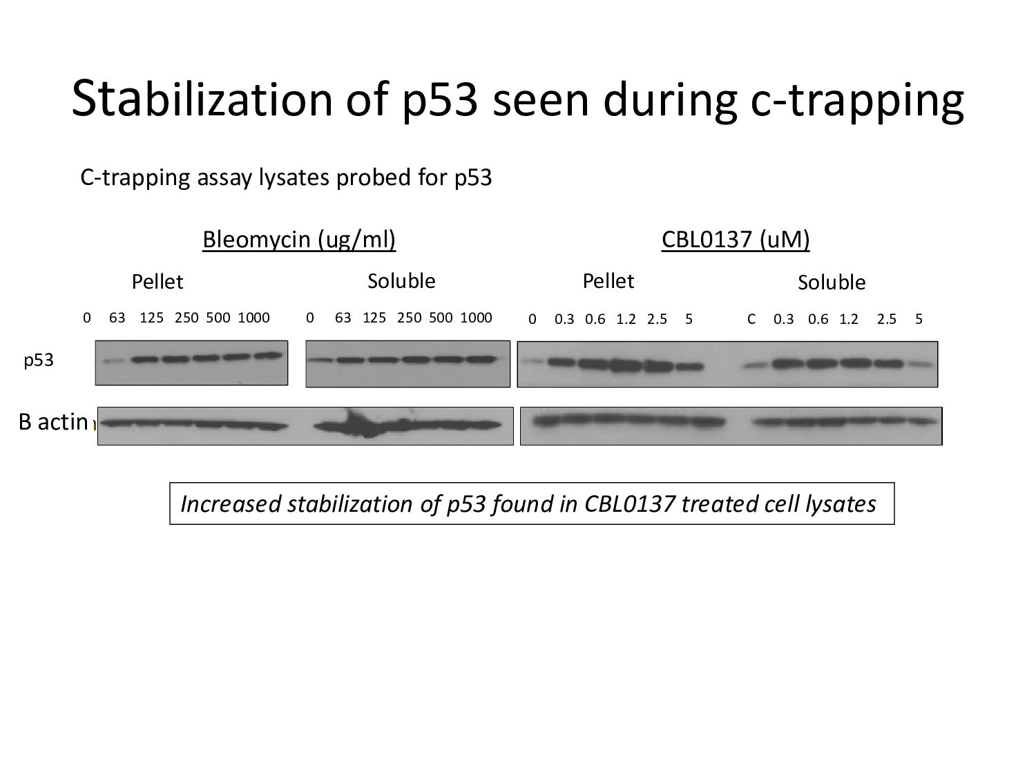
c-trapping assay This assay involves probing for FACT in nuclear and cytoplasmic cell fractions to assess its distribution. Since C-trapping causes FACT to appear in chromatin fraction, it helps to detect chromatin damage. 62.5, 125, 250, 500, 1000 0.3, 0.6, 1.2, 2.5, 5.0 Bleomycin CBL0137 Immunoblotting to probe for FACT subunits HT1080 16hrs Chromatin- Insoluble pellet fraction Soluble cellular fraction
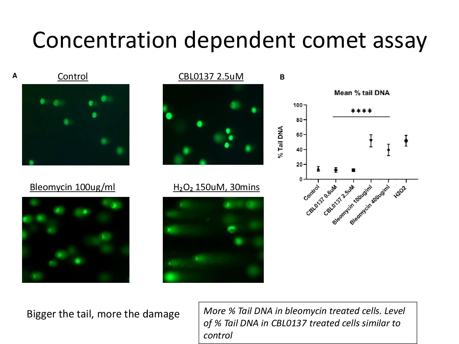
More % Tail DNA in bleomycin treated cells. Level of % Tail DNA in CBL0137 treated cells similar to control Bigger the tail, more the damage Concentration dependent comet assay
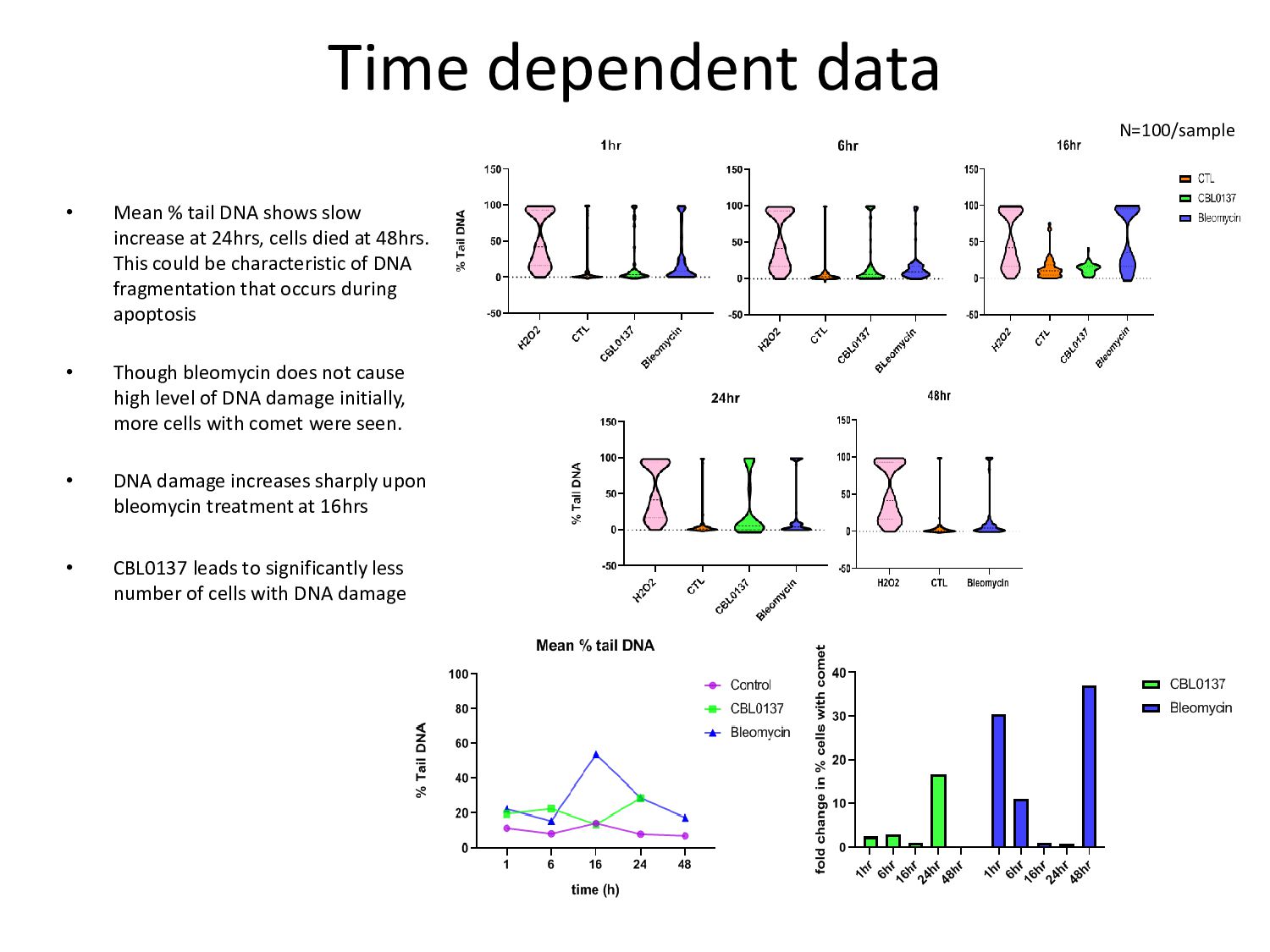
increase at 24hrs, cells died at 48hrs. This could be characteristic of DNA fragmentation that occurs during apoptosis • Though bleomycin does not cause high level of DNA damage initially, more cells with comet were seen. • DNA damage increases sharply upon bleomycin treatment at 16hrs • CBL0137 leads to significantly less number of cells with DNA damage N=100/sample
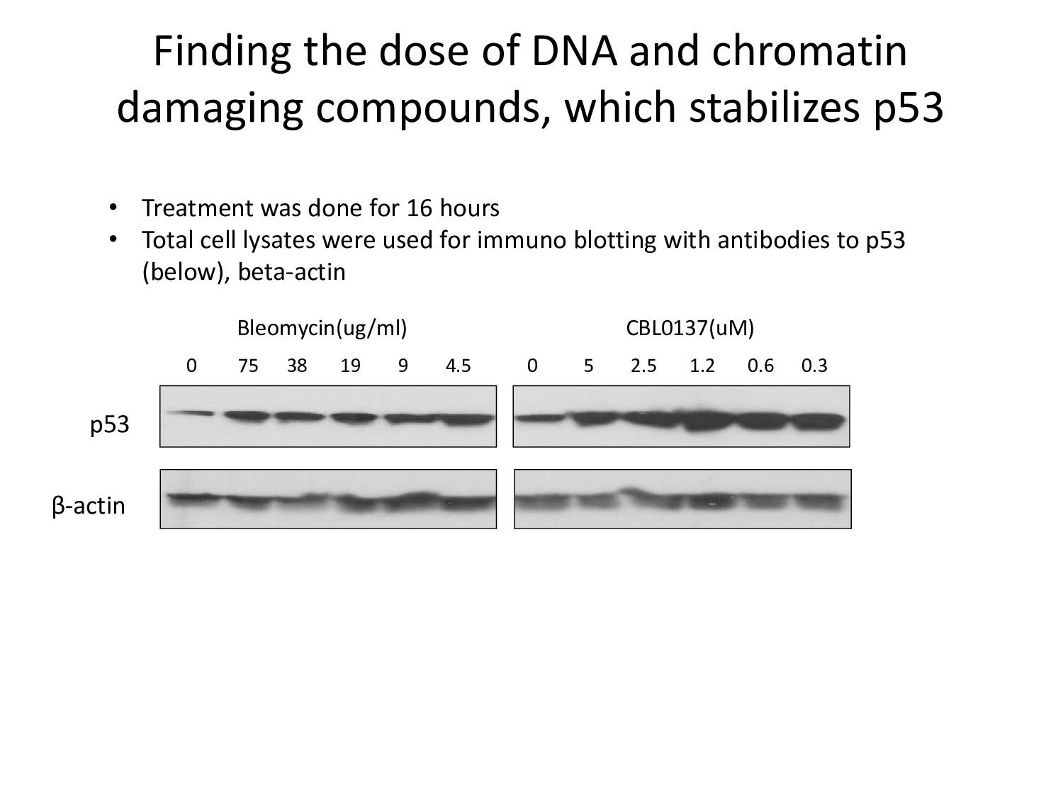
2.5 1.2 0.6 0.3 p53 β-actin Finding the dose of DNA and chromatin damaging compounds, which stabilizes p53 • Treatment was done for 16 hours • Total cell lysates were used for immuno blotting with antibodies to p53 (below), beta-actin
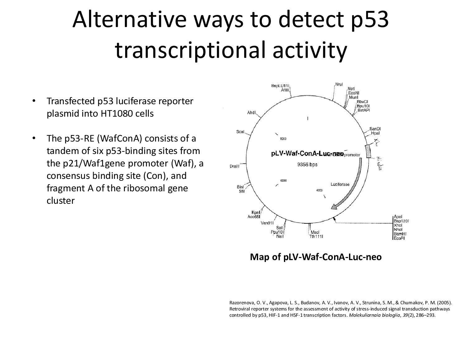
• Transfected p53 luciferase reporter plasmid into HT1080 cells • The p53-RE (WafConA) consists of a tandem of six p53-binding sites from the p21/Waf1gene promoter (Waf), a consensus binding site (Con), and fragment A of the ribosomal gene cluster Razorenova, O. V., Agapova, L. S., Budanov, A. V., Ivanov, A. V., Strunina, S. M., & Chumakov, P. M. (2005). Retroviral reporter systems for the assessment of activity of stress-induced signal transduction pathways controlled by p53, HIF-1 and HSF-1 transcription factors. Molekuliarnaia biologiia, 39(2), 286–293.
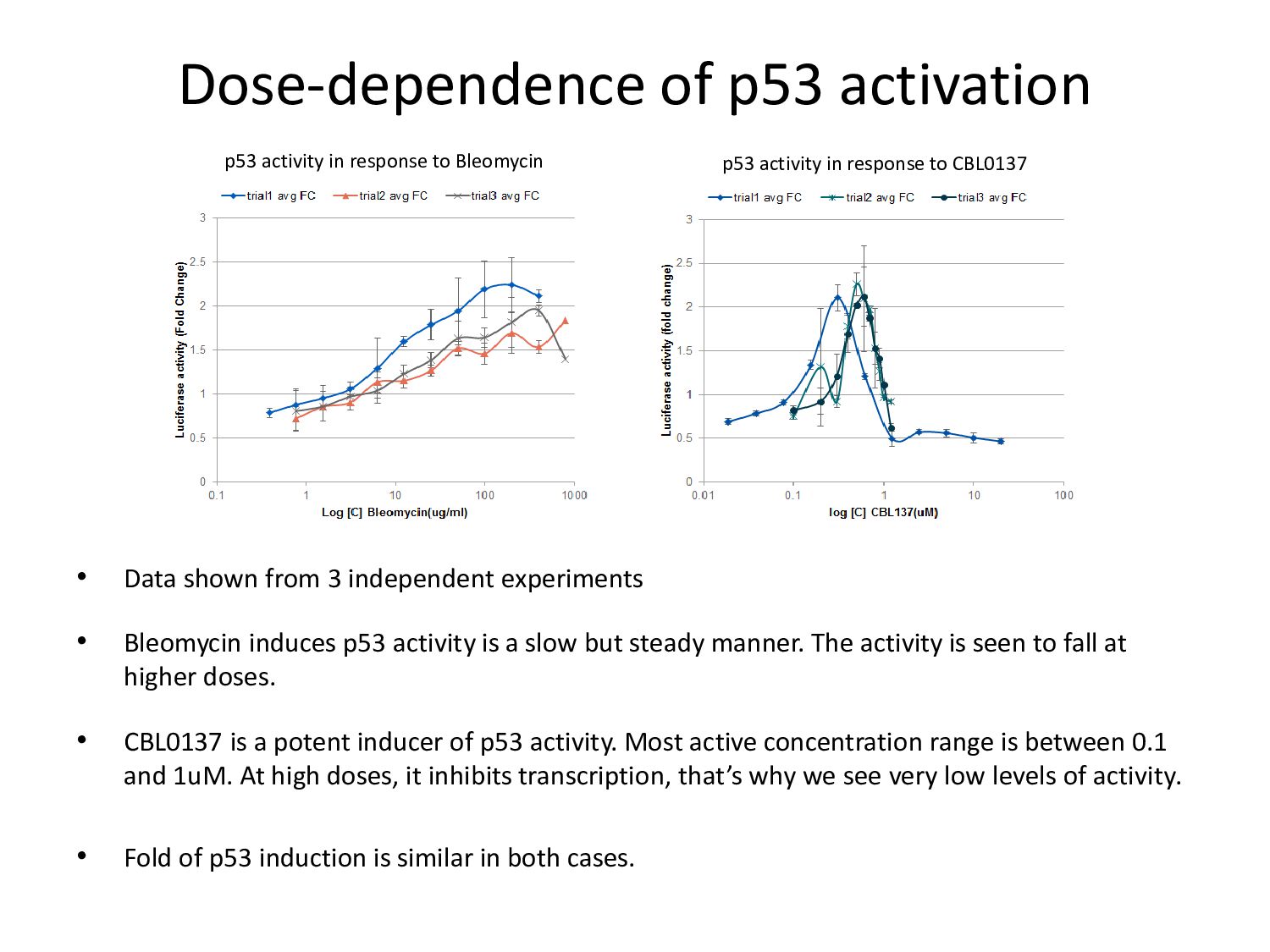
p53 activity is a slow but steady manner. The activity is seen to fall at higher doses. • CBL0137 is a potent inducer of p53 activity. Most active concentration range is between 0.1 and 1uM. At high doses, it inhibits transcription, that’s why we see very low levels of activity. • Fold of p53 induction is similar in both cases. Dose-dependence of p53 activation p53 activity in response to Bleomycin p53 activity in response to CBL0137
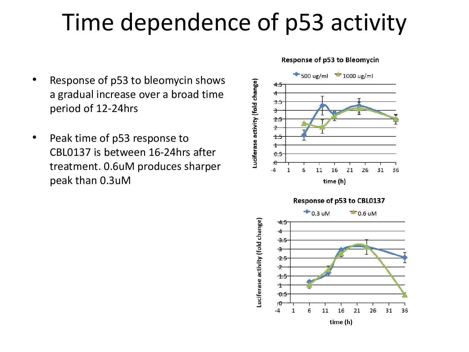
bleomycin shows a gradual increase over a broad time period of 12-24hrs • Peak time of p53 response to CBL0137 is between 16-24hrs after treatment. 0.6uM produces sharper peak than 0.3uM
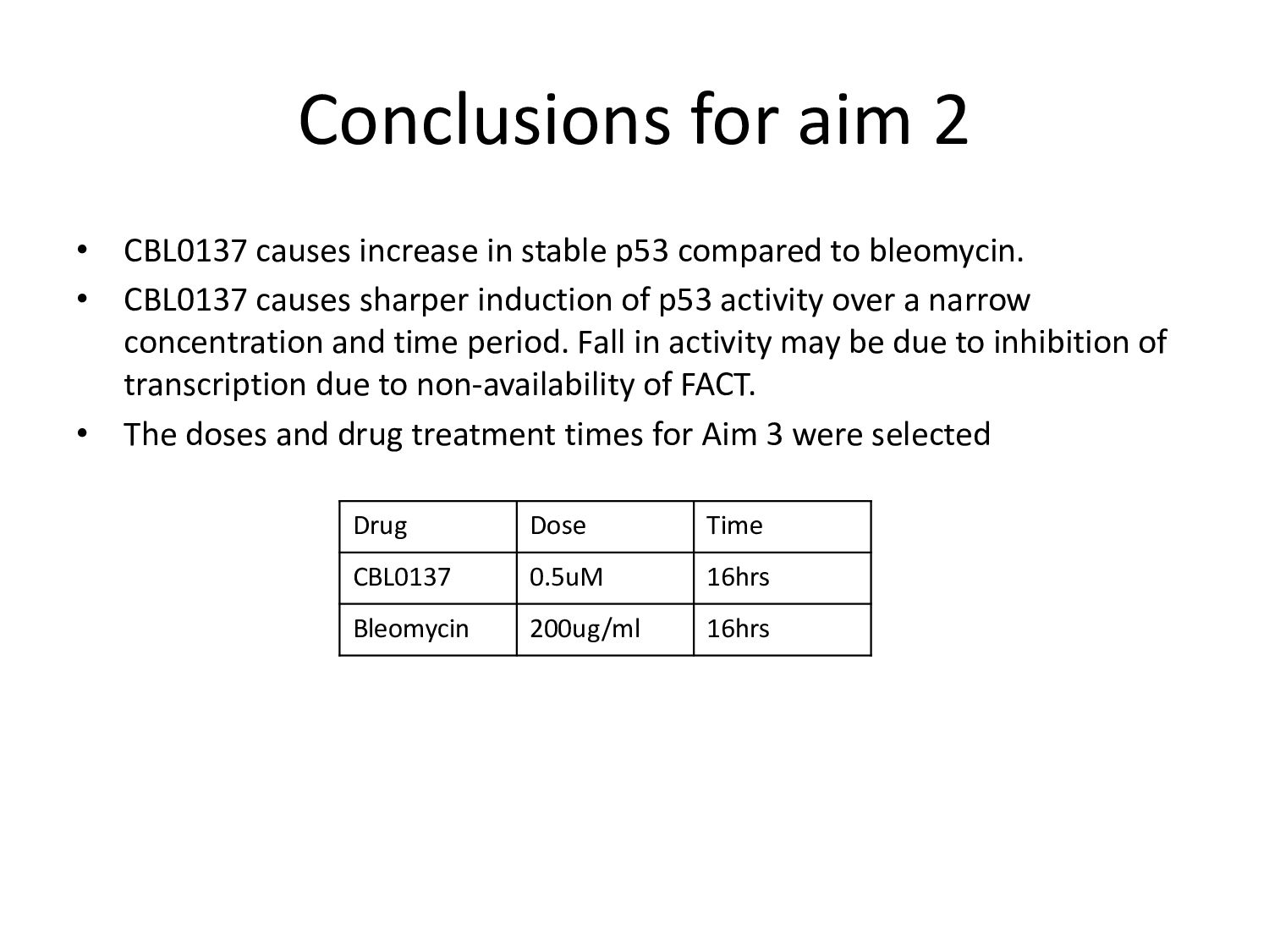
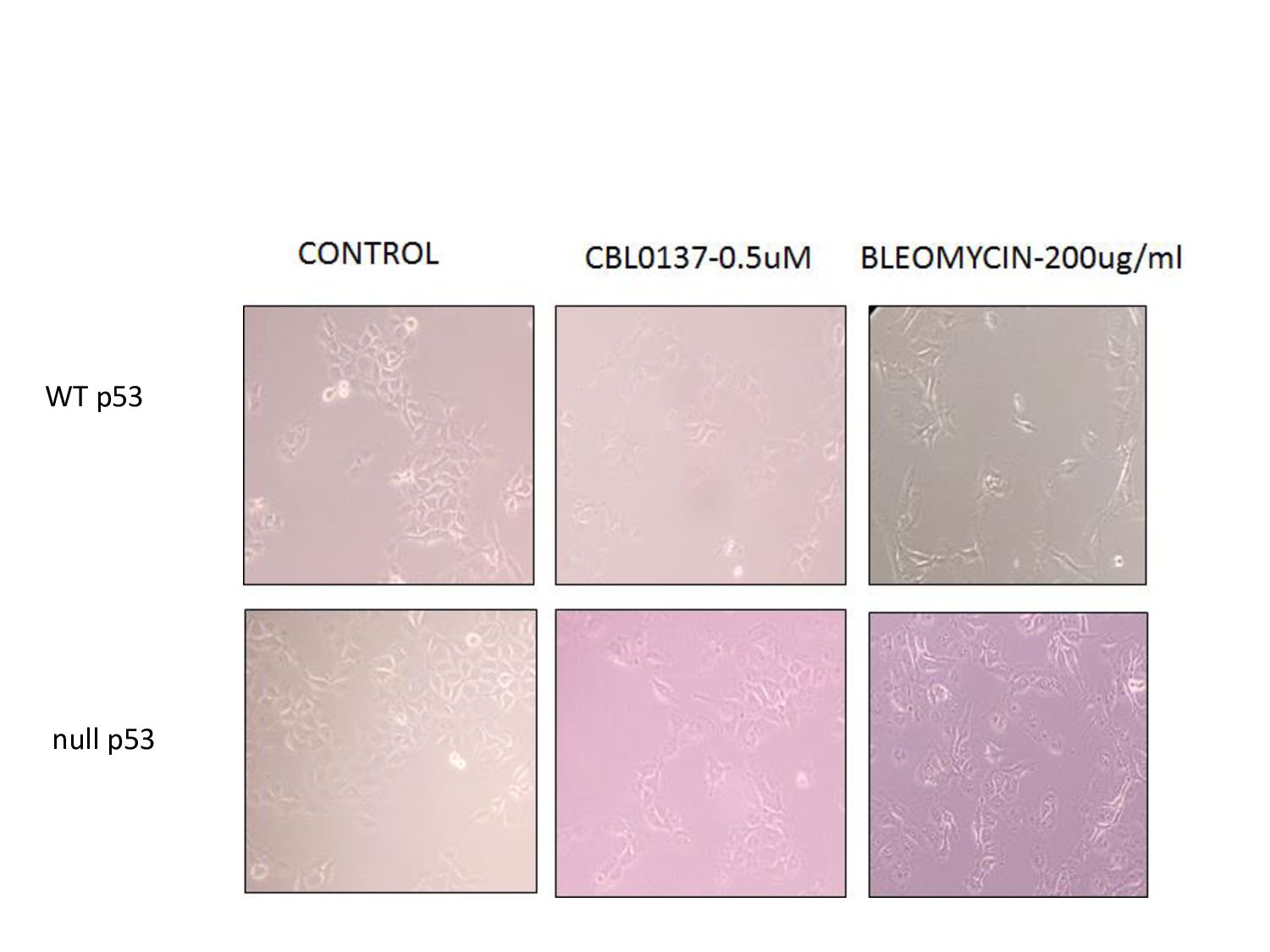
p53 compared to bleomycin. • CBL0137 causes sharper induction of p53 activity over a narrow concentration and time period. Fall in activity may be due to inhibition of transcription due to non-availability of FACT. • The doses and drug treatment times for Aim 3 were selected Drug Dose Time CBL0137 0.5uM 16hrs Bleomycin 200ug/ml 16hrs
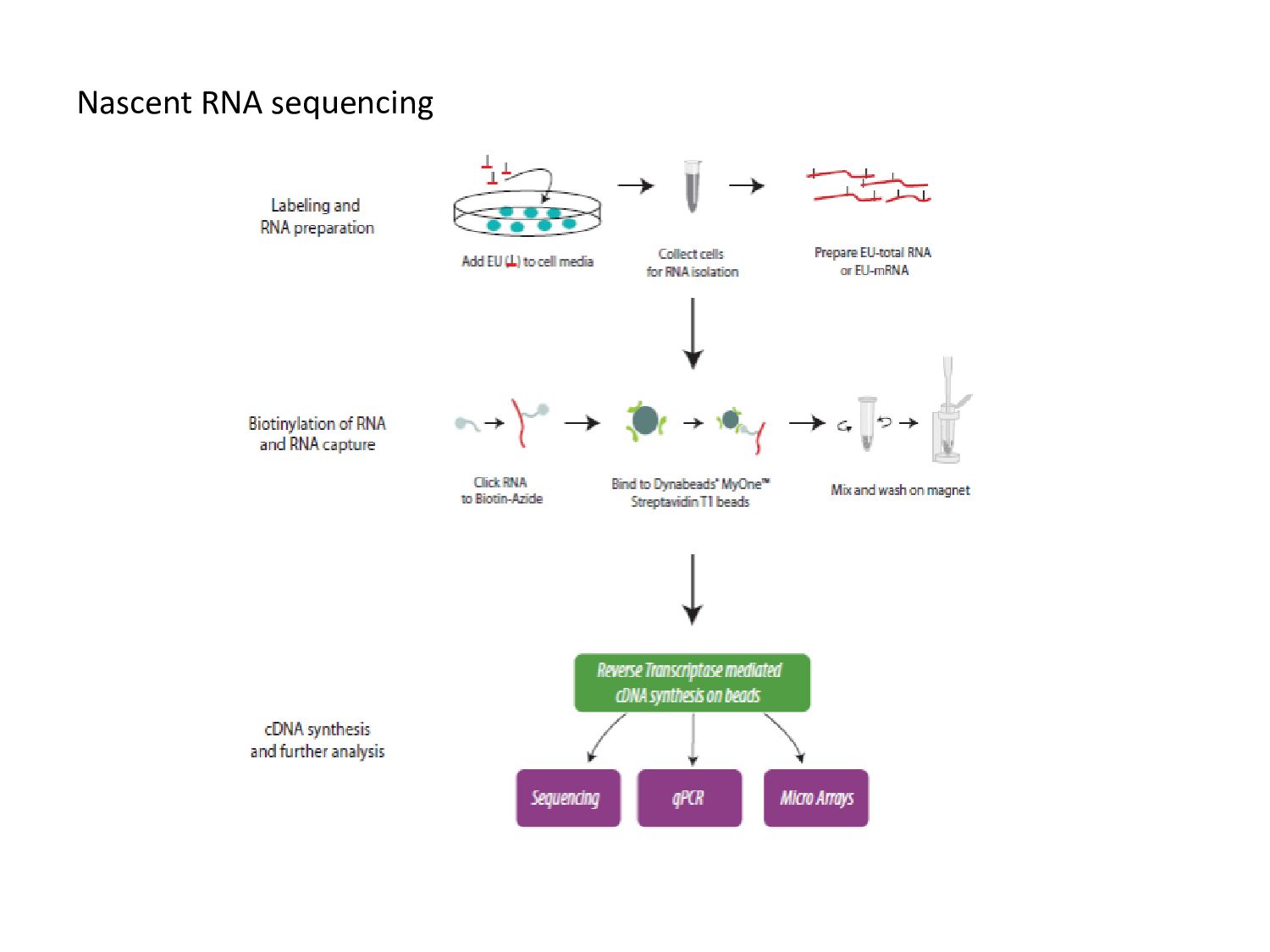
group of uridine Cu biotin bound EU HT1080 Ctl bleomycin CBL137 200ug/ml 0.5uM 16hr incubation Treatment with 1mM EU for 20mins RNA isolation Click reaction-to biotinylate EU cDNA synthesis
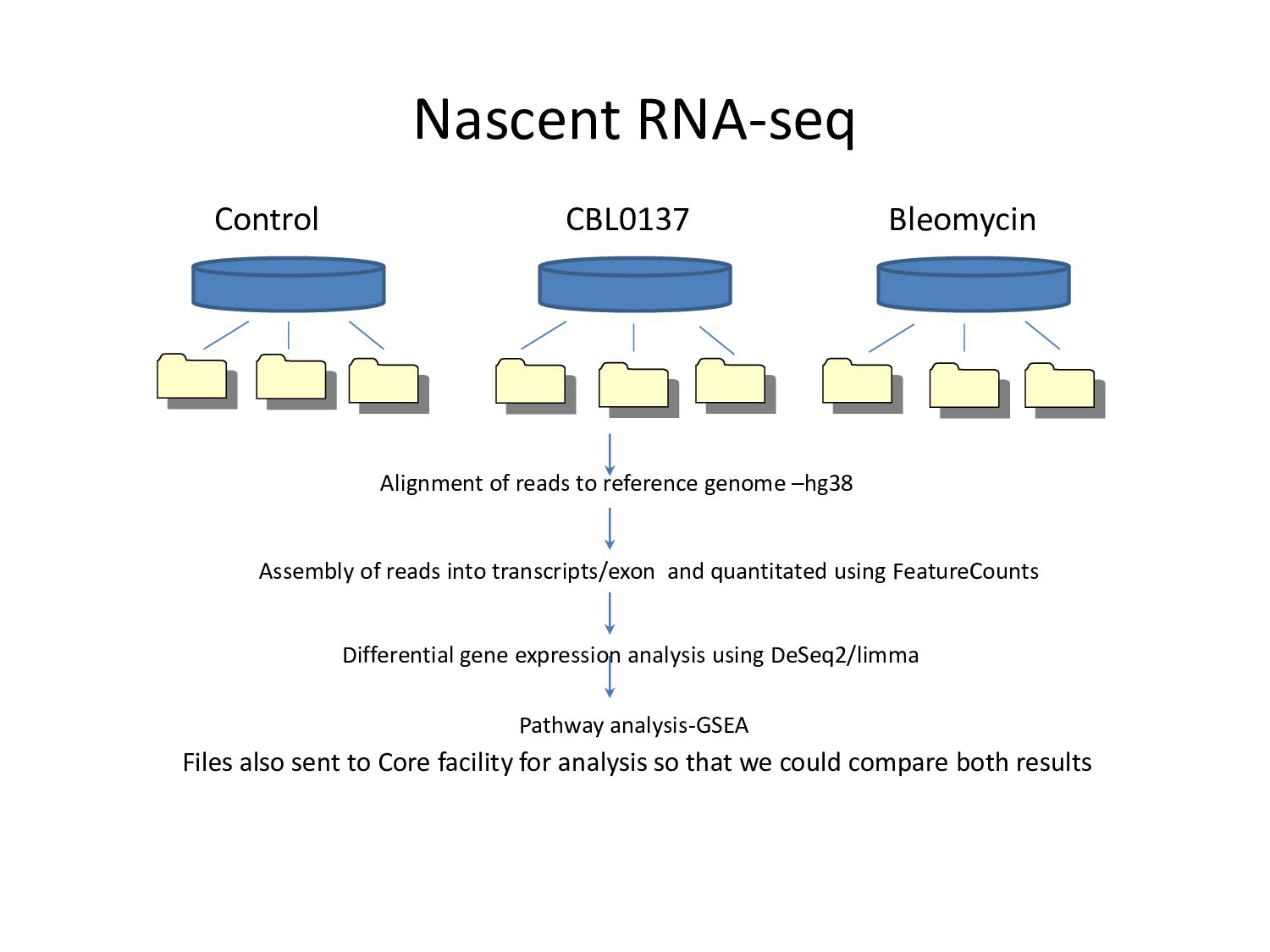
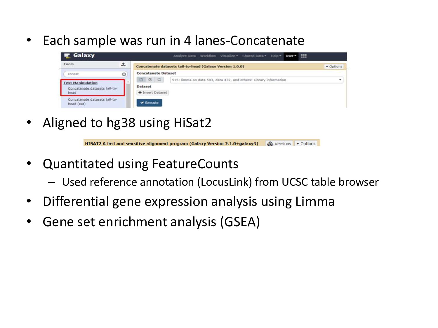
genome –hg38 Assembly of reads into transcripts/exon and quantitated using FeatureCounts Differential gene expression analysis using DeSeq2/limma Pathway analysis-GSEA Files also sent to Core facility for analysis so that we could compare both results
to hg38 using HiSat2 • Quantitated using FeatureCounts – Used reference annotation (LocusLink) from UCSC table browser • Differential gene expression analysis using Limma • Gene set enrichment analysis (GSEA)
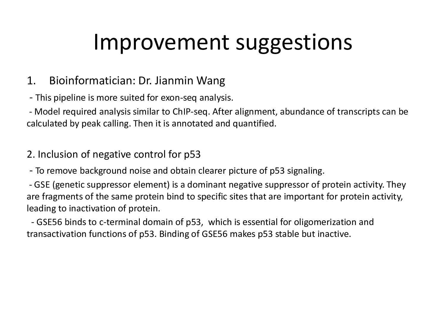
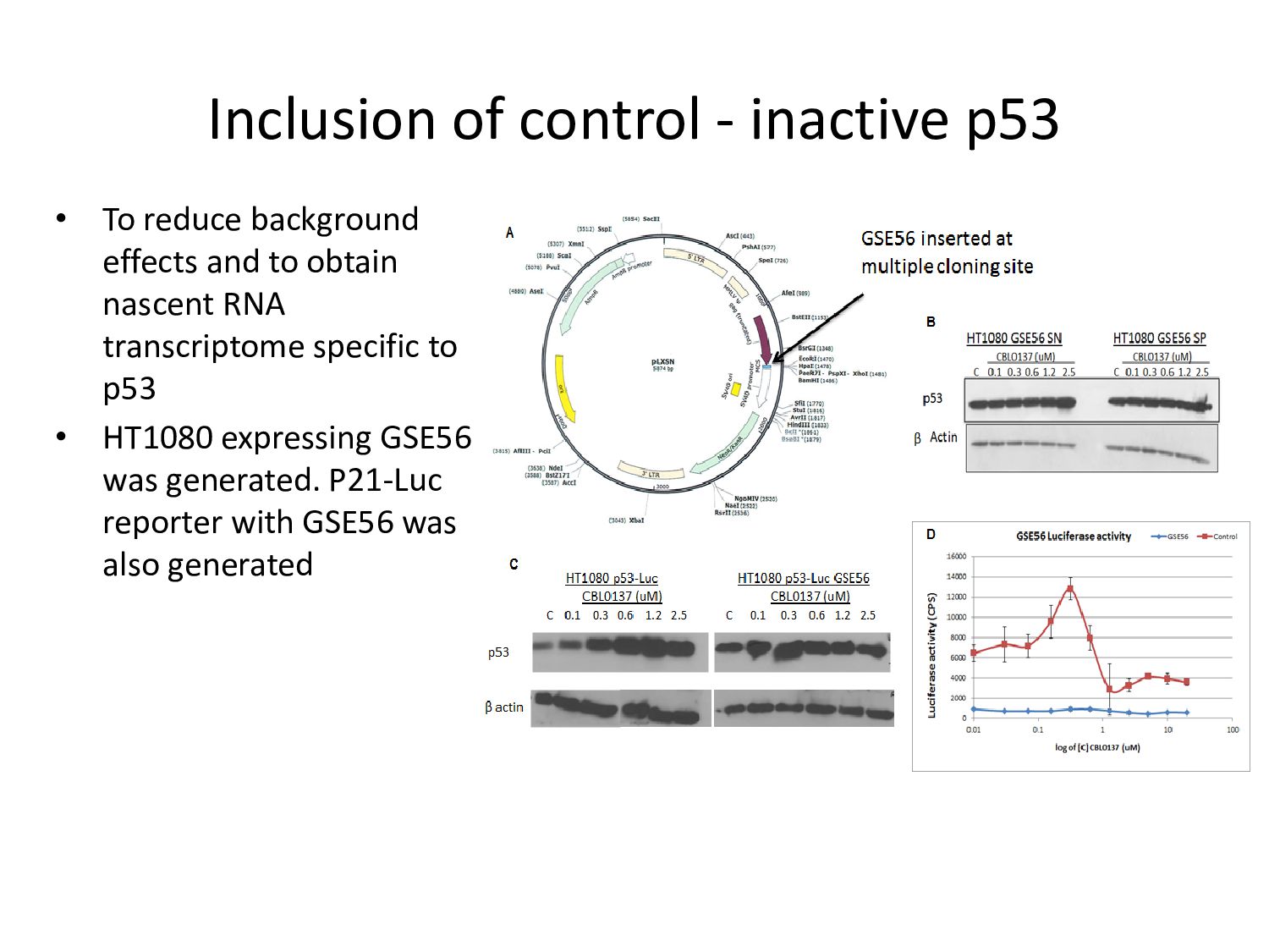
is more suited for exon-seq analysis. - Model required analysis similar to ChIP-seq. After alignment, abundance of transcripts can be calculated by peak calling. Then it is annotated and quantified. 2. Inclusion of negative control for p53 - To remove background noise and obtain clearer picture of p53 signaling. - GSE (genetic suppressor element) is a dominant negative suppressor of protein activity. They are fragments of the same protein bind to specific sites that are important for protein activity, leading to inactivation of protein. - GSE56 binds to c-terminal domain of p53, which is essential for oligomerization and transactivation functions of p53. Binding of GSE56 makes p53 stable but inactive.
effects and to obtain nascent RNA transcriptome specific to p53 • HT1080 expressing GSE56 was generated. P21-Luc reporter with GSE56 was also generated
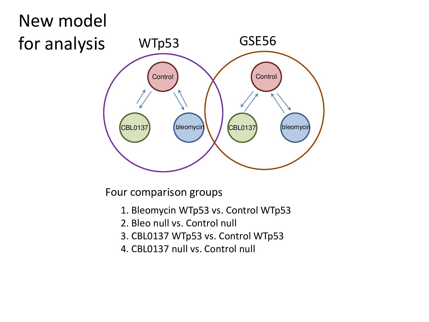
Control WTp53 2. Bleo null vs. Control null 3. CBL0137 WTp53 vs. Control WTp53 4. CBL0137 null vs. Control null Control Control CBL0137 CBL0137 bleomycin bleomycin Four comparison groups
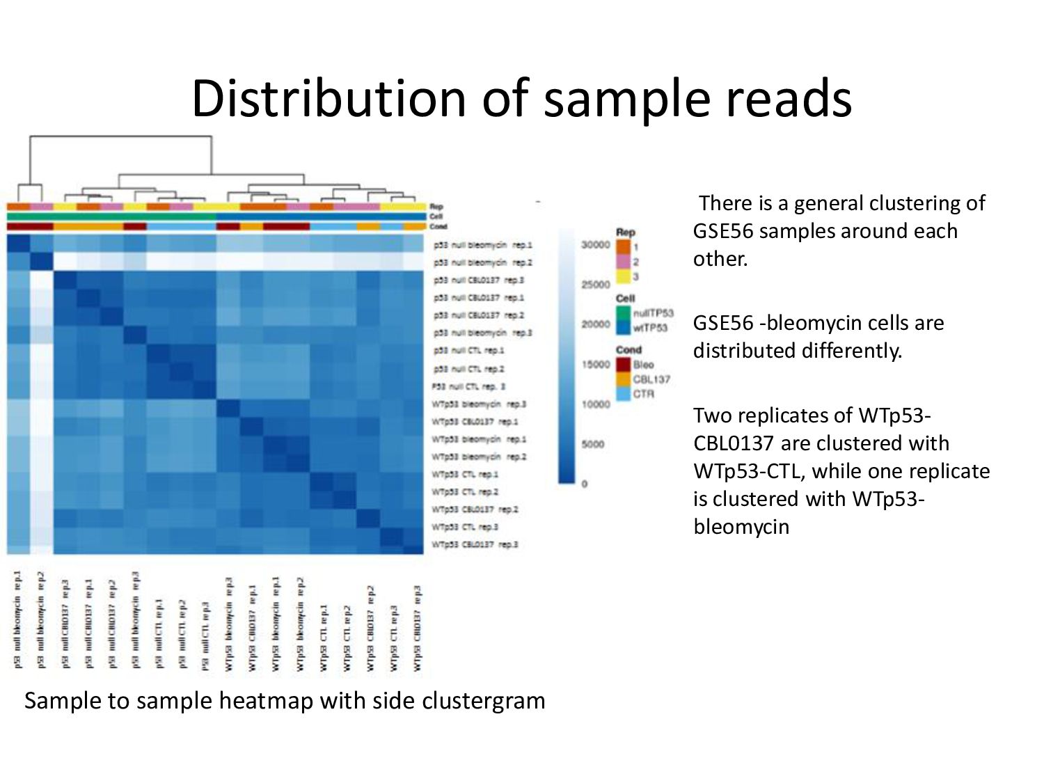
GSE56 samples around each other. GSE56 -bleomycin cells are distributed differently. Two replicates of WTp53- CBL0137 are clustered with WTp53-CTL, while one replicate is clustered with WTp53- bleomycin Sample to sample heatmap with side clustergram
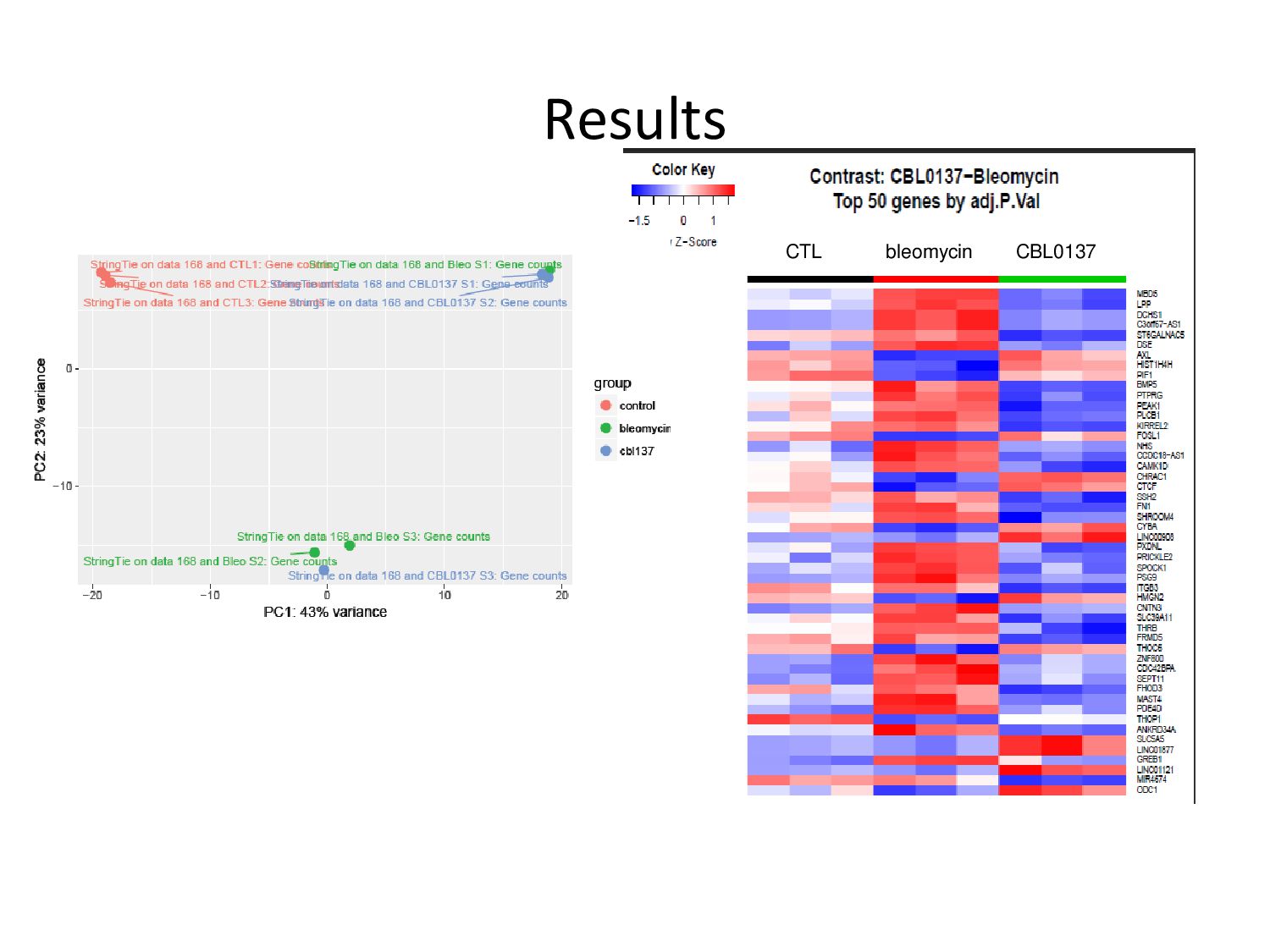
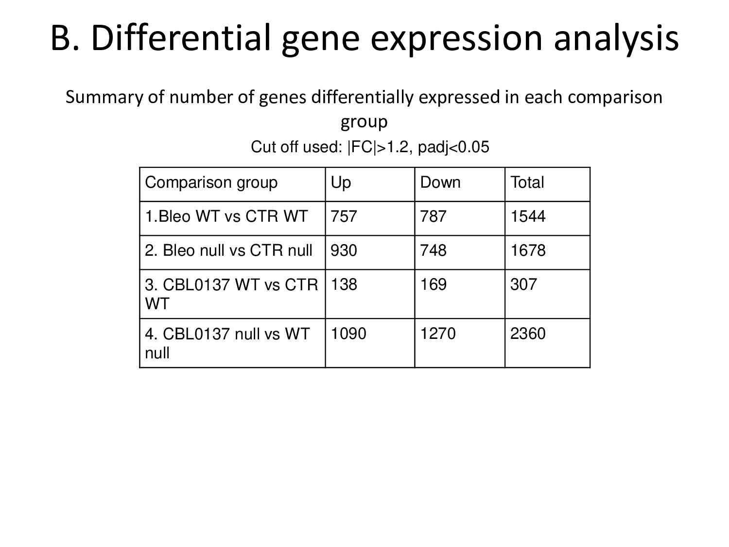
differentially expressed in each comparison group Comparison group Up Down Total 1.Bleo WT vs CTR WT 757 787 1544 2. Bleo null vs CTR null 930 748 1678 3. CBL0137 WT vs CTR WT 138 169 307 4. CBL0137 null vs WT null 1090 1270 2360 Cut off used: |FC|>1.2, padj<0.05
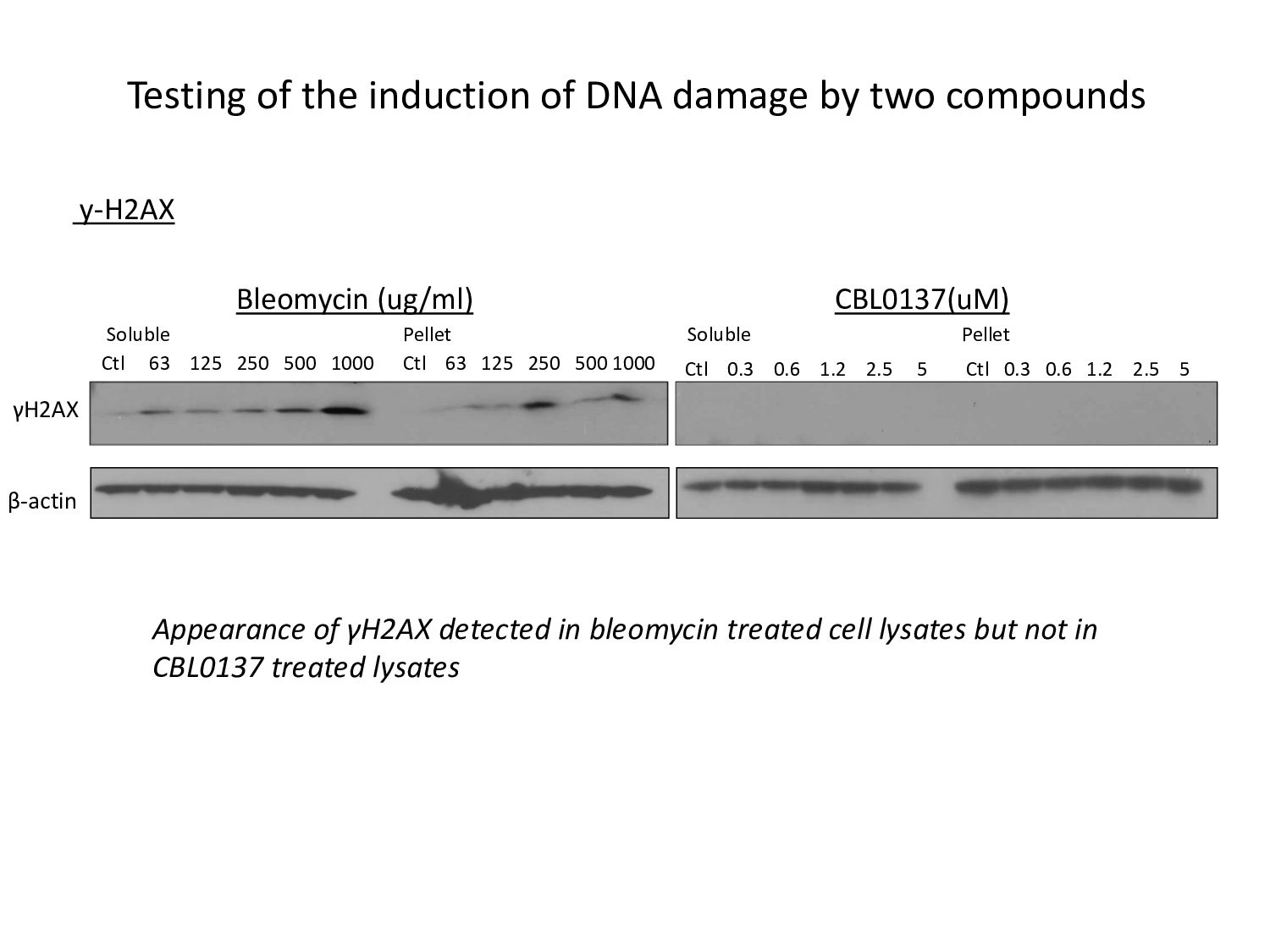
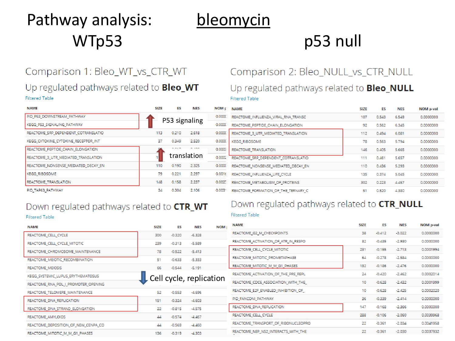
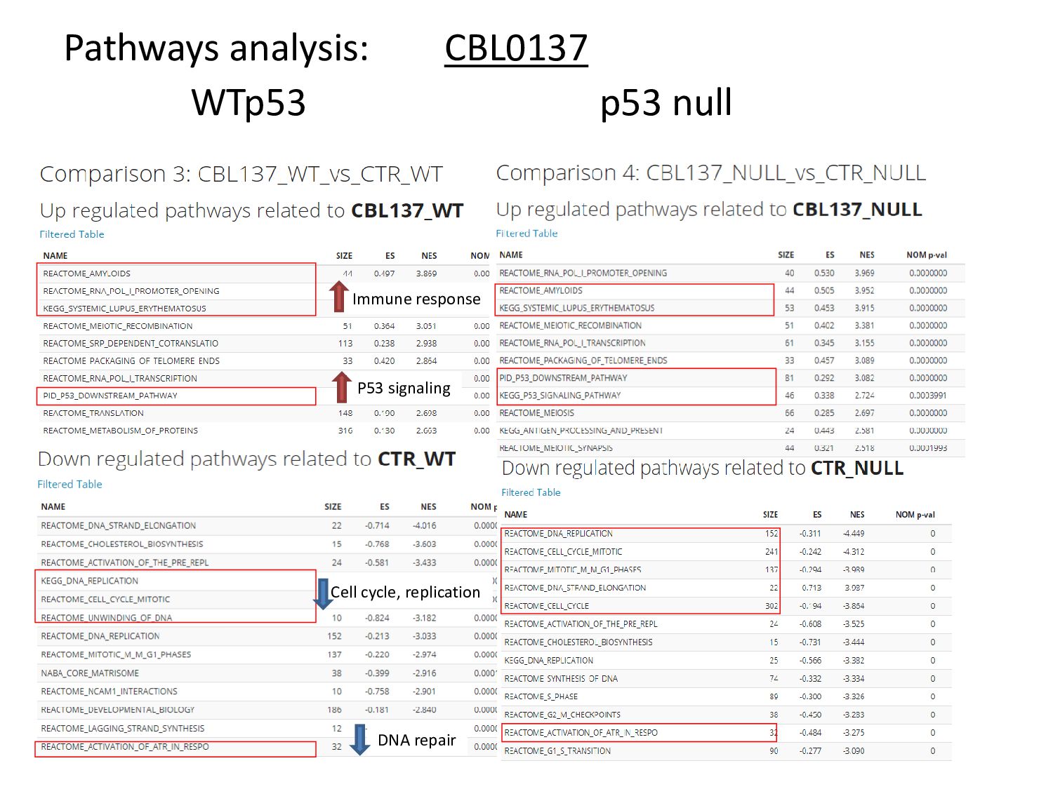
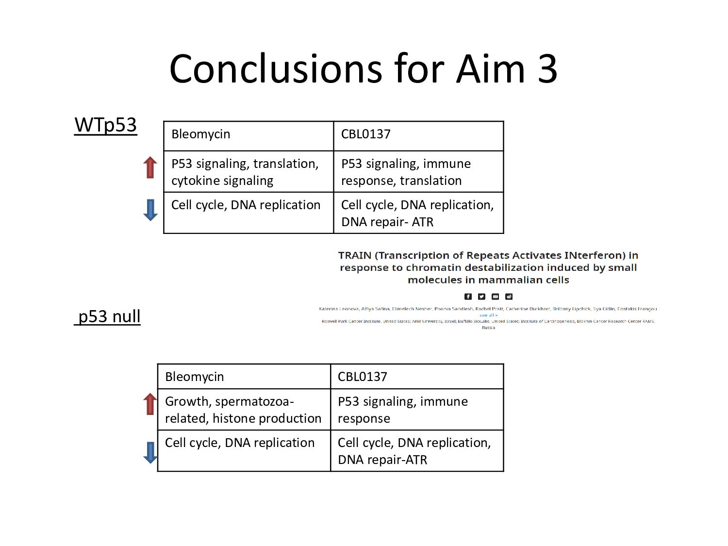
weakly, DNA damage. Bleomycin is not an inducer of chromatin damage but induces DNA damage. • Both agents cause stabilization and activity of p53. However the extent of stabilization and activity is slightly more in case of CBL0137 treatment. • CBL0137 treated samples cluster around the control, in both WT p53 and null cases. There in not much increase in newly synthesized transcripts upon curaxin treatment- downregulation of transcription. – It could be due to unavailability of FACT to aid in transcription, as it gets trapped in chromatin in the presence of curaxins.
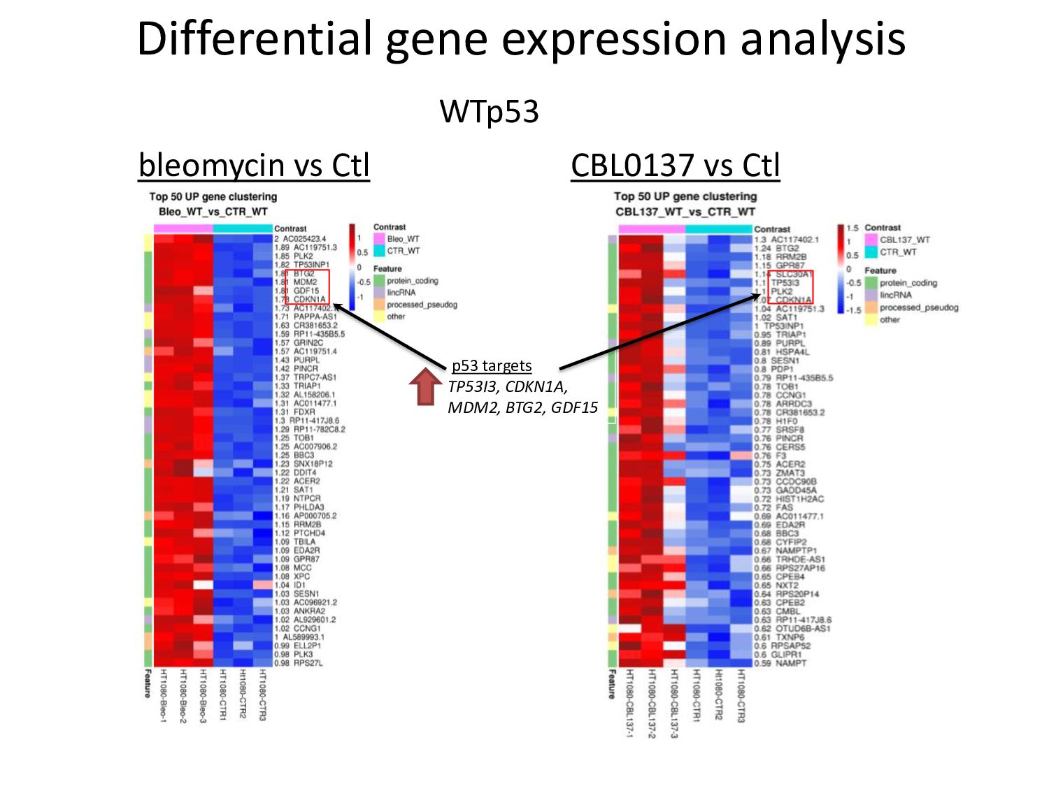
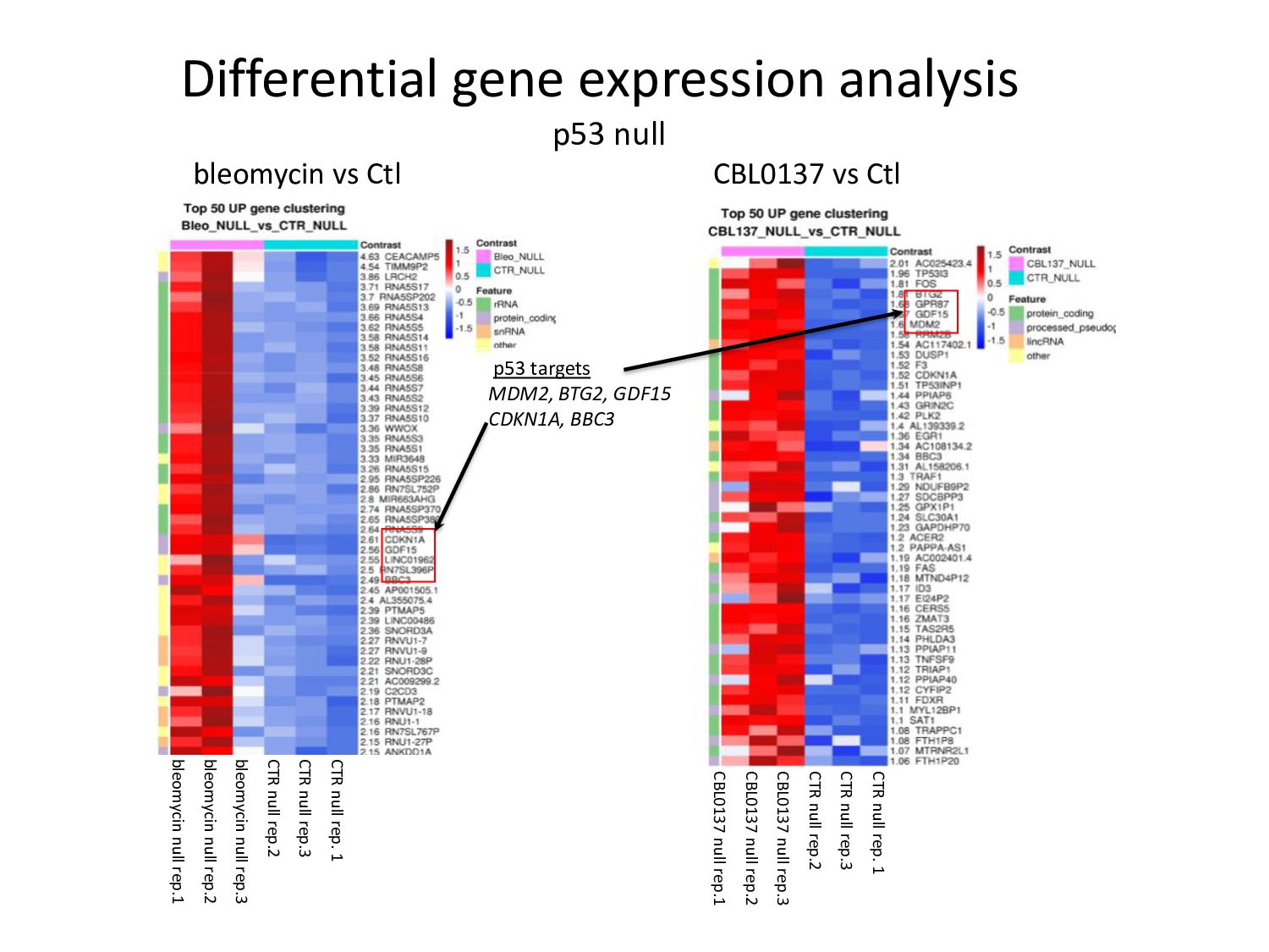
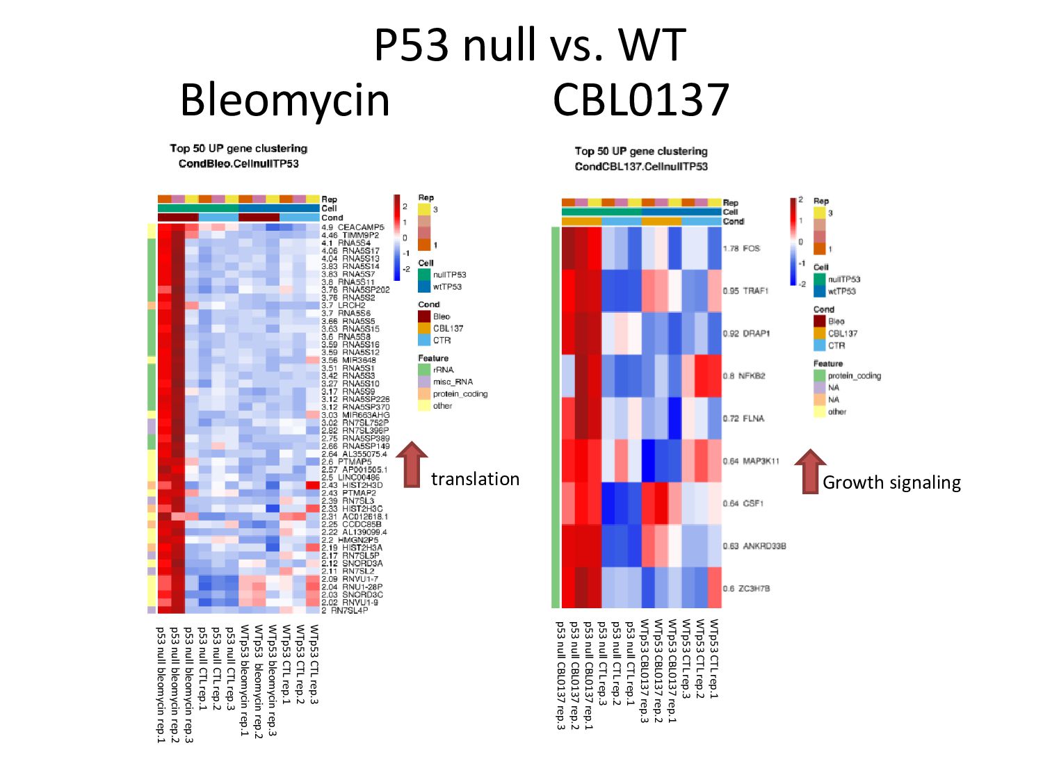
treated WT p53 cells is 308 with padj<0.05 and 1.2 < |FC| < 2.0. Whereas in bleomycin treated WT p53 cells, the number is 1544, with the same cutoffs • The number of differentially expressed genes rises to 2360 in CBL0137 treated p53 null cells while the number hasn't changed significantly in bleomycin treated p53 null cells (1678). • It is evident that increased transcription is taking place in absence of p53 upon chromatin damage. Hence p53 could have a role in suppression of transcription, when a cell undergoes chromatin damage. • Consistent with the established studies claiming that absence of p53 leads to unrestricted transcription of heterochromatin (retroviral elements, SINEs, non coding regions) when p53 null cells are treated with a DNA demethylating agent.
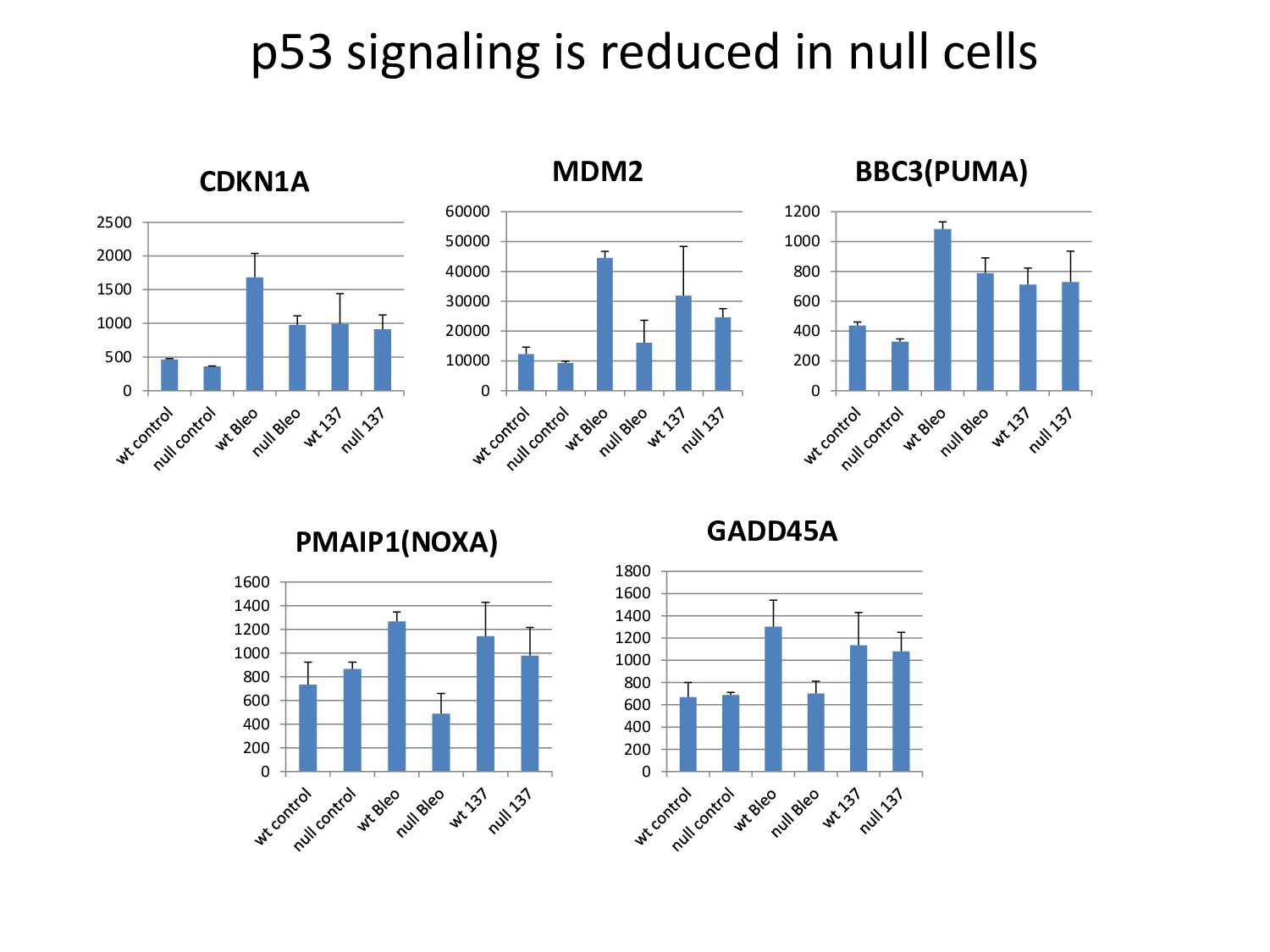
and bleomycin treated p53 null cells • The heatmaps show upregulation in p53 downstream targets like CDKN1A, BBC3, GDF15 in bleomycin treated null cells and in TP53I3, MDM2, CDKN1A in CBL0137 treated null cells. • Perform further experiments to validate inactivation of p53 in null cells
by GSE56 – western blotting of WT p53 and null cells treated under the same conditions is being carried out. The lysates are being probed for p53, p21 and MDM2. – Introduction of shp53 into HT1080. Understand how much it permits p53 activity • Analyze RNA-seq data more carefully – Characterizing downstream players, comparing their function in untreated vs treated conditions • To optimize genetic tools for induction of chromatin and DNA damage – Develop genetic models of DNA and chromatin damage
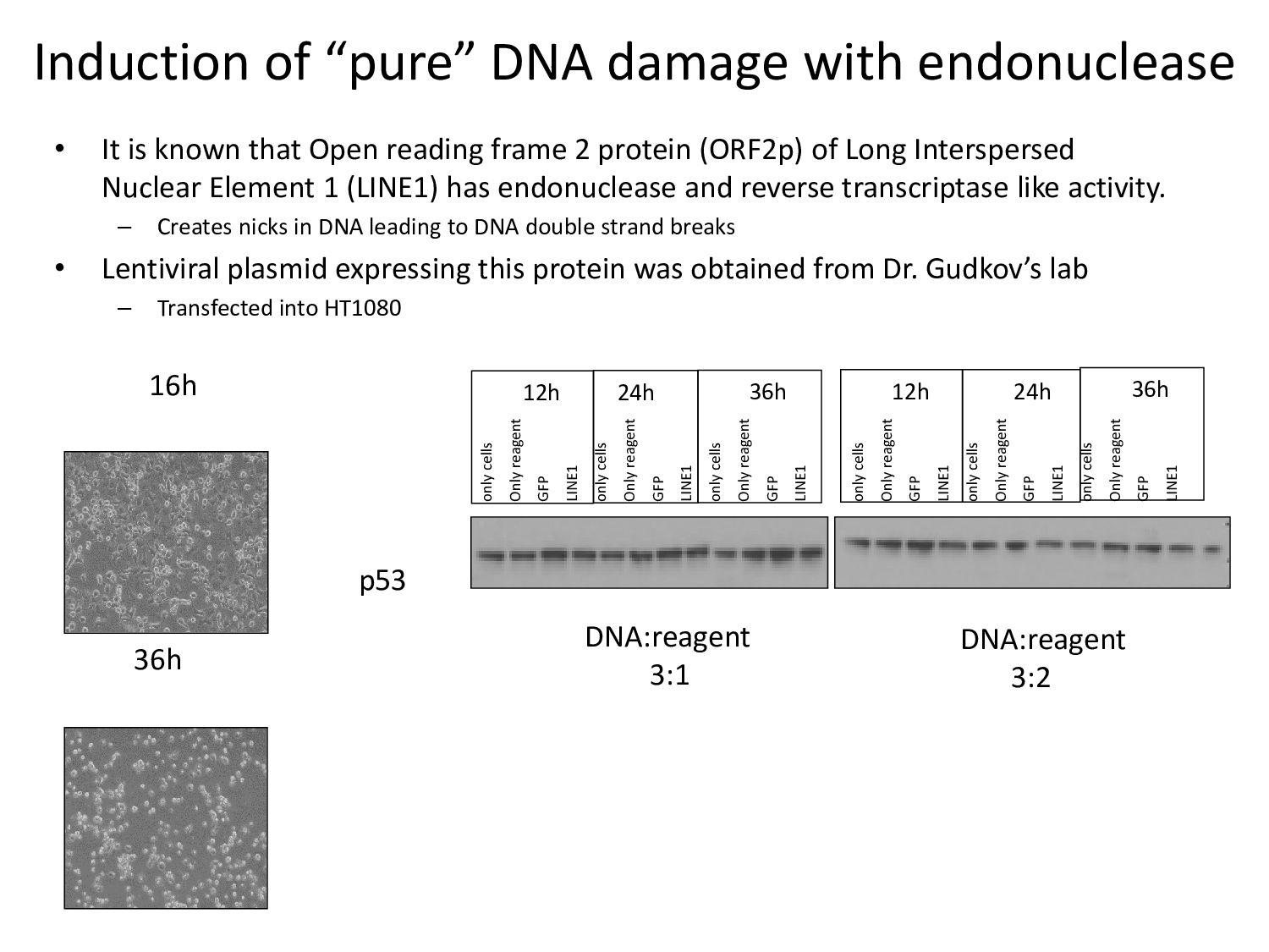
known that Open reading frame 2 protein (ORF2p) of Long Interspersed Nuclear Element 1 (LINE1) has endonuclease and reverse transcriptase like activity. – Creates nicks in DNA leading to DNA double strand breaks • Lentiviral plasmid expressing this protein was obtained from Dr. Gudkov’s lab – Transfected into HT1080 16h 36h only cells Only reagent GFP LINE1 only cells Only reagent GFP LINE1 only cells Only reagent GFP LINE1 only cells Only reagent GFP LINE1 only cells Only reagent GFP LINE1 only cells Only reagent GFP LINE1 12h 24h 36h 36h 24h 12h DNA:reagent 3:1 DNA:reagent 3:2 p53
Poorva Sandlesh Ms. Laura Prendergast Ms. Imon Goswami Committee: Dr. Thomas Melendy Labs: Gudkov, Smiraglia, Dasgupta, Paragh Departments: Cell stress biology Education Core Facility: Cell line generation Bioinformatics Supply Center Family and friends