Upgrade to Pro — share decks privately, control downloads, hide ads and more …

Cellvizio Lab Product

sowon.
April 16, 2012

Cellvizio Lab Product

In vivo fibered confocal microscopy for small animal research.

sowon.

April 16, 2012
Tweet

More Decks by sowon.

Other Decks in Science

Transcript

  1. MKEA_CORP v0.9 © 2011 Mauna Kea Technologies Our Strength, Our

    Mission • We find our strength in our solid background in multidisciplinary astrophysics • Our mission is to improve the standard- of-care thanks to our unique probe- based endomicroscopy technology 2
  2. A fast growing Medical Device company Leader in the endomicroscopy

    field • Cellvizio: a unique probe-based confocal laser endomicroscopy solution Based in Paris and Palo Alto (California), presence in Europe and Asia. Listed on NYSE-Euronext, compartment B under the ticker MKEA. Mauna Kea Technologies 3
  3. : Confocal Laser endo-microscope 5 is ? Empower your experiments

    with s the smallest video-microscope. gh-resolution, confocal imaging and n vivo & in situ imaging. Deep brain imaging in freely-moving mice ! VENESS ICAL BREAKTHROUGH E, TURN-KEY SOLUTION anning Unit oscope m excitation beam n detection (APD) ey, easy-to use ProFlex™ MiCROprobes Designed for di erent applications High Resolution: up to 1.4 ȝm Thin diameter: down to 300 ȝm LIGHTEST IMPLANTS Only 0,3g LOW INVASIVE MICROPROBE Only 350ȝm in diameter* A FULL SOLUTION Dedicated set of tools Fits the workflow Works with any stereotaxic set-up 3 2 2
  4. : Confocal Laser endo-microscope 5 is ? Empower your experiments

    with s the smallest video-microscope. gh-resolution, confocal imaging and n vivo & in situ imaging. Deep brain imaging in freely-moving mice ! VENESS ICAL BREAKTHROUGH E, TURN-KEY SOLUTION anning Unit oscope m excitation beam n detection (APD) ey, easy-to use ProFlex™ MiCROprobes Designed for di erent applications High Resolution: up to 1.4 ȝm Thin diameter: down to 300 ȝm LIGHTEST IMPLANTS Only 0,3g LOW INVASIVE MICROPROBE Only 350ȝm in diameter* A FULL SOLUTION Dedicated set of tools Fits the workflow Works with any stereotaxic set-up 3 2 2 ProFlex™ Microprobes •Designed for different applications •High Resolution: up to 1.4 µm •Thin Diameter: down to 300 µm Laser Scanning Unit •Confocal Microscope •488 or 660 nm excitation beam •Single-photon detection (APD) •Handy, turn-key, easy-to-use ImageCell™ Software •Real-Time image processing •Quantification features •Framerate up to 200 fps •LSU control Foot Pedal •ImageCell™ remote control Connecting the Computer • Plug the power cord to the wall outlet • Connect the FireWire cable • Attach the keyboard, the mouse and the screen Connecting the Laser Scanning Unit •!Plug the main power cord •!Connect the FireWire cable from the computer •!Connect the foot pedal cable •! Foot pedal •! ProFlex Holder Connecting the Cellvizio system Connecting the Computer • Plug the power cord to the wall outlet • Connect the FireWire cable • Attach the keyboard, the mouse and the screen Connecting the Laser Scanning Unit •!Plug the main power cord •!Connect the FireWire cable from the computer •!Connect the foot pedal cable •! Set of ProFlex flexible imaging microprobes •! Power Mac G5 computer •! Color flat screen •! Keyboard and mouse •! Foot pedal •! ProFlex Holder
  5. : Confocal Laser endo-microscope is ? Empower your experiments with

    s the smallest video-microscope. gh-resolution, confocal imaging and n vivo & in situ imaging. Deep brain imaging in freely-moving mice ! VENESS ICAL BREAKTHROUGH E, TURN-KEY SOLUTION anning Unit oscope m excitation beam n detection (APD) ey, easy-to use ProFlex™ MiCROprobes Designed for di erent applications High Resolution: up to 1.4 ȝm Thin diameter: down to 300 ȝm LIGHTEST IMPLANTS Only 0,3g LOW INVASIVE MICROPROBE Only 350ȝm in diameter* A FULL SOLUTION Dedicated set of tools Fits the workflow Works with any stereotaxic set-up 3 2 2 Each fiber acts as a point source and a point detector or pinhole « » Confocal Fluorescence detection system
  6. : Confocal Laser endo-microscope is ? Empower your experiments with

    s the smallest video-microscope. gh-resolution, confocal imaging and n vivo & in situ imaging. Deep brain imaging in freely-moving mice ! VENESS ICAL BREAKTHROUGH E, TURN-KEY SOLUTION anning Unit oscope m excitation beam n detection (APD) ey, easy-to use ProFlex™ MiCROprobes Designed for di erent applications High Resolution: up to 1.4 ȝm Thin diameter: down to 300 ȝm LIGHTEST IMPLANTS Only 0,3g LOW INVASIVE MICROPROBE Only 350ȝm in diameter* A FULL SOLUTION Dedicated set of tools Fits the workflow Works with any stereotaxic set-up 3 2 2 High technology •Simultaneous injection and collection •Patented technology •High sensitivity Each fiber acts as a point source and a point detector or pinhole « » Confocal Fluorescence detection system
  7. 7 ProFlex™ Microprobes is ? Empower your experiments with s

    the smallest video-microscope. gh-resolution, confocal imaging and n vivo & in situ imaging. Deep brain imaging in freely-moving mice ! VENESS ICAL BREAKTHROUGH E, TURN-KEY SOLUTION anning Unit oscope m excitation beam n detection (APD) ey, easy-to use ProFlex™ MiCROprobes Designed for di erent applications High Resolution: up to 1.4 ȝm Thin diameter: down to 300 ȝm LIGHTEST IMPLANTS Only 0,3g LOW INVASIVE MICROPROBE Only 350ȝm in diameter* A FULL SOLUTION Dedicated set of tools Fits the workflow Works with any stereotaxic set-up 3 2 2
  8. 7 Working Distance WD WD+ WD++ ProFlex™ Microprobes is ?

    Empower your experiments with s the smallest video-microscope. gh-resolution, confocal imaging and n vivo & in situ imaging. Deep brain imaging in freely-moving mice ! VENESS ICAL BREAKTHROUGH E, TURN-KEY SOLUTION anning Unit oscope m excitation beam n detection (APD) ey, easy-to use ProFlex™ MiCROprobes Designed for di erent applications High Resolution: up to 1.4 ȝm Thin diameter: down to 300 ȝm LIGHTEST IMPLANTS Only 0,3g LOW INVASIVE MICROPROBE Only 350ȝm in diameter* A FULL SOLUTION Dedicated set of tools Fits the workflow Works with any stereotaxic set-up 3 2 2
  9. 7 OS+ OS++ Optical Sectioning OS+ Working Distance WD WD+

    WD++ ProFlex™ Microprobes is ? Empower your experiments with s the smallest video-microscope. gh-resolution, confocal imaging and n vivo & in situ imaging. Deep brain imaging in freely-moving mice ! VENESS ICAL BREAKTHROUGH E, TURN-KEY SOLUTION anning Unit oscope m excitation beam n detection (APD) ey, easy-to use ProFlex™ MiCROprobes Designed for di erent applications High Resolution: up to 1.4 ȝm Thin diameter: down to 300 ȝm LIGHTEST IMPLANTS Only 0,3g LOW INVASIVE MICROPROBE Only 350ȝm in diameter* A FULL SOLUTION Dedicated set of tools Fits the workflow Works with any stereotaxic set-up 3 2 2
  10. 8 Model Applications Tip Diameter (mm) Lateral Resolution (µm) Optical

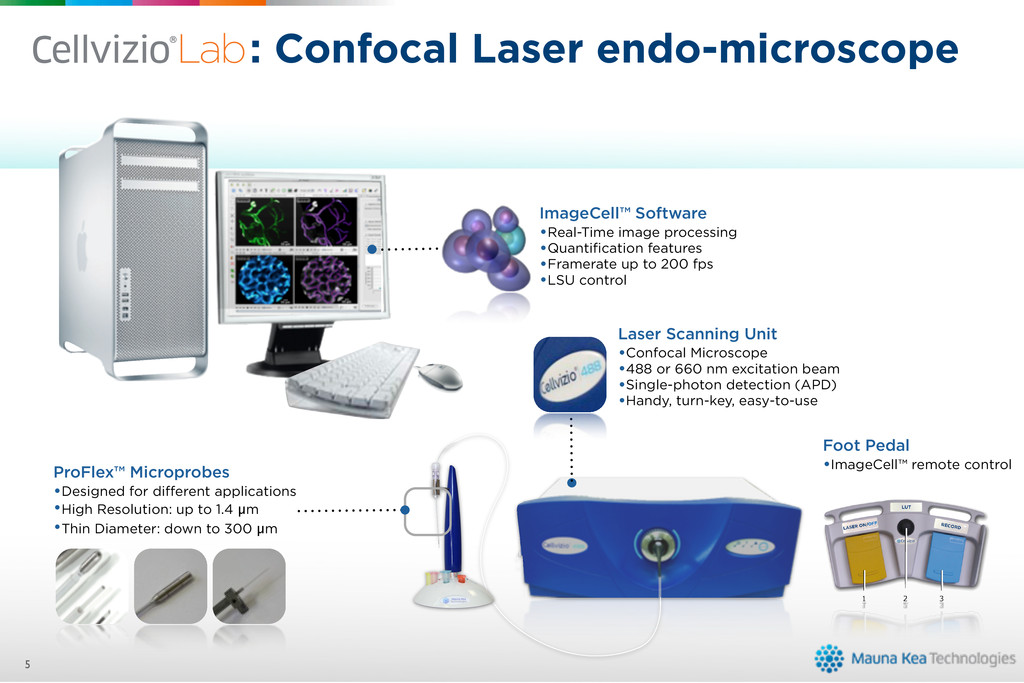
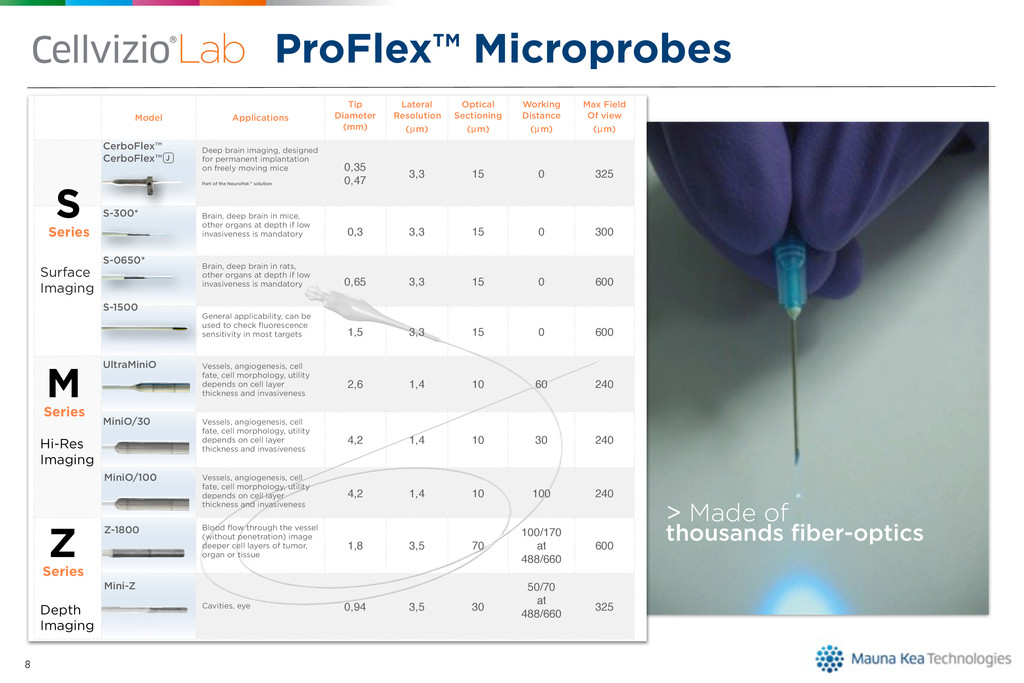
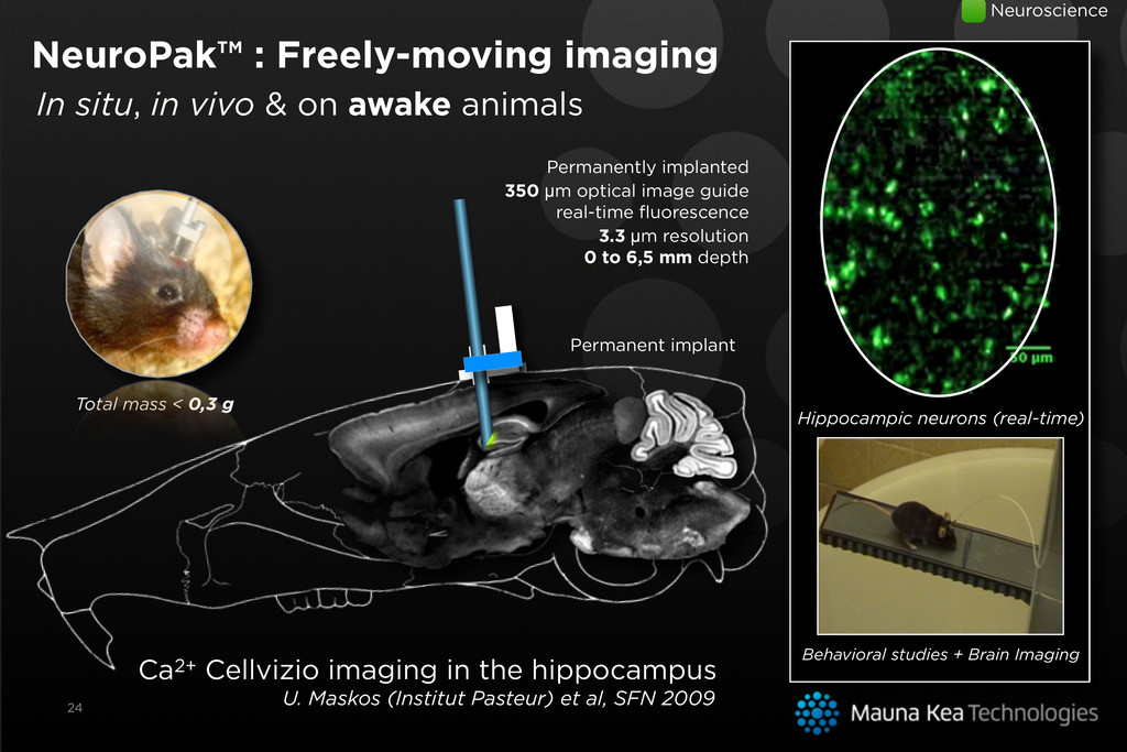
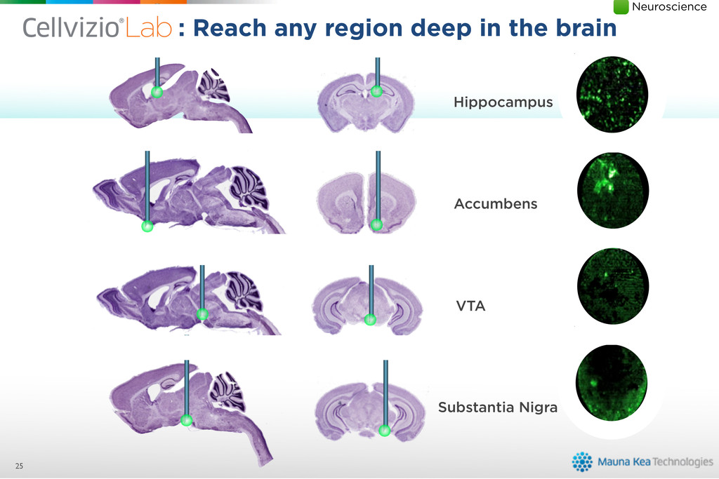
    Sectioning (µm) Working Distance (µm) Max Field Of view (µm) Deep brain imaging, designed for permanent implantation on freely moving mice Part of the NeuroPak™ solution 0,35 0,47 3,3 15 0 325 Brain, deep brain in mice, other organs at depth if low invasiveness is mandatory 0,3 3,3 15 0 300 Surface Imaging Brain, deep brain in rats, other organs at depth if low invasiveness is mandatory 0,65 3,3 15 0 600 General applicability, can be used to check fluorescence sensitivity in most targets 1,5 3,3 15 0 600 Vessels, angiogenesis, cell fate, cell morphology, utility depends on cell layer thickness and invasiveness 2,6 1,4 10 60 240 Hi-Res Imaging Vessels, angiogenesis, cell fate, cell morphology, utility depends on cell layer thickness and invasiveness 4,2 1,4 10 30 240 Imaging Vessels, angiogenesis, cell fate, cell morphology, utility depends on cell layer thickness and invasiveness 4,2 1,4 10 100 240 Blood flow through the vessel (without penetration) image deeper cell layers of tumor, organ or tissue 1,8 3,5 70 100/170 at 488/660 600 Depth Imaging Cavities, eye 0,94 3,5 30 50/70 at 488/660 325 S Series M Series S-300* S-0650* S-1500 UltraMiniO MiniO/30 MiniO/100 Z-1800 Mini-Z Z Series CerboFlex™ CerboFlex™ J ProFlex™ Microprobes is ? Empower your experiments with s the smallest video-microscope. gh-resolution, confocal imaging and n vivo & in situ imaging. Deep brain imaging in freely-moving mice ! VENESS ICAL BREAKTHROUGH E, TURN-KEY SOLUTION anning Unit oscope m excitation beam n detection (APD) ey, easy-to use ProFlex™ MiCROprobes Designed for di erent applications High Resolution: up to 1.4 ȝm Thin diameter: down to 300 ȝm LIGHTEST IMPLANTS Only 0,3g LOW INVASIVE MICROPROBE Only 350ȝm in diameter* A FULL SOLUTION Dedicated set of tools Fits the workflow Works with any stereotaxic set-up 3 2 2 > Made of thousands fiber-optics
  11. Microscopic Resolution Noninvasive Invasive Macroscopic Resolution Confocal 2-photon STED CARS

    Where does fit? Ultrasound MRI PET/CT Fluo/Visible Near IR What is ? Empower you with Cellvizio® is the smallest video-microscope. It o ers High-resolution, confocal imaging and provides in vivo & in situ imaging. Deep brain imaging in freely-moving mice ! LOW INVASIVENESS TECHNOLOGICAL BREAKTHROUGH EASY-TO-USE, TURN-KEY SOLUTION Laser Scanning Unit Confocal Microscope 488 or 660 nm excitation beam Single-photon detection (APD) Handy, turn-key, easy-to use ProFlex™ MiCROprobes Designed for di erent applications High Resolution: up to 1.4 ȝm Thin diameter: down to 300 ȝm LIGHTEST IMPLANTS On 1 1 3 2 2
  12. Microscopic Resolution Noninvasive Invasive Macroscopic Resolution Confocal 2-photon STED CARS

    Where does fit? Ultrasound MRI PET/CT Fluo/Visible Near IR What is ? Cellvizio® is the smallest video-micros It o ers High-resolution, confocal ima provides in vivo & in situ imaging. LOW INVASIVENESS TECHNOLOGICAL BREAKTHROUGH EASY-TO-USE, TURN-KEY SOLUTION Laser Scanning Unit Confocal Microscope 488 or 660 nm excitation beam Single-photon detection (APD) Handy, turn-key, easy-to use ProFlex™ Designed for High Resoluti Thin diameter 1 1 3 2 2 High resolution Micro-invasive What is ? Empower you with Cellvizio® is the smallest video-microscope. It o ers High-resolution, confocal imaging and provides in vivo & in situ imaging. Deep brain imaging in freely-moving mice ! LOW INVASIVENESS TECHNOLOGICAL BREAKTHROUGH EASY-TO-USE, TURN-KEY SOLUTION Laser Scanning Unit Confocal Microscope 488 or 660 nm excitation beam Single-photon detection (APD) Handy, turn-key, easy-to use ProFlex™ MiCROprobes Designed for di erent applications High Resolution: up to 1.4 ȝm Thin diameter: down to 300 ȝm LIGHTEST IMPLANTS On 1 1 3 2 2
  13. ImageCell™: Quantification tools • Advanced Mosaïcing™ • Enhanced resolution •

    Bigger field of view • Follow the probe’s track • Run Kinetic analysis • Quantify the fluorescence over time-charter • Manage multiple ROIs • Analyze and export • Segment vessels, measure neoangiogenesis • Assess the effects of anti-angiogenic compounds • in vivo tracking of vascular changes over time in longitudinal studies on the same animal 10 Vessel Detection®
  14. A Wealth of Applications Neuro- mus. Junction Vascularization Lymph Node

    Lung Brain Cornea Heart Liver Bladder Kidney GI tract Muscle Nerve
  15. Cellvizio®Lab for Cancer Research 13 Applications • In vivo histology

    at cellular resolution • Molecular Characterization • Angiogenesis Quantification • Treatment evaluation (Cytotoxic/antiangiogenic/cell therapy) • Multidrug resistance • Animal evaluation
  16. Angiogenesis 14 Vascularisation of normal and tumoral mammary glands Automated

    quantification of vascular density and blood vessel diameter with Cellvizio Vessel Detection® Oncology 9 weeks 10 weeks 11 weeks Modification of tumoral vascularisation under chemotherapy Longitudinal analysis of tumor vascularization in PyMT mice • Longitudinal study of molecular and microvascular events • In vivo monitoring of changes in vascular density, diameter and morphology What is ? Empower y with Cellvizio® is the smallest video-microscope. It o ers High-resolution, confocal imaging and provides in vivo & in situ imaging. Deep brain imagin in freely-moving m LOW INVASIVENESS TECHNOLOGICAL BREAKTHROUGH EASY-TO-USE, TURN-KEY SOLUTION Laser Scanning Unit Confocal Microscope 488 or 660 nm excitation beam Single-photon detection (APD) Handy, turn-key, easy-to use ImageCell™ Software Real-time image processing Quantification features Framerate up to 200 fps LSU control ProFlex™ MiCROprobes Designed for di erent applications High Resolution: up to 1.4 ȝm Thin diameter: down to 300 ȝm LIGHTEST IMPLAN LOW INVASIVE MIC *Available in 350 ȝm or 470 ȝm 1 1 3 3 2 2
  17. : Dynamic in vivo histology, in real time is ?

    Empower your experiments with s the smallest video-microscope. gh-resolution, confocal imaging and n vivo & in situ imaging. Deep brain imaging in freely-moving mice ! VENESS ICAL BREAKTHROUGH E, TURN-KEY SOLUTION anning Unit oscope m excitation beam n detection (APD) ey, easy-to use ProFlex™ MiCROprobes Designed for di erent applications High Resolution: up to 1.4 ȝm Thin diameter: down to 300 ȝm LIGHTEST IMPLANTS Only 0,3g LOW INVASIVE MICROPROBE Only 350ȝm in diameter* A FULL SOLUTION Dedicated set of tools Fits the workflow Works with any stereotaxic set-up 3 2 2 Oncology Janssen et al, 2006 Normal colonic mucosa • Round Crypts • Regular size. Tubulous adenoma • Larger Crypts • Irregular size. Protocol • Colon washing (water) • Syringe + catheter insertion • Topical dye application Protocol • ProFlex™ insertion • MiniZ or S-1500 • Endoscope combination possible Longitudinal studies made easy Mouse colon acriflavine stained
  18. : Dynamic in vivo histology, in real time is ?

    Empower your experiments with s the smallest video-microscope. gh-resolution, confocal imaging and n vivo & in situ imaging. Deep brain imaging in freely-moving mice ! VENESS ICAL BREAKTHROUGH E, TURN-KEY SOLUTION anning Unit oscope m excitation beam n detection (APD) ey, easy-to use ProFlex™ MiCROprobes Designed for di erent applications High Resolution: up to 1.4 ȝm Thin diameter: down to 300 ȝm LIGHTEST IMPLANTS Only 0,3g LOW INVASIVE MICROPROBE Only 350ȝm in diameter* A FULL SOLUTION Dedicated set of tools Fits the workflow Works with any stereotaxic set-up 3 2 2 Oncology D’Hallewin et al, 2005 Rat Bladder Rhodamine 123 stained Protocol • Tumor inoculation • Topical Rhodamine 123 application (100 µM) Protocol • Bladder catheterized with 18 gauge cannula • Transurethral S-1500 ProFlex™ insertion Umbrella cells Basal cells tumor Longitudinal studies made easy intermediate cells Different structures can be visualized with other Microprobes
  19. In vivo microcirculation 17 Oncology Cortex vessels: • C57 Bl/6

    mouse • FITC Dextran 70kDa • S-1500 ProFlex • 3,3 µm lateral resolution Mesenteric vessels: • C57 Bl/6 mouse • FITC Dextran 70kDa • Z-1800 ProFlex • 100 µm Optical sectioning • 3,5 µm lateral resolution What is ? Empowe with Cellvizio® is the smallest video-microscope. It o ers High-resolution, confocal imaging and provides in vivo & in situ imaging. Deep brain ima in freely-movin LOW INVASIVENESS TECHNOLOGICAL BREAKTHROUGH EASY-TO-USE, TURN-KEY SOLUTION Laser Scanning Unit Confocal Microscope 488 or 660 nm excitation beam Single-photon detection (APD) Handy, turn-key, easy-to use ImageCell™ Software Real-time image processing Quantification features Framerate up to 200 fps LSU control ProFlex™ MiCROprobes Designed for di erent applications High Resolution: up to 1.4 ȝm Thin diameter: down to 300 ȝm LIGHTEST IMPL LOW INVASIVE *Available in 350 ȝm or 47 1 1 3 3 2 2 Pancreas vascularization Normal PDAC Pancreas Ductal Adeno Carcinoma
  20. Cellvizio®Lab for Neuroscience 18 Applications • In vivo Deep brain

    imaging • Neuronal activity assessment • Freely-moving animal : Associate behavioral studies to neuronal activation • Epilepsy, Alzheimer, Parkinson, Addiction... • Animal model evaluation
  21. 19 Investigation of nerve regeneration after crush injury of the

    Saphenous Nerve for PNS is ? Empower your experiments with s the smallest video-microscope. gh-resolution, confocal imaging and n vivo & in situ imaging. Deep brain imaging in freely-moving mice ! VENESS ICAL BREAKTHROUGH E, TURN-KEY SOLUTION anning Unit oscope m excitation beam n detection (APD) ey, easy-to use ProFlex™ MiCROprobes Designed for di erent applications High Resolution: up to 1.4 ȝm Thin diameter: down to 300 ȝm LIGHTEST IMPLANTS Only 0,3g LOW INVASIVE MICROPROBE Only 350ȝm in diameter* A FULL SOLUTION Dedicated set of tools Fits the workflow Works with any stereotaxic set-up 3 2 2 Neuroscience
  22. 19 Ex vivo : Periodic control epifluorescence microscopy can only

    be performed on fixed tissue and with multiple animals. Investigation of nerve regeneration after crush injury of the Saphenous Nerve Post-crush outgrowth measurement, 4 days after injury for PNS is ? Empower your experiments with s the smallest video-microscope. gh-resolution, confocal imaging and n vivo & in situ imaging. Deep brain imaging in freely-moving mice ! VENESS ICAL BREAKTHROUGH E, TURN-KEY SOLUTION anning Unit oscope m excitation beam n detection (APD) ey, easy-to use ProFlex™ MiCROprobes Designed for di erent applications High Resolution: up to 1.4 ȝm Thin diameter: down to 300 ȝm LIGHTEST IMPLANTS Only 0,3g LOW INVASIVE MICROPROBE Only 350ȝm in diameter* A FULL SOLUTION Dedicated set of tools Fits the workflow Works with any stereotaxic set-up 3 2 2 Neuroscience
  23. 19 Ex vivo : Periodic control epifluorescence microscopy can only

    be performed on fixed tissue and with multiple animals. In vivo & in situ : Investigation of nerve regeneration after crush injury of the Saphenous Nerve Post-crush outgrowth measurement, 4 days after injury for PNS is ? Empower your experiments with s the smallest video-microscope. gh-resolution, confocal imaging and n vivo & in situ imaging. Deep brain imaging in freely-moving mice ! VENESS ICAL BREAKTHROUGH E, TURN-KEY SOLUTION anning Unit oscope m excitation beam n detection (APD) ey, easy-to use ProFlex™ MiCROprobes Designed for di erent applications High Resolution: up to 1.4 ȝm Thin diameter: down to 300 ȝm LIGHTEST IMPLANTS Only 0,3g LOW INVASIVE MICROPROBE Only 350ȝm in diameter* A FULL SOLUTION Dedicated set of tools Fits the workflow Works with any stereotaxic set-up 3 2 2 Neuroscience Control Crushed Nerve Adapted from Wong et al., 2009 Thy1.2-YFP16 mouse
  24. 19 Ex vivo : Periodic control epifluorescence microscopy can only

    be performed on fixed tissue and with multiple animals. In vivo & in situ : Investigation of nerve regeneration after crush injury of the Saphenous Nerve Post-crush outgrowth measurement, 4 days after injury for PNS is ? Empower your experiments with s the smallest video-microscope. gh-resolution, confocal imaging and n vivo & in situ imaging. Deep brain imaging in freely-moving mice ! VENESS ICAL BREAKTHROUGH E, TURN-KEY SOLUTION anning Unit oscope m excitation beam n detection (APD) ey, easy-to use ProFlex™ MiCROprobes Designed for di erent applications High Resolution: up to 1.4 ȝm Thin diameter: down to 300 ȝm LIGHTEST IMPLANTS Only 0,3g LOW INVASIVE MICROPROBE Only 350ȝm in diameter* A FULL SOLUTION Dedicated set of tools Fits the workflow Works with any stereotaxic set-up 3 2 2 Neuroscience Control Crushed Nerve Adapted from Wong et al., 2009 Thy1.2-YFP16 mouse 4 days
  25. 500 µm To date, an immense domain remains unreachable to

    cellular in vivo and in situ imaging Mouse brain 2-photon Microscopy Limitation Addiction Adult neurogenesis Alzheimer Parkinson Huntington : Microscopy brought into the Brain is ? Empower your experiments with s the smallest video-microscope. gh-resolution, confocal imaging and n vivo & in situ imaging. Deep brain imaging in freely-moving mice ! VENESS ICAL BREAKTHROUGH E, TURN-KEY SOLUTION anning Unit oscope m excitation beam n detection (APD) ey, easy-to use ProFlex™ MiCROprobes Designed for di erent applications High Resolution: up to 1.4 ȝm Thin diameter: down to 300 ȝm LIGHTEST IMPLANTS Only 0,3g LOW INVASIVE MICROPROBE Only 350ȝm in diameter* A FULL SOLUTION Dedicated set of tools Fits the workflow Works with any stereotaxic set-up 3 2 2 Neuroscience
  26. 500 µm To date, an immense domain remains unreachable to

    cellular in vivo and in situ imaging Mouse brain 2-photon Microscopy Limitation Addiction Adult neurogenesis Alzheimer Parkinson Huntington What is ? Image behavior in REALTIME Empower your experiments with Cellvizio® is the smallest video-microscope. It o ers High-resolution, confocal imaging and provides in vivo & in situ imaging. Neuron activity as it happens in vivo ! Deep network activity monitoring. Real-time imaging of neurons in animals achieving basic behavioral tasks. Deep brain imaging in freely-moving mice ! LOW INVASIVENESS TECHNOLOGICAL BREAKTHROUGH EASY-TO-USE, TURN-KEY SOLUTION Laser Scanning Unit Confocal Microscope 488 or 660 nm excitation beam Single-photon detection (APD) Handy, turn-key, easy-to use ImageCell™ Software Real-time image processing Quantification features Framerate up to 200 fps LSU control ProFlex™ MiCROprobes Designed for di erent applications High Resolution: up to 1.4 ȝm Thin diameter: down to 300 ȝm CONSCIOUS, FREELY-MOVING MICE Functional Fluorescence imaging LONGITUDINAL STUDIES Make your results more relevant Image more with fewer animals www.cellviziolab.com LIGHTEST IMPLANTS Only 0,3g LOW INVASIVE MICROPROBE Only 350ȝm in diameter* *Available in 350 ȝm or 470 ȝm (hardened tip) A FULL SOLUTION Dedicated set of tools Fits the workflow Works with any stereotaxic set-up 1 1 3 3 2 2 : Microscopy brought into the Brain is ? Empower your experiments with s the smallest video-microscope. gh-resolution, confocal imaging and n vivo & in situ imaging. Deep brain imaging in freely-moving mice ! VENESS ICAL BREAKTHROUGH E, TURN-KEY SOLUTION anning Unit oscope m excitation beam n detection (APD) ey, easy-to use ProFlex™ MiCROprobes Designed for di erent applications High Resolution: up to 1.4 ȝm Thin diameter: down to 300 ȝm LIGHTEST IMPLANTS Only 0,3g LOW INVASIVE MICROPROBE Only 350ȝm in diameter* A FULL SOLUTION Dedicated set of tools Fits the workflow Works with any stereotaxic set-up 3 2 2 Neuroscience
  27. Neuron Direction Specificity in Cat Visual Cortex Images acquired in

    layer 4 of the cat visual cortex Cells labeled with Oregon Green BAPTA-1 by local microinjection after sterotaxic surgery Visual stimulation with drifting orientated patterns and grey screen in-between Courtesy of Aaron Kerlin, Kenichi Ohki, Clay Reid Harvard Medical School, Boston, MA FOV = Φ = 300 µm Grid orientation : Monitor Neuronal activity is ? Empower your experiments with s the smallest video-microscope. gh-resolution, confocal imaging and n vivo & in situ imaging. Deep brain imaging in freely-moving mice ! VENESS ICAL BREAKTHROUGH E, TURN-KEY SOLUTION anning Unit oscope m excitation beam n detection (APD) ey, easy-to use ProFlex™ MiCROprobes Designed for di erent applications High Resolution: up to 1.4 ȝm Thin diameter: down to 300 ȝm LIGHTEST IMPLANTS Only 0,3g LOW INVASIVE MICROPROBE Only 350ȝm in diameter* A FULL SOLUTION Dedicated set of tools Fits the workflow Works with any stereotaxic set-up 3 2 2 Neuroscience
  28. Ultrasound triggered BBB disruption 22 Application: Local Drug Delivery BBB

    Disruption: MiniZ probe FITC-Dextran 30, 50, 70, 150 kDa range Focalized Ultrasound application SonoVue® Microbubbles injection
  29. NeuroPak™ : Freely-moving imaging! 23 ? Image behavior in REALTIME

    Empower your experiement with microscope. al imaging and g. Neuron activity as it happens in vivo ! Deep network activity monitoring. Real-time imaging of neurons in animals achieving basic behavioral tasks. Deep brain imaging in freely-moving mice ! H ON LEX™ MICROPROBES ned for di erent cations Resolution: up to 1.4 !m diameter: down to 300 !m CONSIOUS, FREELY-MOVING MICE Functional «Fluorescence» imaging LONGITUDINAL STUDIES Make your results more relevance Image more with the fewer animals www.cellviziolab.com LIGHTEST IMPLANTS Only 0,3g LOW INVASIVE MICROPROBE Only 350!m in diameter* *Available in 350 !m or 470 !m (hardened tip) A FULLY PACKAGED SOLUTION Dedicated set of tools Fits the researcher workflow Adapted to any steretaxic set-up 3 Deep Brain imaging in freely moving animals • A set of tools, including a stereotaxic holder • Small and Light implants: only 0,3g • Reach the neuron activity: Live! • Monitor the brain activity of mice achieving behavioral tasks • NeuroPak or NeuroPak J Same animal can be imaged several times Neuroscience
  30. NeuroPak™ : Freely-moving imaging! 23 ? Image behavior in REALTIME

    Empower your experiement with microscope. al imaging and g. Neuron activity as it happens in vivo ! Deep network activity monitoring. Real-time imaging of neurons in animals achieving basic behavioral tasks. Deep brain imaging in freely-moving mice ! H ON LEX™ MICROPROBES ned for di erent cations Resolution: up to 1.4 !m diameter: down to 300 !m CONSIOUS, FREELY-MOVING MICE Functional «Fluorescence» imaging LONGITUDINAL STUDIES Make your results more relevance Image more with the fewer animals www.cellviziolab.com LIGHTEST IMPLANTS Only 0,3g LOW INVASIVE MICROPROBE Only 350!m in diameter* *Available in 350 !m or 470 !m (hardened tip) A FULLY PACKAGED SOLUTION Dedicated set of tools Fits the researcher workflow Adapted to any steretaxic set-up 3 Deep Brain imaging in freely moving animals • A set of tools, including a stereotaxic holder • Small and Light implants: only 0,3g • Reach the neuron activity: Live! • Monitor the brain activity of mice achieving behavioral tasks • NeuroPak or NeuroPak J Same animal can be imaged several times Neuroscience
  31. 24 In situ, in vivo & on awake animals NeuroPak™

    : Freely-moving imaging Neuroscience
  32. 24 In situ, in vivo & on awake animals NeuroPak™

    : Freely-moving imaging Neuroscience
  33. 24 In situ, in vivo & on awake animals Permanent

    implant NeuroPak™ : Freely-moving imaging Neuroscience
  34. Permanently implanted 350 µm optical image guide real-time fluorescence 3.3

    µm resolution 0 to 6,5 mm depth 24 In situ, in vivo & on awake animals Permanent implant NeuroPak™ : Freely-moving imaging Neuroscience
  35. Permanently implanted 350 µm optical image guide real-time fluorescence 3.3

    µm resolution 0 to 6,5 mm depth 24 In situ, in vivo & on awake animals Permanent implant NeuroPak™ : Freely-moving imaging Neuroscience
  36. Ca2+ Cellvizio imaging in the hippocampus U. Maskos (Institut Pasteur)

    et al, SFN 2009 Permanently implanted 350 µm optical image guide real-time fluorescence 3.3 µm resolution 0 to 6,5 mm depth Behavioral studies + Brain Imaging Hippocampic neurons (real-time) 24 In situ, in vivo & on awake animals Permanent implant NeuroPak™ : Freely-moving imaging Neuroscience Total mass < 0,3 g
  37. Substantia Nigra VTA Accumbens Hippocampus 25 : Reach any region

    deep in the brain is ? Empower your experiments with s the smallest video-microscope. gh-resolution, confocal imaging and n vivo & in situ imaging. Deep brain imaging in freely-moving mice ! VENESS ICAL BREAKTHROUGH E, TURN-KEY SOLUTION anning Unit oscope m excitation beam n detection (APD) ey, easy-to use ProFlex™ MiCROprobes Designed for di erent applications High Resolution: up to 1.4 ȝm Thin diameter: down to 300 ȝm LIGHTEST IMPLANTS Only 0,3g LOW INVASIVE MICROPROBE Only 350ȝm in diameter* A FULL SOLUTION Dedicated set of tools Fits the workflow Works with any stereotaxic set-up 3 2 2 Neuroscience
  38. 26 : a Revolution in Neurosciences is ? Empower your

    experiments with s the smallest video-microscope. gh-resolution, confocal imaging and n vivo & in situ imaging. Deep brain imaging in freely-moving mice ! VENESS ICAL BREAKTHROUGH E, TURN-KEY SOLUTION anning Unit oscope m excitation beam n detection (APD) ey, easy-to use ProFlex™ MiCROprobes Designed for di erent applications High Resolution: up to 1.4 ȝm Thin diameter: down to 300 ȝm LIGHTEST IMPLANTS Only 0,3g LOW INVASIVE MICROPROBE Only 350ȝm in diameter* A FULL SOLUTION Dedicated set of tools Fits the workflow Works with any stereotaxic set-up 3 2 2 Neuroscience
  39. Anesthetized animals • Networking via Ca2+ imaging • Cell physiology

    • Brain activities 26 : a Revolution in Neurosciences is ? Empower your experiments with s the smallest video-microscope. gh-resolution, confocal imaging and n vivo & in situ imaging. Deep brain imaging in freely-moving mice ! VENESS ICAL BREAKTHROUGH E, TURN-KEY SOLUTION anning Unit oscope m excitation beam n detection (APD) ey, easy-to use ProFlex™ MiCROprobes Designed for di erent applications High Resolution: up to 1.4 ȝm Thin diameter: down to 300 ȝm LIGHTEST IMPLANTS Only 0,3g LOW INVASIVE MICROPROBE Only 350ȝm in diameter* A FULL SOLUTION Dedicated set of tools Fits the workflow Works with any stereotaxic set-up 3 2 2 Neuroscience
  40. Anesthetized animals • Networking via Ca2+ imaging • Cell physiology

    • Brain activities Freely Moving animals • Learning / Memory • Autism • Schizophrenia • Addiction • Other behavorial studies... Gene Protein Cell Tissue Organ Animal Population 26 : a Revolution in Neurosciences is ? Empower your experiments with s the smallest video-microscope. gh-resolution, confocal imaging and n vivo & in situ imaging. Deep brain imaging in freely-moving mice ! VENESS ICAL BREAKTHROUGH E, TURN-KEY SOLUTION anning Unit oscope m excitation beam n detection (APD) ey, easy-to use ProFlex™ MiCROprobes Designed for di erent applications High Resolution: up to 1.4 ȝm Thin diameter: down to 300 ȝm LIGHTEST IMPLANTS Only 0,3g LOW INVASIVE MICROPROBE Only 350ȝm in diameter* A FULL SOLUTION Dedicated set of tools Fits the workflow Works with any stereotaxic set-up 3 2 2 Neuroscience
  41. Take away messages What is ? Im Empower your experiement

    with Cellvizio® is the smallest video-microscope. It o ers High-resolution, confocal imaging and provides in vivo & in situ imaging. Ne De Re ac Deep brain imaging in freely-moving mice ! LOW INVASIVENESS TECHNOLOGICAL BREAKTHROUGH EASY-TO-USE, TURN-KEY SOLUTION LASER SCANNING UNIT Confocal Microscope 488 or 660 nm excitation beam Single-photon detection (APD) Handy, turn-key, easy-to use IMAGECELL™ SOFTWARE Real-time image processing Quantification features Framerate up to 200 fps LSU control PROFLEX™ MICROPROBES Designed for di erent applications High Resolution: up to 1.4 !m Thin diameter: down to 300 !m C F L M I LIGHTEST IMPLANTS Only 0,3g LOW INVASIVE MICROPROBE Only 350!m in diameter* *Available in 350 !m or 470 !m (hardened tip) A FULLY PACKAGED SOLUTION Dedicated set of tools Fits the researcher workflow Adapted to any steretaxic set-up 1 1 3 3 2 2 What is ? Im Empower your experiement with Cellvizio® is the smallest video-microscope. It o ers High-resolution, confocal imaging and provides in vivo & in situ imaging. Ne De Re ac Deep brain imaging in freely-moving mice ! LOW INVASIVENESS TECHNOLOGICAL BREAKTHROUGH EASY-TO-USE, TURN-KEY SOLUTION LASER SCANNING UNIT Confocal Microscope 488 or 660 nm excitation beam Single-photon detection (APD) Handy, turn-key, easy-to use IMAGECELL™ SOFTWARE Real-time image processing Quantification features Framerate up to 200 fps LSU control PROFLEX™ MICROPROBES Designed for di erent applications High Resolution: up to 1.4 !m Thin diameter: down to 300 !m C L LIGHTEST IMPLANTS Only 0,3g LOW INVASIVE MICROPROBE Only 350!m in diameter* *Available in 350 !m or 470 !m (hardened tip) A FULLY PACKAGED SOLUTION Dedicated set of tools Fits the researcher workflow Adapted to any steretaxic set-up 1 1 3 3 2 2 • is the smallest microscope on the market • It offers High-resolution imaging • designed for in vivo & in situ imaging • Low invasiveness, fewer sacrifices • Easy-to-use, turnkey solution Neuroscience Oncology GI tract Stem Cells Cellvizio Lab main applications What is ? Empower with Cellvizio® is the smallest video-microscope. It o ers High-resolution, confocal imaging and provides in vivo & in situ imaging. Deep brain ima in freely-movin LOW INVASIVENESS TECHNOLOGICAL BREAKTHROUGH EASY-TO-USE, TURN-KEY SOLUTION Laser Scanning Unit Confocal Microscope 488 or 660 nm excitation beam Single-photon detection (APD) Handy, turn-key, easy-to use ProFlex™ MiCROprobes Designed for di erent applications High Resolution: up to 1.4 ȝm Thin diameter: down to 300 ȝm LIGHTEST IMPL LOW INVASIVE 1 1 3 2 2
  42. Take away messages 28 What is ? Im Empower your

    experiement with Cellvizio® is the smallest video-microscope. It o ers High-resolution, confocal imaging and provides in vivo & in situ imaging. Ne De Re ac Deep brain imaging in freely-moving mice ! LOW INVASIVENESS TECHNOLOGICAL BREAKTHROUGH EASY-TO-USE, TURN-KEY SOLUTION LASER SCANNING UNIT Confocal Microscope 488 or 660 nm excitation beam Single-photon detection (APD) Handy, turn-key, easy-to use IMAGECELL™ SOFTWARE Real-time image processing Quantification features Framerate up to 200 fps LSU control PROFLEX™ MICROPROBES Designed for di erent applications High Resolution: up to 1.4 !m Thin diameter: down to 300 !m C F L M I LIGHTEST IMPLANTS Only 0,3g LOW INVASIVE MICROPROBE Only 350!m in diameter* *Available in 350 !m or 470 !m (hardened tip) A FULLY PACKAGED SOLUTION Dedicated set of tools Fits the researcher workflow Adapted to any steretaxic set-up 1 1 3 3 2 2 What is ? Im Empower your experiement with Cellvizio® is the smallest video-microscope. It o ers High-resolution, confocal imaging and provides in vivo & in situ imaging. Ne De Re ac Deep brain imaging in freely-moving mice ! LOW INVASIVENESS TECHNOLOGICAL BREAKTHROUGH EASY-TO-USE, TURN-KEY SOLUTION LASER SCANNING UNIT Confocal Microscope 488 or 660 nm excitation beam Single-photon detection (APD) Handy, turn-key, easy-to use IMAGECELL™ SOFTWARE Real-time image processing Quantification features Framerate up to 200 fps LSU control PROFLEX™ MICROPROBES Designed for di erent applications High Resolution: up to 1.4 !m Thin diameter: down to 300 !m C L LIGHTEST IMPLANTS Only 0,3g LOW INVASIVE MICROPROBE Only 350!m in diameter* *Available in 350 !m or 470 !m (hardened tip) A FULLY PACKAGED SOLUTION Dedicated set of tools Fits the researcher workflow Adapted to any steretaxic set-up 1 1 3 3 2 2 • is the smallest microscope on the market • It offers High-resolution imaging • designed for in vivo & in situ imaging • Low invasiveness, fewer sacrifices • Easy-to-use, turnkey solution Neuroscience Oncology GI tract Stem Cells Cellvizio Lab main applications What is ? Empower with Cellvizio® is the smallest video-microscope. It o ers High-resolution, confocal imaging and provides in vivo & in situ imaging. Deep brain ima in freely-movin LOW INVASIVENESS TECHNOLOGICAL BREAKTHROUGH EASY-TO-USE, TURN-KEY SOLUTION Laser Scanning Unit Confocal Microscope 488 or 660 nm excitation beam Single-photon detection (APD) Handy, turn-key, easy-to use ProFlex™ MiCROprobes Designed for di erent applications High Resolution: up to 1.4 ȝm Thin diameter: down to 300 ȝm LIGHTEST IMPL LOW INVASIVE 1 1 3 2 2
  43. Cellvizio® Impact 29 Am. J. Of Resp. and Critical Care

    Medicine Gastroenterology Clinical Gastro and Hepatology Nature Medicine Nature EMBO reports Translational Oncology International Journal of Cancer Journal of Urology PNAS Cancer Research J. of Cell Biology Chemical Senses Cell Research Neuroimage J. of Vascular Research Stem Cells Endoscopy Official Clinical Practice Journal of the www.cghjournal.org November 2007 Volume 5 Number 11 Issue Highlights Autoimmune Enteropathy Liver Biopsy in ESRD Celiac Disease and Pancreatitis Confocal Microscopy
  44. 90 installed Worldwide hat is ? Empower your experime with

    zio® is the smallest video-microscope. ers High-resolution, confocal imaging and des in vivo & in situ imaging. Deep brain imaging in freely-moving mice ! INVASIVENESS NOLOGICAL BREAKTHROUGH -TO-USE, TURN-KEY SOLUTION ser Scanning Unit cal Microscope r 660 nm excitation beam -photon detection (APD) y, turn-key, easy-to use ProFlex™ MiCROprobes Designed for di erent applications High Resolution: up to 1.4 ȝm Thin diameter: down to 300 ȝm LIGHTEST IMPLANTS Only 0,3g A FULL SOLU Dedicated set o Fits the workflo Works with any stereotaxic set- 3 2 2
  45. 20 10 1 1 3 1 6 3 6 3

    5 4 2 1 1 2 21 21 1 30 90 installed Worldwide hat is ? Empower your experime with zio® is the smallest video-microscope. ers High-resolution, confocal imaging and des in vivo & in situ imaging. Deep brain imaging in freely-moving mice ! INVASIVENESS NOLOGICAL BREAKTHROUGH -TO-USE, TURN-KEY SOLUTION ser Scanning Unit cal Microscope r 660 nm excitation beam -photon detection (APD) y, turn-key, easy-to use ProFlex™ MiCROprobes Designed for di erent applications High Resolution: up to 1.4 ȝm Thin diameter: down to 300 ȝm LIGHTEST IMPLANTS Only 0,3g A FULL SOLU Dedicated set o Fits the workflo Works with any stereotaxic set- 3 2 2 1 10
  46. Imaging of other small animals... 32 REPRODUCTION RESEARCH In vivo

    imaging of in situ motility of fresh and liquid stored ram spermatozoa in the ewe genital tract Xavier Druart, Juliette Cognie ´, Ge ´rard Baril, Fre ´de ´rique Cle ´ment1, Jean-Louis Dacheux and Jean-Luc Gatti UMR 6175 INRA, CNRS-Universite ´ de Tours-Haras Nationaux, Station de Physiologie de la Reproduction et des Comportements, Institut National de la Recherche Agronomique, 37380 Nouzilly, France and 1INRIA Paris-Rocquencourt, Domaine de Voluceau, Rocquencourt BP 105, 78153 Le Chesnay Cedex, France Correspondence should be addressed to X Druart; Email: [email protected] Abstract The fertility of ram semen after cervical insemination is substantially reduced by 24 h of storage in liquid form. The effects of liquid storage on the transit of ram spermatozoa in the ewe genital tract was investigated using a new procedure allowing direct observation of the spermatozoa in the genital tract. Ejaculated ram spermatozoa were double labeled with R18 and MitoTracker Green FM, and used to inseminate ewes in estrus either cervically through the vagina or laparoscopically into the base of the uterine horns. Four hours after insemination, the spermatozoa were directly observed in situ using fibered confocal fluorescence microscopy in the base, middle and tip of the uterine horns, the utero-tubal junction (UTJ) and the oviduct. The high resolution video images obtained with this technique allowed determination of the distribution of spermatozoa and individual motility in the lumen of the ewe’s genital tract. The results showed a gradient of increasing concentration of spermatozoa from the base of the uterus to the UTJ 4 h after intra-uterine insemination into the base of the horns. The UTJ was shown to be a storage region for spermatozoa before their transfer to the oviduct. The in vitro storage of spermatozoa in liquid form decreased their migration through the cervix and reduced the proportion of motile spermatozoa and their straight line velocity at the UTJ and their transit into the oviduct. Reproduction (2009) 138 45–53 Introduction Fertility in the ewe after cervical insemination has been shown to be reduced when semen have been stored in liquid form for several hours, despite any major reduction in semen quality as assessed by standard in vitro parameters such as motility (Salamon & Maxwell 2000). A substantial decrease in fertility was also reported when frozen–thawed semen was used for cervical insemination, and this effect was partially overcome by intra-uterine insemination (Gillan & Maxwell 1999), which suggested that impaired migration of spermatozoa through the cervix caused reduced fertility in ewes inseminated with preserved semen. Indeed, migration through the cervix appeared to be the critical limiting factor for migration of ram spermatozoa from the vagina to the oviducts (Lightfoot & Restall 1971, Hawk & Conley 1975, Hawk 1983). The migration of spermatozoa through the cervix may involve increased contractility of the cervix at estrus (Wergin 1985) because cervical myoelectrical activity was found to vary during the cycle and reach a peak around ovulation (Cavaco-Gonc ¸alves et al. 2006). The morphology of the cervix is very variable, and differences between animals, during the cycle (Kershaw et al. 2005) and between breeds (Kaabi et al. 2006) have been reported. The differences in fertility reported between breeds of sheep after insemination with frozen–thawed semen might be related to differences in sperm transit in the genital tract of the ewe (Fair et al. 2005). The physiological study of sperm transit and quality in the female genital tract has to date been limited by the lack of techniques to observe the behavior of spermatozoa in the genital tract of the living female at a cellular level. A new technique, fibered confocal fluorescence microscopy (FCM), provides direct obser- vation of the microscopic structure of living tissue (Laemmel et al. 2004, Thiberville et al. 2007). The technique is based on the principle of confocal microscopy, which provides an in-focus image of a thin section within a biological sample, where the microscope objective is replaced by a flexible fiber-optic miniprobe. With this technique it is possible to obtain images with endogenous or exogenous tissue fluoro- phores through a fiber-optic probe of 1 mm diameter or less. In this study, we developed a method based on FCM to observe the behavior of individual spermatozoa q 2009 Society for Reproduction and Fertility DOI: 10.1530/REP-09-0108 ISSN 1470–1626 (paper) 1741–7899 (online) Online version via www.reproduction-online.org
  47. One Technology, Two imaging Worlds 33 Features & Benefits •

    in vivo characterization of human tissues • Reduction in tissue sampling and histological examination • Real-time decision for best treatment to the patient • Fully integrated in endoscopy suite • FDA, CE cleared Confocal processor™ • Real-Time image processing • Laser Scanning Unit driving • Patient ID management • Very simple interface Clinical Clinical • Cellvizio 100 Series
  48. Features & Benefits • The smallest microscope for small animal

    imaging • Bring High resolution to in vivo research: up to 1.4 µm • A wealth of applications including Cancer research, Angiogenesis, Deep brain imaging, and more. • Microinvasive: 10 Lab-dedicated microprobes Down to Ø300µm • Longitudinal studies made possible • Plug & Play, Easy-to-use One Technology, Two imaging Worlds 33 Features & Benefits • in vivo characterization of human tissues • Reduction in tissue sampling and histological examination • Real-time decision for best treatment to the patient • Fully integrated in endoscopy suite • FDA, CE cleared Confocal processor™ • Real-Time image processing • Laser Scanning Unit driving • Patient ID management • Very simple interface ImageCell™ Software • Real-Time image processing • Quantification features • Fast acquisition up to 200 fps • Powerful post-processing tools • Numerous video export formats • Intuitive User Interface Clinical Clinical • Cellvizio 100 Series Connecting the Computer • Plug the power cord to the wall outlet • Connect the FireWire cable • Attach the keyboard, the mouse and the screen Connecting the Computer • Plug the power cord to the wall outlet • Connect the FireWire cable • Attach the keyboard, the mouse and the screen Conn •!Plug •!Con •!Con Research Research • Cellvizio LAB Research Easy Transfer from Research to Clinical practice Clinical
  49. and beyond... • Dual • Combine 2 lasers and 2

    detectors in One Cellvizio® 34 t is ? Empower your experiments with is the smallest video-microscope. High-resolution, confocal imaging and in vivo & in situ imaging. Deep brain imaging in freely-moving mice ! SIVENESS OGICAL BREAKTHROUGH USE, TURN-KEY SOLUTION canning Unit icroscope nm excitation beam ton detection (APD) ProFlex™ MiCROprobes Designed for di erent applications High Resolution: up to 1.4 ȝm Thin diameter: down to 300 ȝm LIGHTEST IMPLANTS Only 0,3g A FULL SOLUTION Dedicated set of tools Fits the workflow Works with any stereotaxic set-up 3 2 2
  50. and beyond... • Dual • Combine 2 lasers and 2

    detectors in One Cellvizio® 34 t is ? Empower your experiments with is the smallest video-microscope. High-resolution, confocal imaging and in vivo & in situ imaging. Deep brain imaging in freely-moving mice ! SIVENESS OGICAL BREAKTHROUGH USE, TURN-KEY SOLUTION canning Unit icroscope nm excitation beam ton detection (APD) ProFlex™ MiCROprobes Designed for di erent applications High Resolution: up to 1.4 ȝm Thin diameter: down to 300 ȝm LIGHTEST IMPLANTS Only 0,3g A FULL SOLUTION Dedicated set of tools Fits the workflow Works with any stereotaxic set-up 3 2 2
  51. and beyond... • Dual • Combine 2 lasers and 2

    detectors in One Cellvizio® 34 Release 2013 t is ? Empower your experiments with is the smallest video-microscope. High-resolution, confocal imaging and in vivo & in situ imaging. Deep brain imaging in freely-moving mice ! SIVENESS OGICAL BREAKTHROUGH USE, TURN-KEY SOLUTION canning Unit icroscope nm excitation beam ton detection (APD) ProFlex™ MiCROprobes Designed for di erent applications High Resolution: up to 1.4 ȝm Thin diameter: down to 300 ȝm LIGHTEST IMPLANTS Only 0,3g A FULL SOLUTION Dedicated set of tools Fits the workflow Works with any stereotaxic set-up 3 2 2
  52. MKEA_CORP v0.9 © 2011 Mauna Kea Technologies Thanks! Merci! 감사합니다

    ͋Γ͕ͱ͏ 35 www.maunakeatech.com For More on Cellvizio®