Upgrade to Pro — share decks privately, control downloads, hide ads and more …

Aneurysms

daiju_ueda
April 16, 2018
240

 Aneurysms

Mammography

daiju_ueda

April 16, 2018
Tweet

Transcript

  1. Development and Validation of Deep Learning Algorithms for Detecting Cerebral

    Aneurysms in MR angiography. [1] Department of Diagnostic and Interventional Radiology, Osaka City University, Graduate School of Medicine [2] LPixel Inc. [3] Department of Premier Preventive Medicine, Osaka City University, Graduate School of Medicine Daiju Ueda, Akira Yamamoto, Masataka Nishimori, Taro Shimono, Satoshi Doishita, Akitoshi Shimazaki, Shinichi Tsutsumi, Shinya Fukumoto, Choppin Antoine, Yuki Shimahara, Yukio Miki [1] [1] [2] [1] [1] [3] [1] [1] [1] [2] [2]
  2. Results: 1) FROCs 0 10 20 30 40 50 60

    70 80 90 100 0 1 2 3 4 5 6 7 8 9 10 Hospital test-dataset Clinic test-dataset Number of false positive per case Sensitivity[%] Number of false positive per case 1 2 3 4 5 6 7 8 9 10 Hospital test-dataset [%] 61.1 78.0 84.6 87.5 90.0 90.3 90.8 90.9 91.1 91.1 Clinic test-dataset [%] 66.2 81.2 88.8 90.0 92.5 92.5 92.5 92.5 92.5 92.5
  3. Results: 2) Sensitivities and characteristics Characteristics Hospital test-dataset Clinics test-dataset

    No. of aneurysms 592/649 (91.2%) 74/80 (92.5%) Size of aneurysms [mm] <3 125/141(88.7%) 10/10 (100%) 3-4.9 336/354 (94.9%) 48/52 (92.3%) 5-9.9 107/125 (85.6%) 9/10 (90.0%) ≧10 24/29 (82.8%) 4/4 (100%) Aneurysms location Internal carotid artery area 336/358 (94.4%) 44/47 (93.6%) Middle cerebral artery area 111/129 (86.0%) 14/15 (93.3%) Anterior cerebral artery area 79/87 (90.8%) 9/9 (100%) Posterior cerebral artery area 3/3 (100%) 0 Basilar artery area 40/43 (93.0%) 4/4 (100%) Vertebral artery area 23/29 (79.3%) 3/5 (60%) No. of postoperative cases 25/28 (89.3%) 4/4 (100%) Magnetic field strength 1.5T 275/303 (90.1%) 74/80 (92.5%) 3.0T 317/346 (91.6%) 0
  4. Results: 3) Characteristic of newly detected aneurysms Characteristics Hospital test-dataset

    Clinic test-dataset No. of new aneurysms 31 10 Size of aneurysms [mm] <3 15 2 3-4.9 12 7 5-9.9 4 1 ≧10 0 0 Aneurysms location Internal carotid artery area 18 4 Middle cerebral artery area 4 2 Anterior cerebral artery area 1 1 Posterior cerebral artery area 0 0 Basilar artery area 3 3 Vertebral artery area 5 0 Magnetic field strength 1.5T 10 10 3.0T 21 0
  5. Examples: Aneurysms missed by the algorithm 15-mm aneurysm at the

    bifurcation of the middle cerebral artery of a 67-year-old woman. 10-mm aneurysm in the vertebral artery of a 30-year-old woman.
  6. Examples: Aneurysms newly detected by the algorithm 3-mm aneurysm in

    the A4 segment of the anterior cerebral artery of an 82-year-old woman. 2-mm aneurysm in the vertebral artery of a 79-year-old man.
  7. Discussionᶅ Radiologists may not overlook aneurysms missed by algorithm because

    of their size and irregularity The algorithm could support to detect ɹɹɹ aneurysms in less common locations. ᶃ ᶄ ʴ This might show that radiologists and this algorithm were complementary to each other.
  8. Discussionᶆ No Algorithm Sentitivity Number of test-aneurysms Size [1] [2]

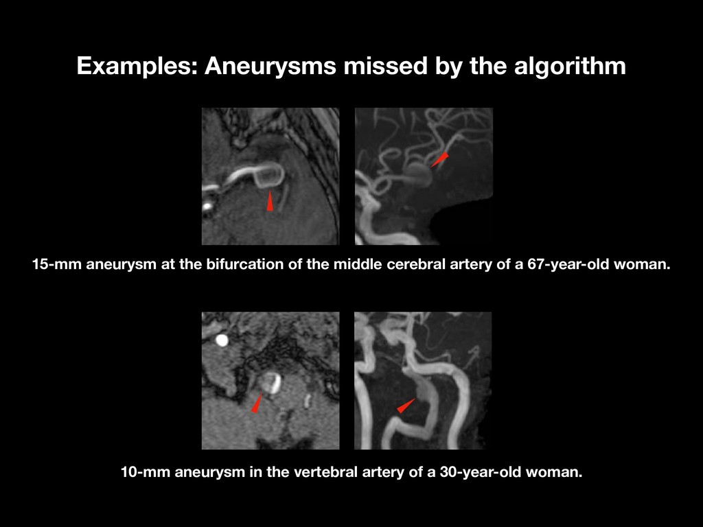
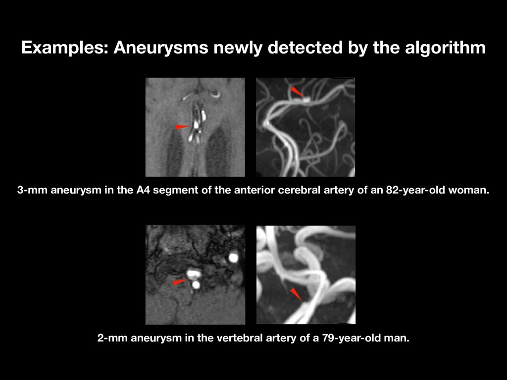
    [3] 84% 100% 61 36 3–26 mm (mean 6.6 mm) 3–26 mm (mean 7.1 mm) [4] 96% 147 ≥5 mm (62.5%) [5] 91% 11 mean 3.12 mm [6] [7] 82% (top-3) 203 relatively small (about 3 mm) [8] 94% 100 relatively small (about 3 mm) This study 91.1% 92.5% 521 + 67 [1] Hirai T, Korogi Y, Arimura H, et al. Intracranial aneurysms at MR angiography: effect of computer-aided diagnosis on radiologists' detection performance. Radiology. 2005;237(2):605-10. [2] Kakeda S, Korogi Y, Arimura H, et al. Diagnostic accuracy and reading time to detect intracranial aneurysms on MR angiography using a computer-aided diagnosis system. American Journal of Roentgenology. 2008;190(2):459-65. [3] Arimura H, Li Q, Korogi Y, et al. Automated computerized scheme for detection of unruptured intracranial aneurysms in three-dimensional magnetic resonance angiography. Acad Radiol. 2004;11(10):1093-104. [4] Yang X, Blezek DJ, Cheng LT, Ryan WJ, Kallmes DF, Erickson BJ. Computer-aided detection of intracranial aneurysms in MR angiography. J Digit Imaging. 2011;24(1):86-95. [5] Štepán-Buksakowska I, Accurso J, Diehn F, et al. Computer-aided diagnosis improves detection of small intracranial aneurysms on MRA in a clinical setting. American Journal of Neuroradiology. 2014;35(10):1897-902. [6] Miki S, Hayashi N, Masutani Y, et al. Computer-assisted detection of cerebral aneurysms in MR angiography in a routine image-reading environment: effects on diagnosis by radiologists. American Journal of Neuroradiology. 2016;37(6):1038-43. [7] Nomura Y, Masutani Y, Miki S, et al. Performance improvement in computerized detection of cerebral aneurysms by retraining classifier using feedback data collected in routine reading environment. Journal of Biomedical Graphics and Computing. 2014;4(4):12. [8] Nakao T, Hanaoka S, Nomura Y, et al. Deep neural network-based computer-assisted detection of cerebral aneurysms in MR angiography. J Magn Reson Imaging. 2017. Comparison to prior Research