FNA procedure is highly operator dependent. operator dependent. Physician performing FNA should receive adequate Physician performing FNA should receive adequate training in: training in: 1. 1. Selection Selection of cases for FNA. of cases for FNA. 1. 1. Selection Selection of cases for FNA. of cases for FNA. 2. 2. Sample collection & preparation. Sample collection & preparation. 20 20- -30 supervised cases; after that, available 30 supervised cases; after that, available supervision. supervision. To maintain privilege of doing FNA, To maintain privilege of doing FNA, <20% insufficient <20% insufficient sample rate sample rate should be maintained. should be maintained.
where required. Number of needle passes depends on Number of needle passes depends on size of lump. size of lump. If If cystic, cystic, perform a perform a diagnostic and therapeutic diagnostic and therapeutic FNA FNA (evacuation of cyst); if contents are thin, (evacuation of cyst); if contents are thin, watery, greenish gray & no residual mass watery, greenish gray & no residual mass after evacuation after evacuation - - fluid can be discarded.* fluid can be discarded.* * * Some laboratories routinely process the fluid for Some laboratories routinely process the fluid for cytology. cytology.
re re- -aspirate. aspirate. If If sanguinous sanguinous fluid (not due to trauma) fluid (not due to trauma) - - careful cytologic & clinical assessment. careful cytologic & clinical assessment. If cystic and solid mass, obtain material from If cystic and solid mass, obtain material from If cystic and solid mass, obtain material from If cystic and solid mass, obtain material from both components of the lump. both components of the lump. Use of fine needle capillary sampling Use of fine needle capillary sampling technique ( technique (FNC) technique FNC) technique for small or highly for small or highly vascular lesions. vascular lesions.
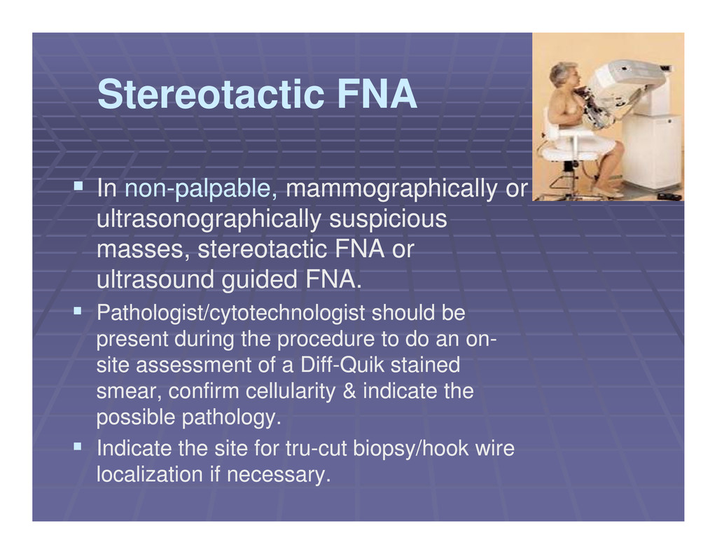
mammographically or mammographically or ultrasonographically suspicious ultrasonographically suspicious masses, stereotactic FNA or masses, stereotactic FNA or ultrasound guided FNA. ultrasound guided FNA. ultrasound guided FNA. ultrasound guided FNA. Pathologist/cytotechnologist should be Pathologist/cytotechnologist should be present during the procedure to do an on present during the procedure to do an on- - site assessment of a Diff site assessment of a Diff- -Quik stained Quik stained smear, confirm cellularity & indicate the smear, confirm cellularity & indicate the possible pathology. possible pathology. Indicate the site for tru Indicate the site for tru- -cut biopsy/hook wire cut biopsy/hook wire localization if necessary. localization if necessary.
solid lesion solid lesion No standard rule. No standard rule. Depends on the clinico Depends on the clinico- -cytologic assessment: cytologic assessment: Opinion of the aspirator that the Opinion of the aspirator that the cytologic cytologic Opinion of the aspirator that the Opinion of the aspirator that the cytologic cytologic features features and diagnosis are and diagnosis are consistent consistent with the with the clinical findings. clinical findings. Opinion of the pathologist that the slides do not Opinion of the pathologist that the slides do not have any have any artifacts or distortion artifacts or distortion and can be and can be interpreted safely. interpreted safely.
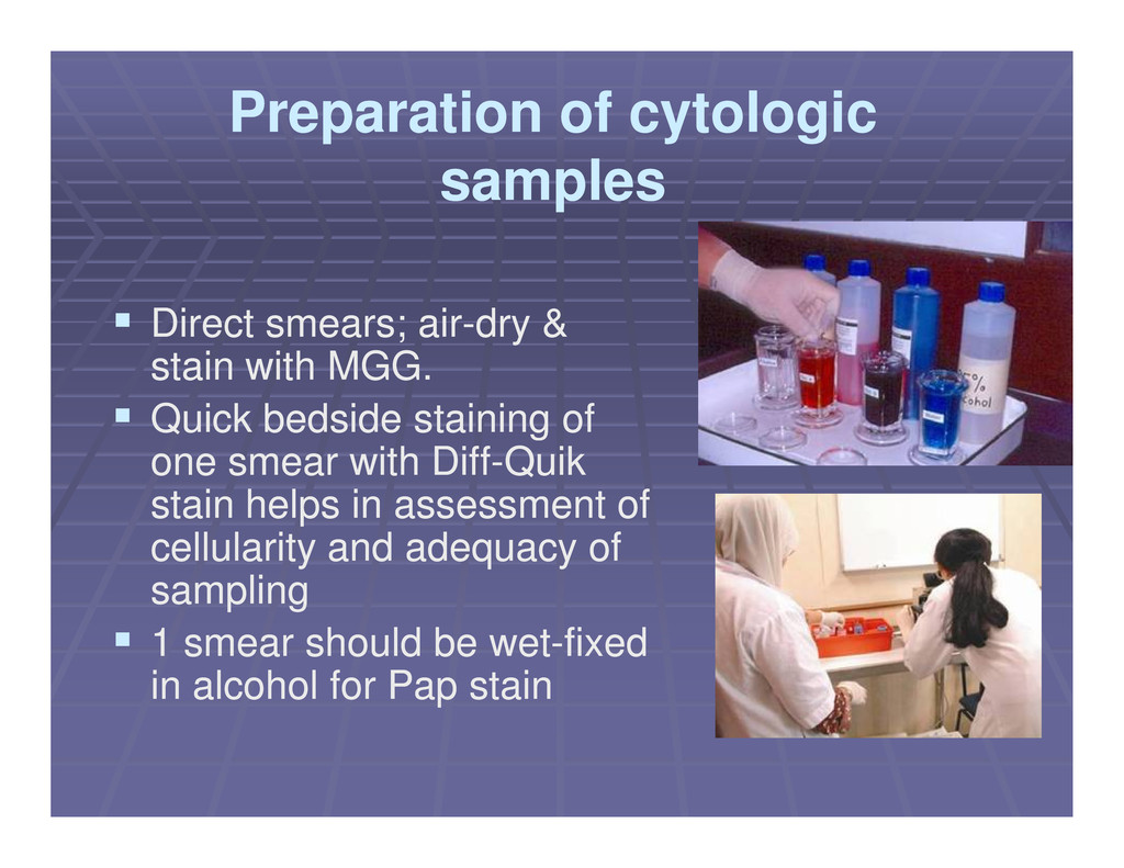
air Direct smears; air- -dry & dry & stain with MGG. stain with MGG. Quick bedside staining of Quick bedside staining of Quick bedside staining of Quick bedside staining of one smear with Diff one smear with Diff- -Quik Quik stain helps in assessment of stain helps in assessment of cellularity and adequacy of cellularity and adequacy of sampling sampling 1 smear should be wet 1 smear should be wet- -fixed fixed in alcohol for Pap stain in alcohol for Pap stain
Ancillary studies Ancillary studies (ER/PR; special stains; (ER/PR; special stains; immunostains) immunostains) - - can be done on ethanol can be done on ethanol- - fixed smears or cell blocks. fixed smears or cell blocks. Microbiologic studies Microbiologic studies - - smears for smears for microbiologic stains and pus/fluid for microbiologic stains and pus/fluid for culture culture
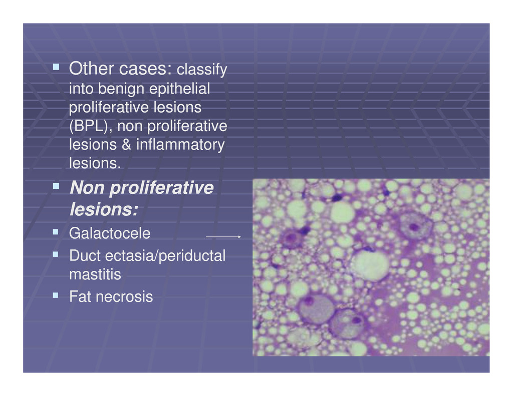
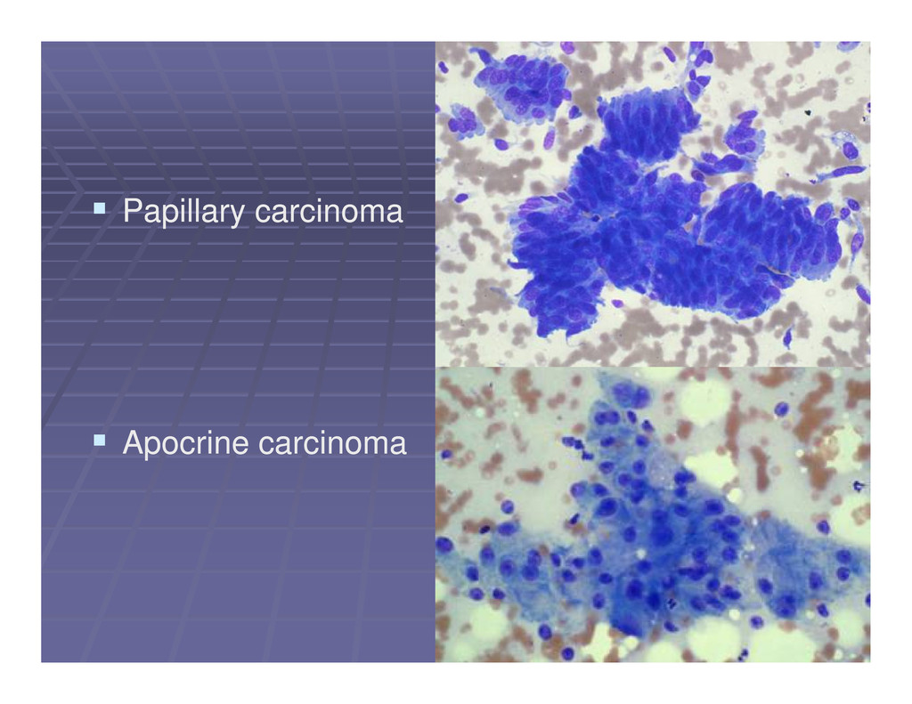
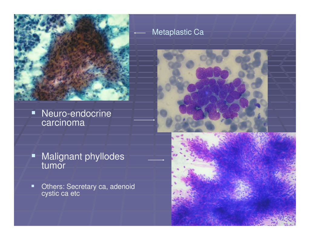
benign & malignant categorization of benign & malignant lesions lesions Nomenclature similar to histological Nomenclature similar to histological nomenclature as far as possible so that nomenclature as far as possible so that management protocols can be standardized. management protocols can be standardized. management protocols can be standardized. management protocols can be standardized. Benign proliferative lesions: Benign proliferative lesions: Exact categorization Exact categorization possible with use of histological nomenclature in possible with use of histological nomenclature in the following: the following: Fibroadenoma , fibrocystic condition, Fibroadenoma , fibrocystic condition, gynecomastia, some phyllodes tumors gynecomastia, some phyllodes tumors
is not possible, the the lesion is not possible, the cytology report should include the cytology report should include the following: following: Summary of clinical findings Summary of clinical findings Summary of clinical findings Summary of clinical findings Precise location of the aspirated Precise location of the aspirated lesion (laterality, quadrant/o’clock lesion (laterality, quadrant/o’clock position, distance from nipple). position, distance from nipple).
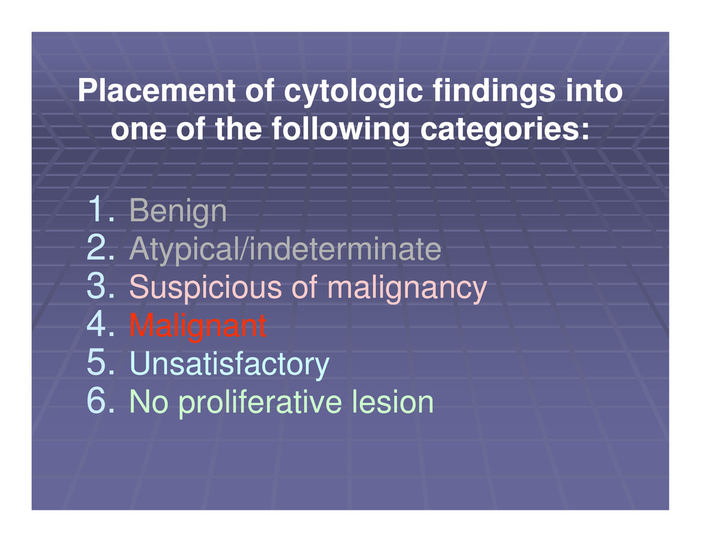
Suspicious of malignancy Suspicious of malignancy Placement of cytologic findings into Placement of cytologic findings into one of the following categories: one of the following categories: 3. 3. Suspicious of malignancy Suspicious of malignancy 4. 4. Malignant Malignant 5. 5. Unsatisfactory Unsatisfactory 6. 6. No proliferative lesion No proliferative lesion
Comment on adequacy. adequacy. Recommendations Recommendations for correlation of for correlation of Recommendations Recommendations for correlation of for correlation of cytologic diagnosis with clinical and cytologic diagnosis with clinical and imaging findings and the need for imaging findings and the need for clinical follow clinical follow- -up. up.
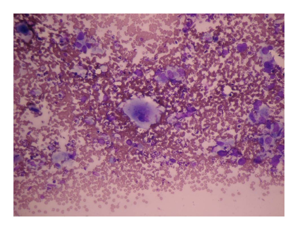
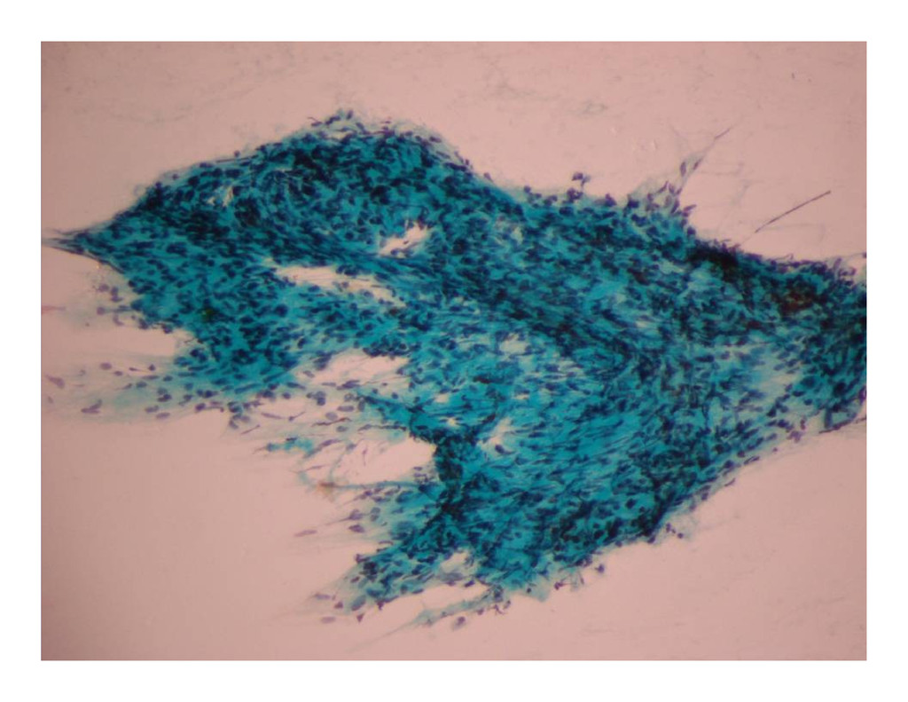
Lump in left breast Case 1 Case 1 Lump in left breast Lump in left breast FNA yielded 20 ml of altered blood FNA yielded 20 ml of altered blood After attempted evacuation, a After attempted evacuation, a residual lump was felt residual lump was felt
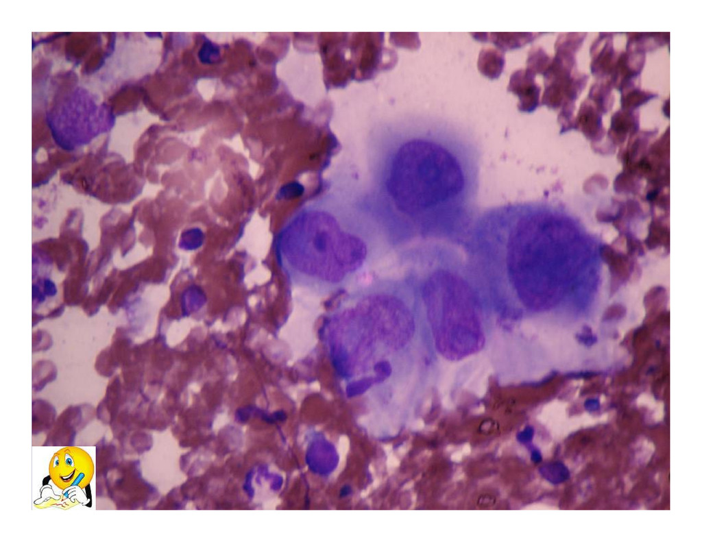
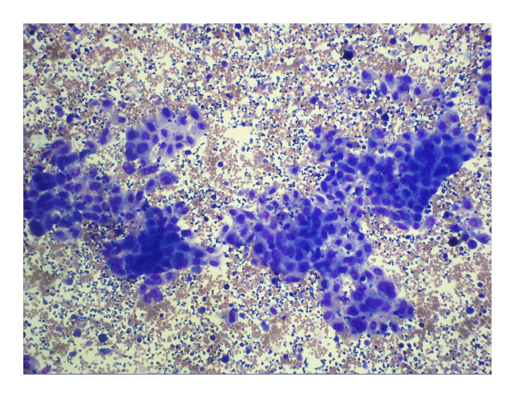
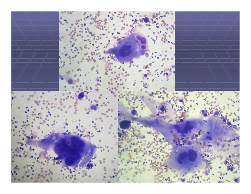
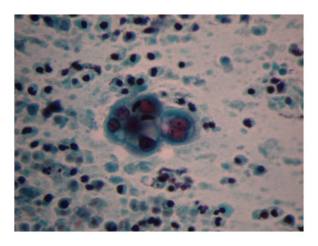
old female with lump in the right breast of 3 days duration right breast of 3 days duration Case 2 Case 2 right breast of 3 days duration right breast of 3 days duration Pain in the right breast Pain in the right breast FNA yielded 8 ml of dull greenish FNA yielded 8 ml of dull greenish fluid. fluid.
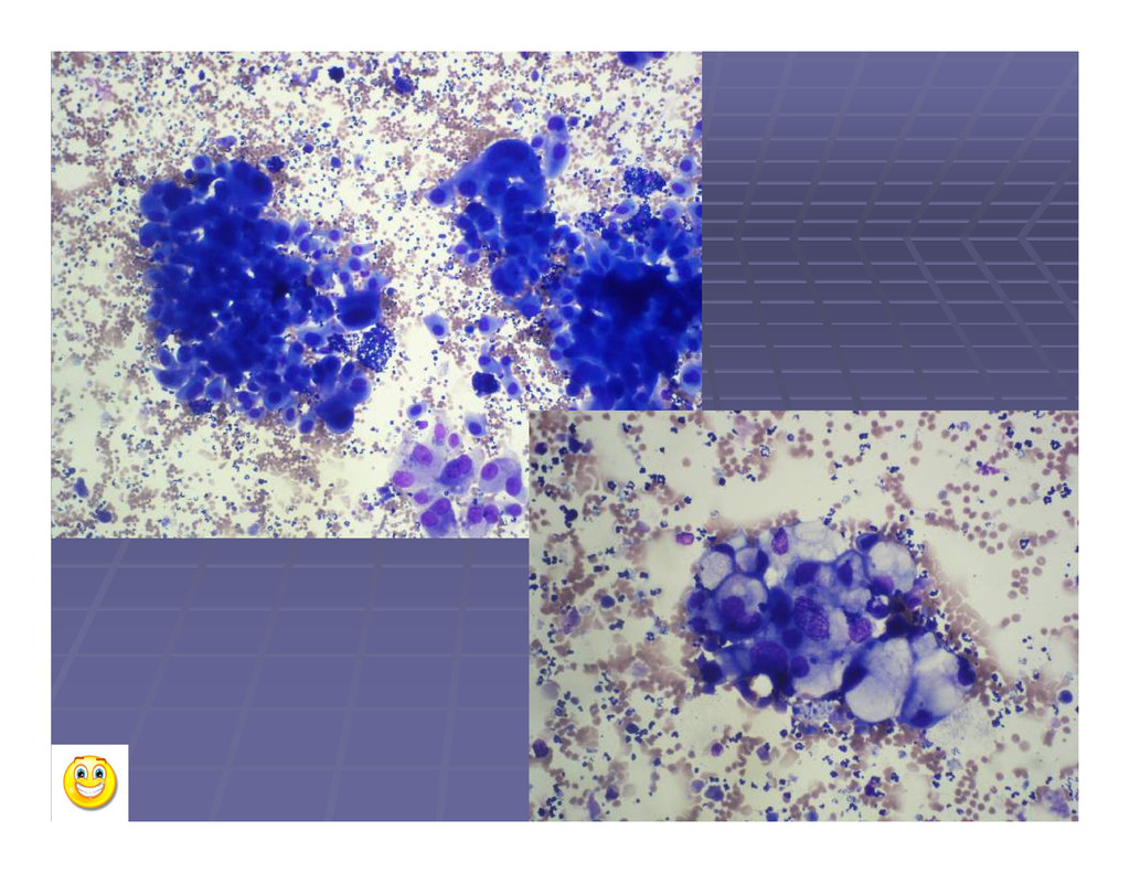
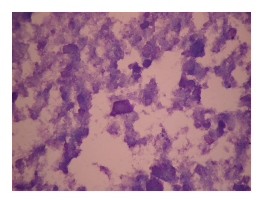
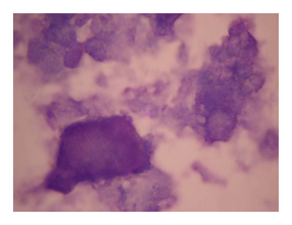
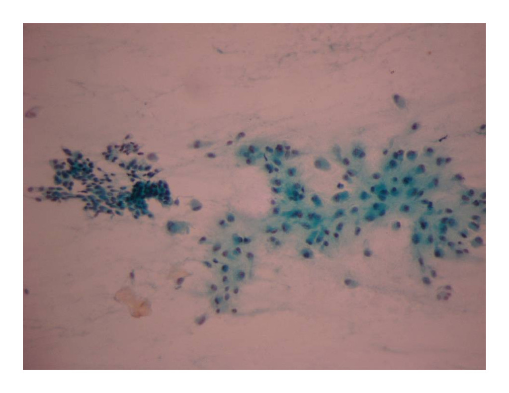
Right breast lump: 1 year Right breast lump: 1 year Para 1 gravida 2 Para 1 gravida 2 On examination: 4 cm sized mobile lump On examination: 4 cm sized mobile lump right lateral quadrant right lateral quadrant LCB: 18 months ago LCB: 18 months ago
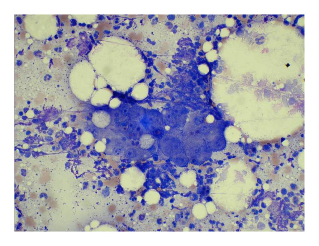
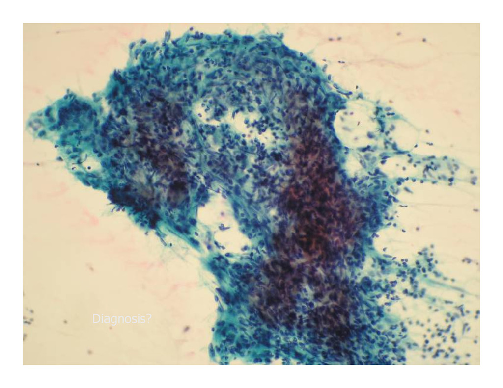
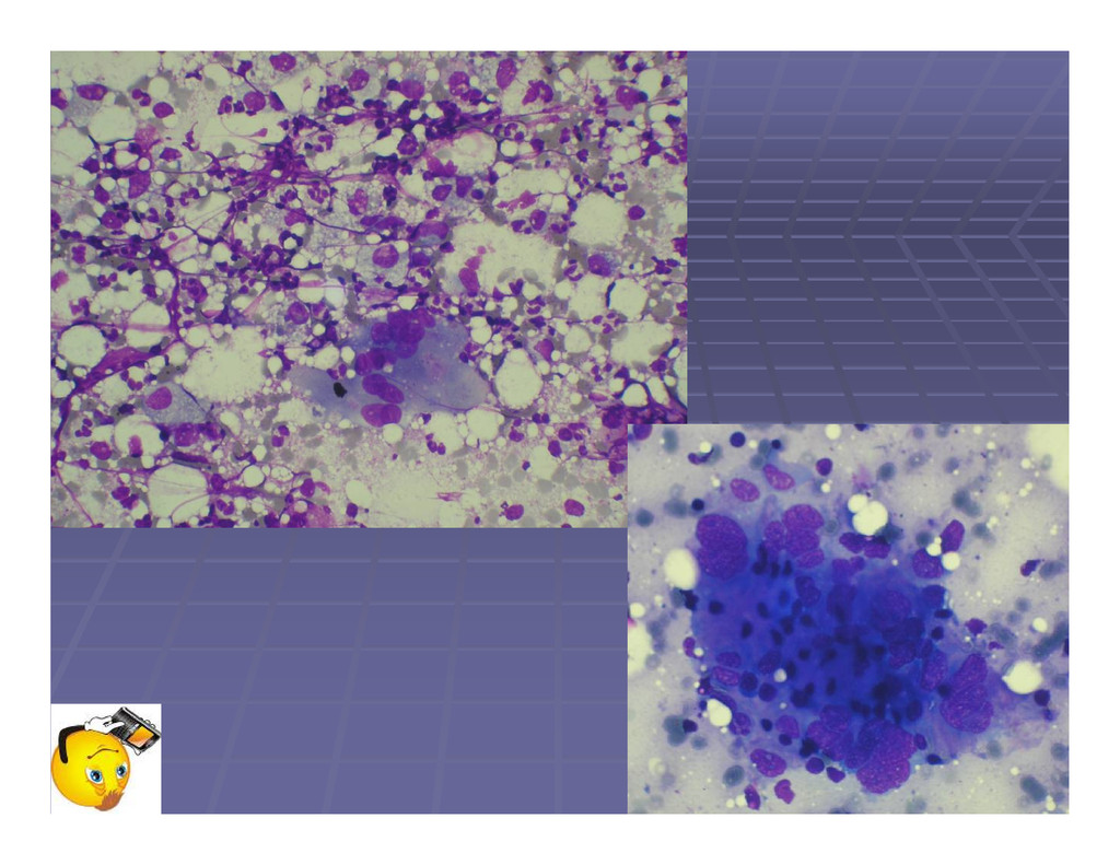
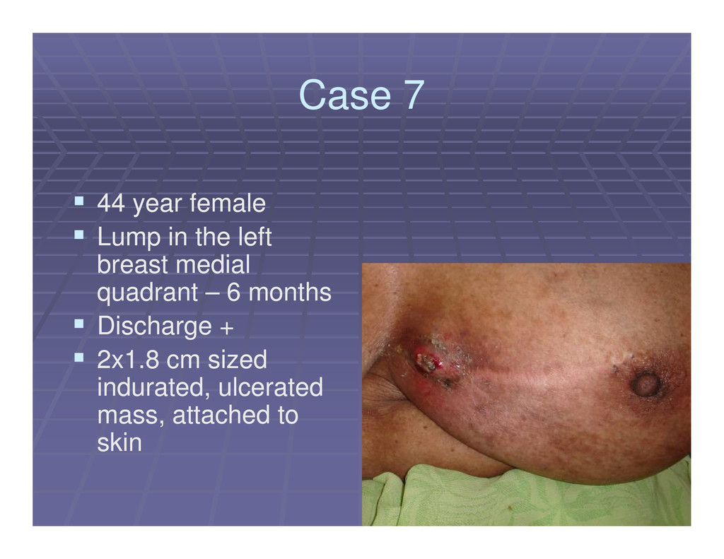
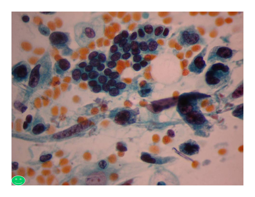
Lump in the left Lump in the left breast medial breast medial quadrant quadrant – – 6 months 6 months quadrant quadrant – – 6 months 6 months Discharge + Discharge + 2x1.8 cm sized 2x1.8 cm sized indurated, ulcerated indurated, ulcerated mass, attached to mass, attached to skin skin
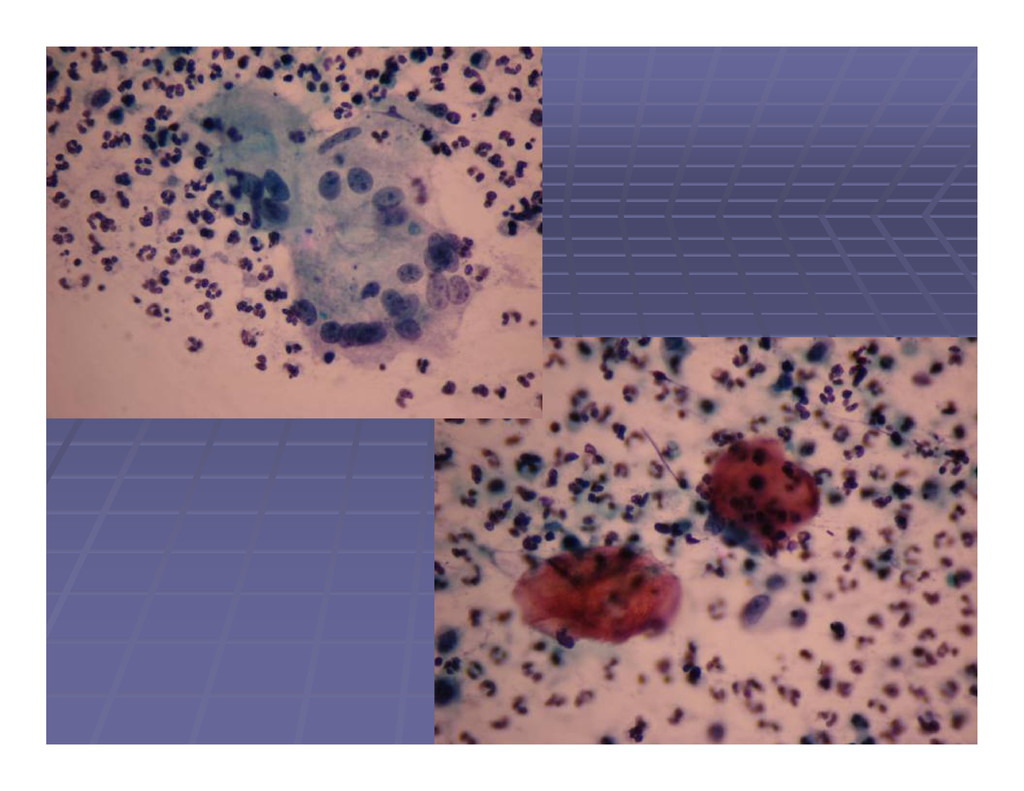
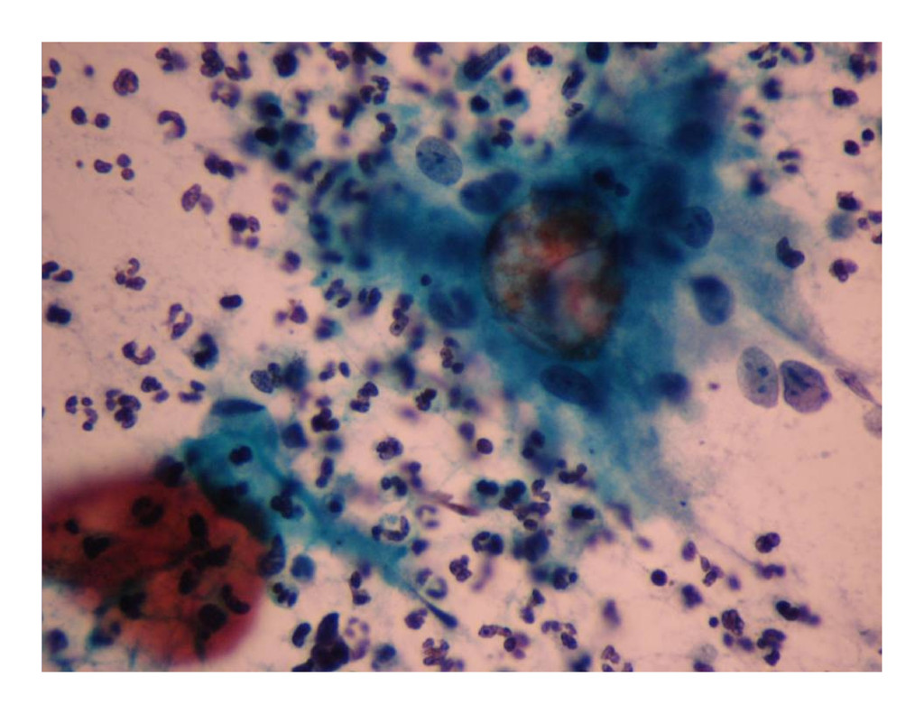
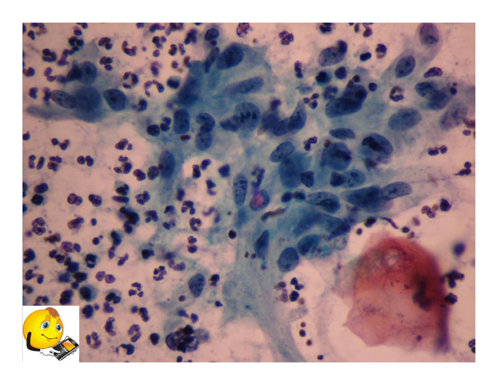
breast lump – – 1 year 1 year Case 8 Case 8 History of open biopsy done 6 months History of open biopsy done 6 months ago, reported as benign ago, reported as benign FNA smears sent from outside FNA smears sent from outside
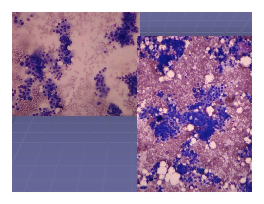
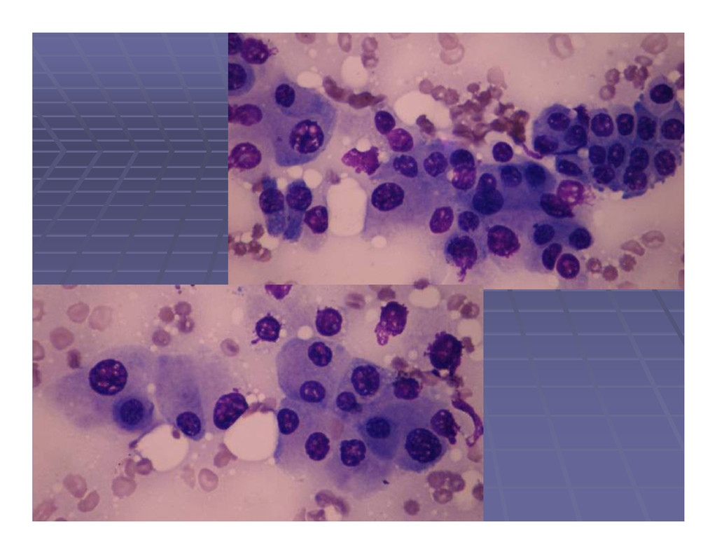
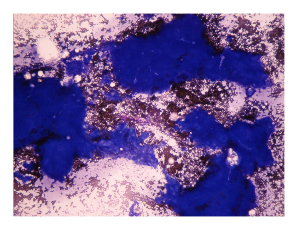
FNA requested FNA requested Patient sent to Lab for FNA Patient sent to Lab for FNA On examination, 1.5 cm sized, mobile, soft On examination, 1.5 cm sized, mobile, soft On examination, 1.5 cm sized, mobile, soft On examination, 1.5 cm sized, mobile, soft mass felt in the lateral quadrant of left mass felt in the lateral quadrant of left breast. breast. FNA done FNA done – – yielded sanguinous fluid yielded sanguinous fluid
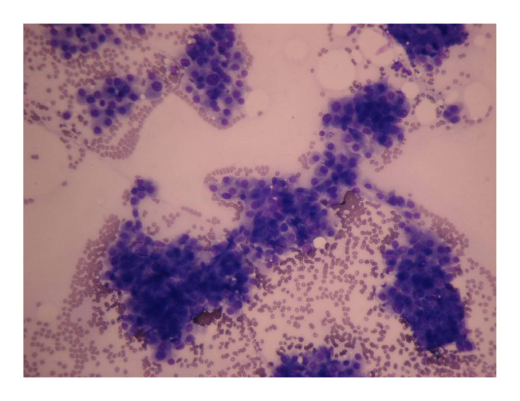
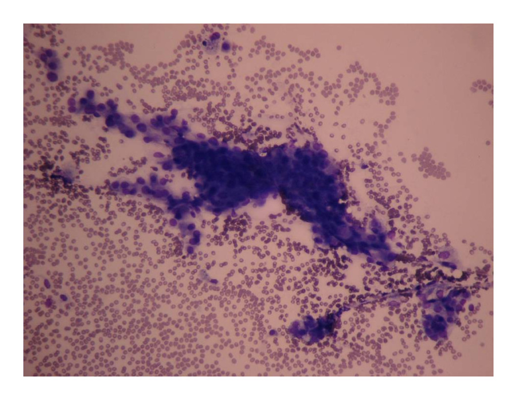
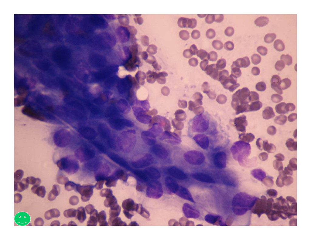
Left breast lump Left breast lump – – 3 years 3 years Left breast lump Left breast lump – – 3 years 3 years O/E: 5 cm, firm, mobile lump in the O/E: 5 cm, firm, mobile lump in the central and adjacent medial central and adjacent medial quadrants of left breast, partly retro quadrants of left breast, partly retro- - areolar areolar