England Rugby CPD: Imaging of the Developing Rugby Shoulder
England Rugby held a fantastic CPD day as part of the annual Wellington Festival at Wellington College. This is the presentation I gave on developing rugby shoulder injuries.
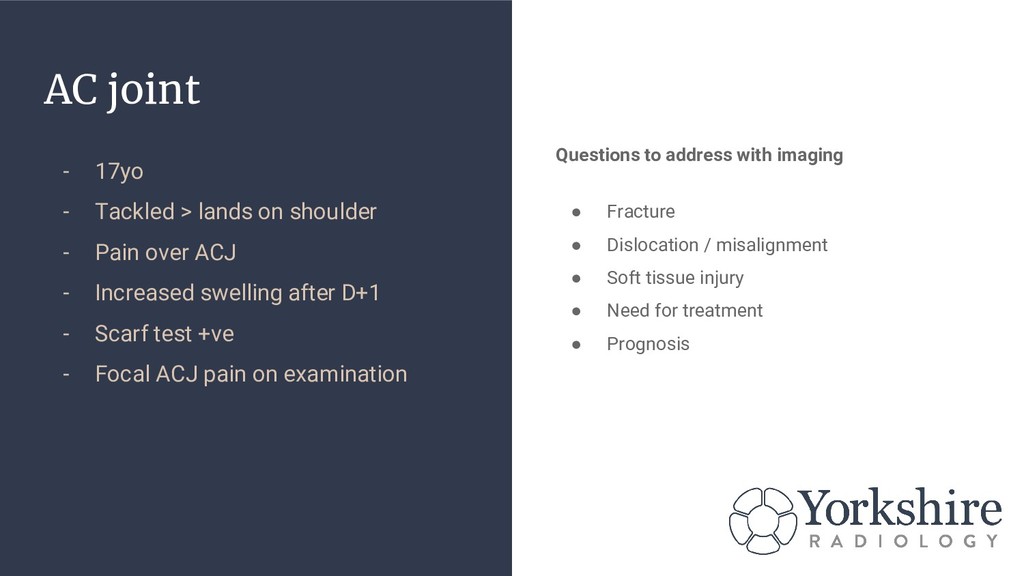
- Pain over ACJ - Increased swelling after D+1 - Scarf test +ve - Focal ACJ pain on examination Questions to address with imaging • Fracture • Dislocation / misalignment • Soft tissue injury • Need for treatment • Prognosis
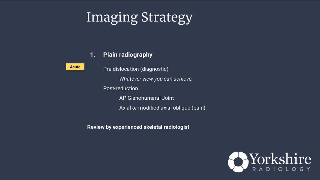
b. Dislocations c. Soft tissue swelling (clues) d. Anatomical variation / morphology Views Shoulder / ACJ AP view Clavicle series (supplement above with angled view) Glenohumeral axial Glenohumeral joint (Grashey view) Acute Normal AP Shoulder 11yo Normal Axial
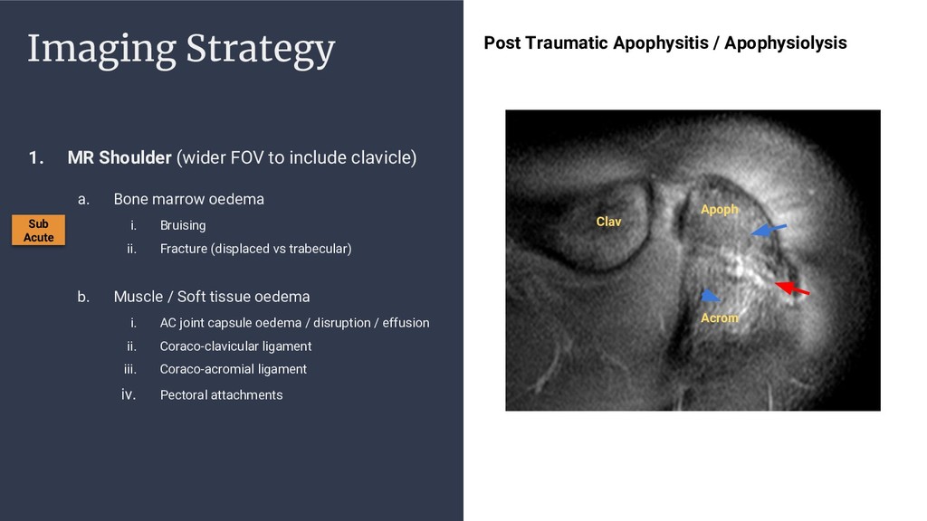
a. Bone marrow oedema i. Bruising ii. Fracture (displaced vs trabecular) b. Muscle / Soft tissue oedema i. AC joint capsule oedema / disruption / effusion ii. Coraco-clavicular ligament iii. Coraco-acromial ligament iv. Pectoral attachments Sub Acute
a. Bone marrow oedema i. Bruising ii. Fracture (displaced vs trabecular) b. Muscle / Soft tissue oedema i. AC joint capsule oedema / disruption / effusion ii. Coraco-clavicular ligament iii. Coraco-acromial ligament iv. Pectoral attachments Sub Acute
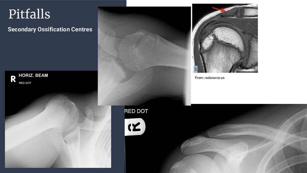
a. Bone marrow oedema i. Bruising ii. Fracture (displaced vs trabecular) b. Muscle / Soft tissue oedema i. AC joint capsule oedema / disruption / effusion ii. Coraco-clavicular ligament iii. Coraco-acromial ligament iv. Pectoral attachments Sub Acute Clav Apoph Acrom Post Traumatic Apophysitis / Apophysiolysis
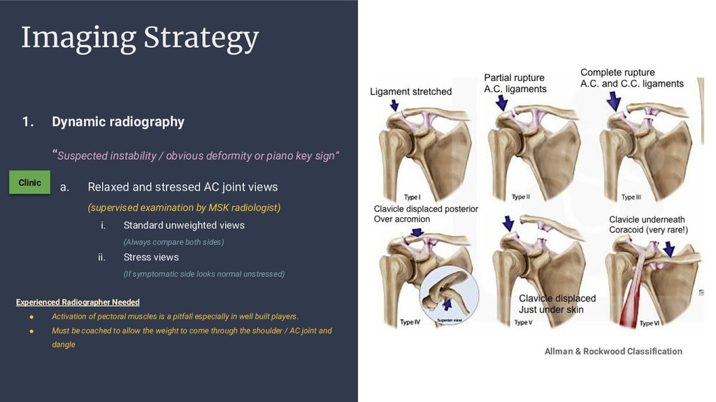
or piano key sign” a. Relaxed and stressed AC joint views (supervised examination by MSK radiologist) i. Standard unweighted views (Always compare both sides) ii. Stress views (If symptomatic side looks normal unstressed) Experienced Radiographer Needed • Activation of pectoral muscles is a pitfall especially in well built players. • Must be coached to allow the weight to come through the shoulder / AC joint and dangle Clinic Allman & Rockwood Classification
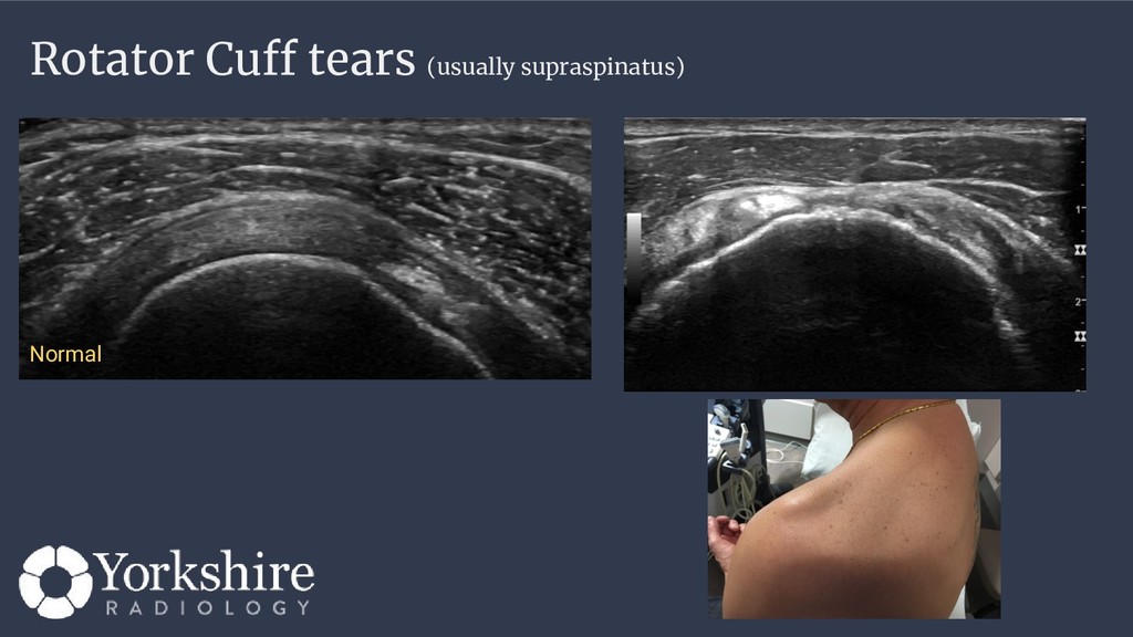
You might pick up ACJ injury during a shoulder USS - Interventionally useful Injections for ACJ capsulitis Slow to settle… No fracture / displacement shown USS swollen capsule and Doppler vascularity
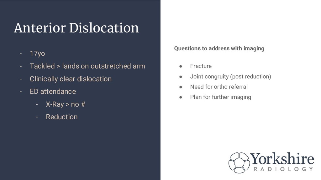
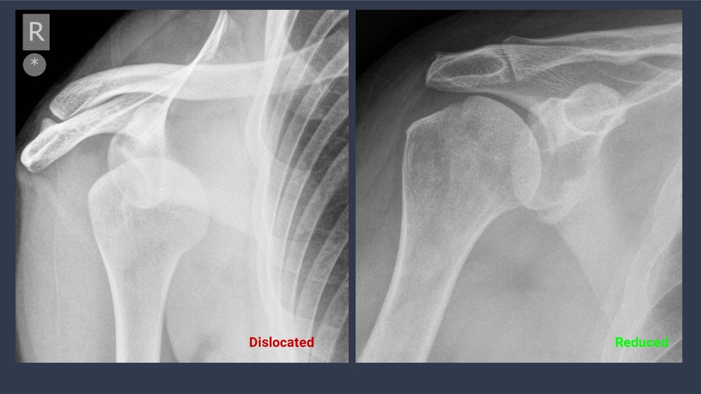
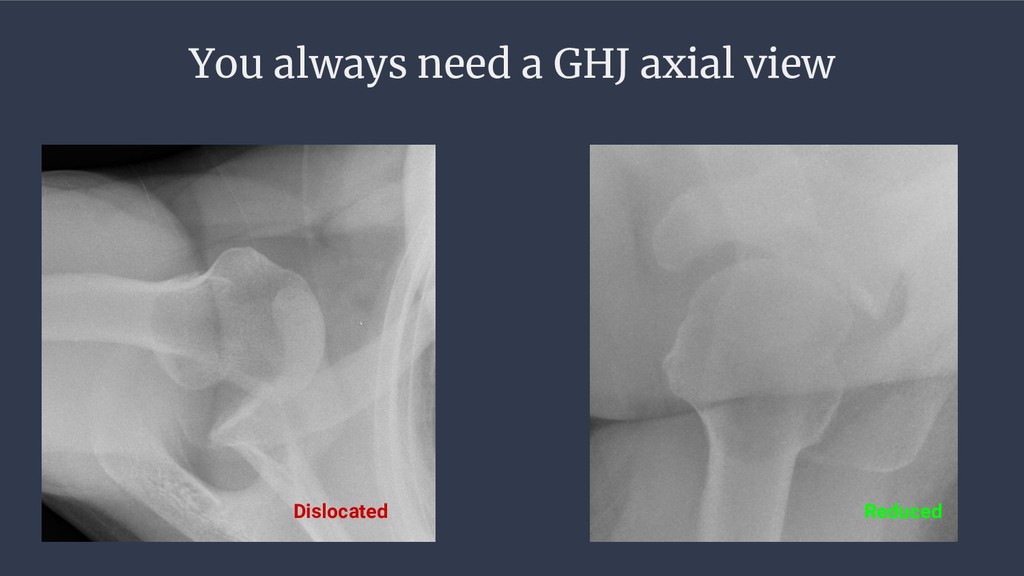
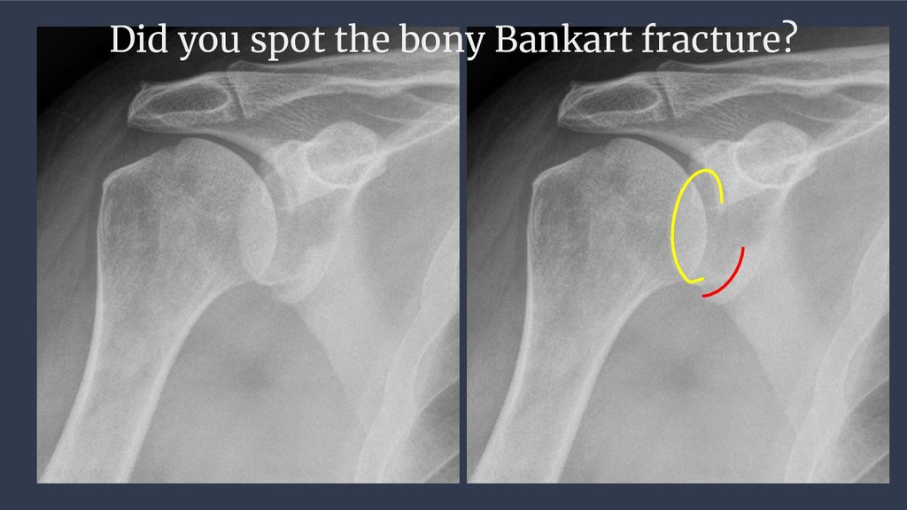
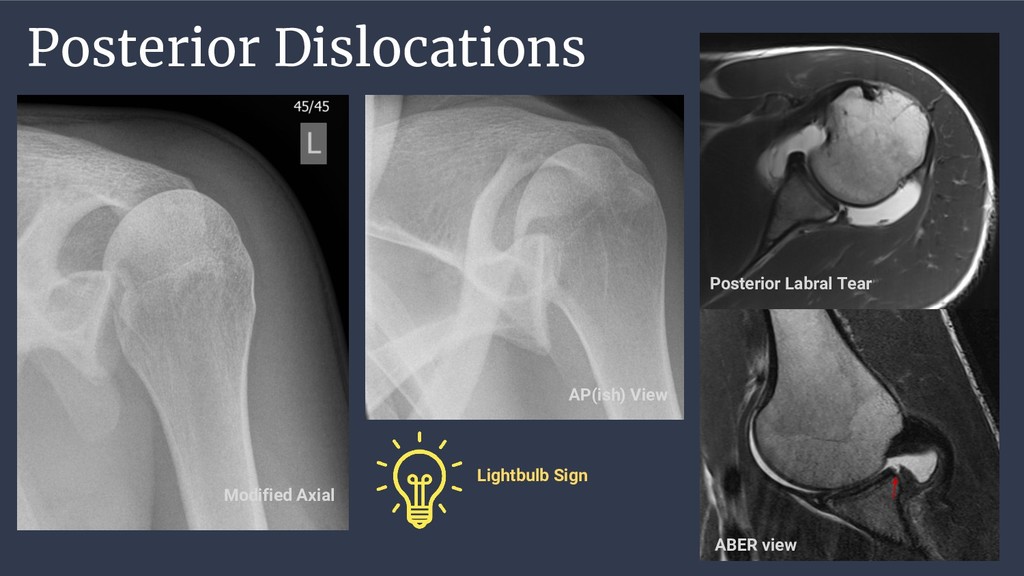
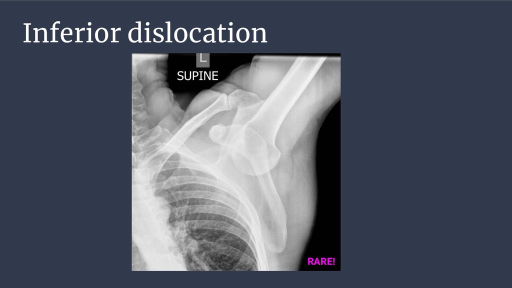
arm - Clinically clear dislocation - ED attendance - X-Ray > no # - Reduction Questions to address with imaging • Fracture • Joint congruity (post reduction) • Need for ortho referral • Plan for further imaging
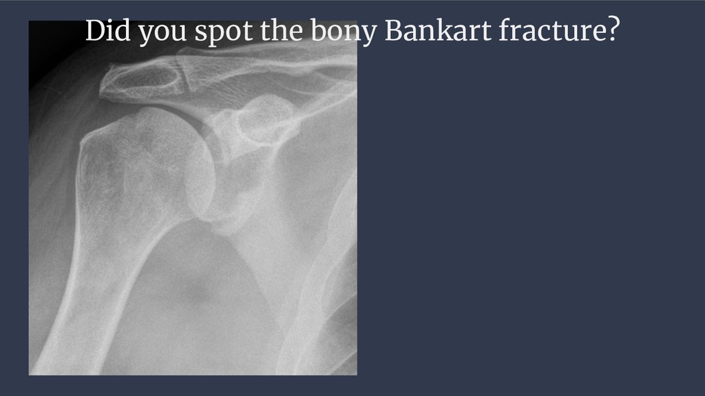
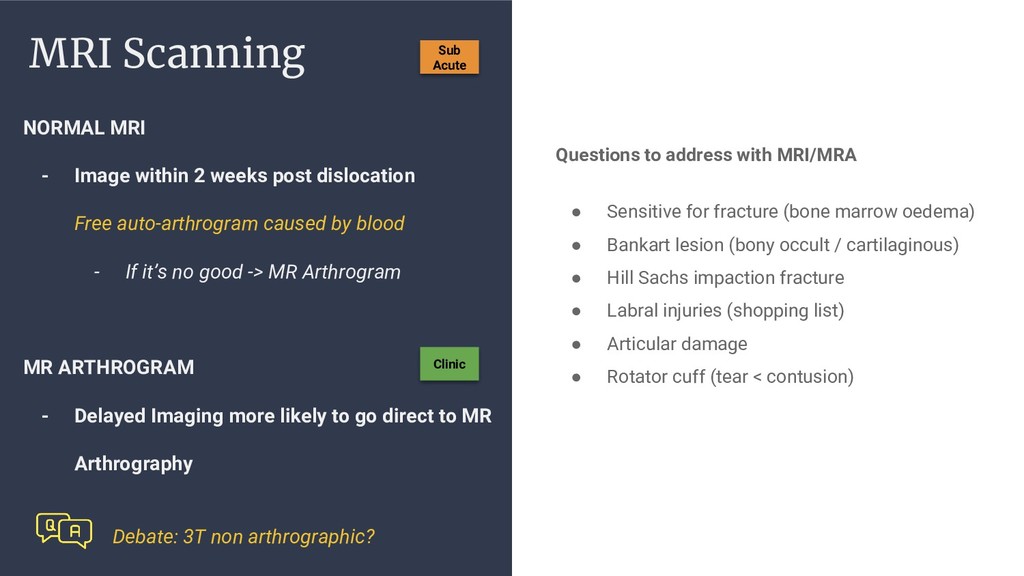
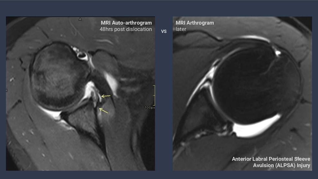
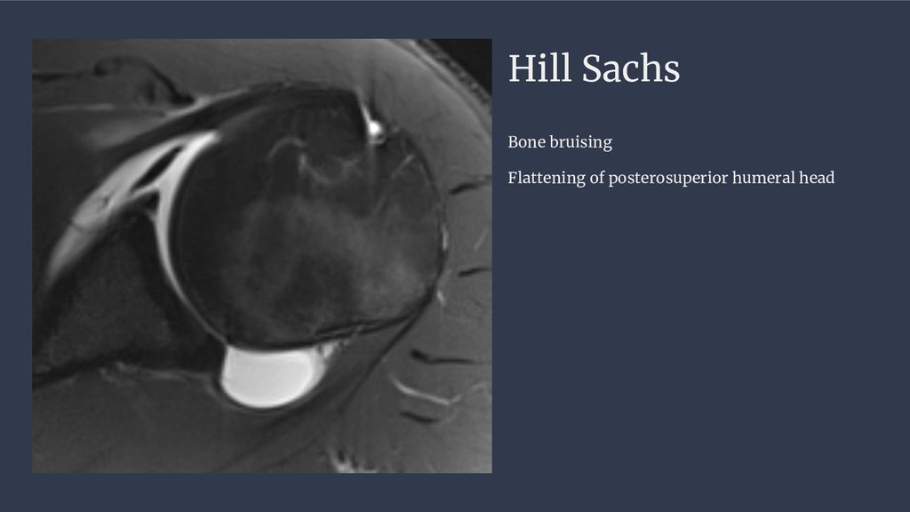
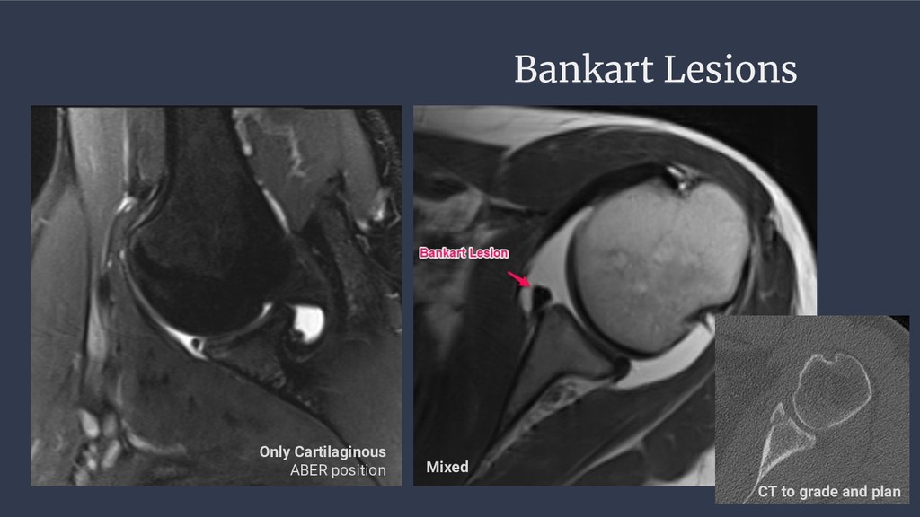
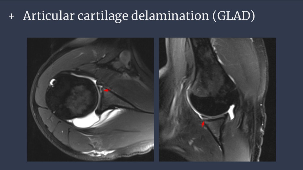
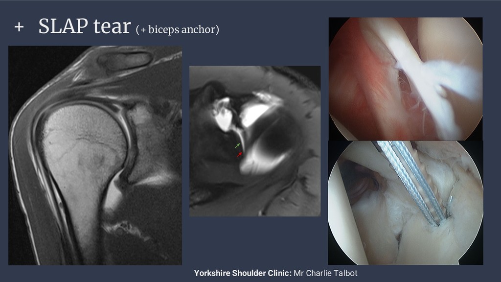
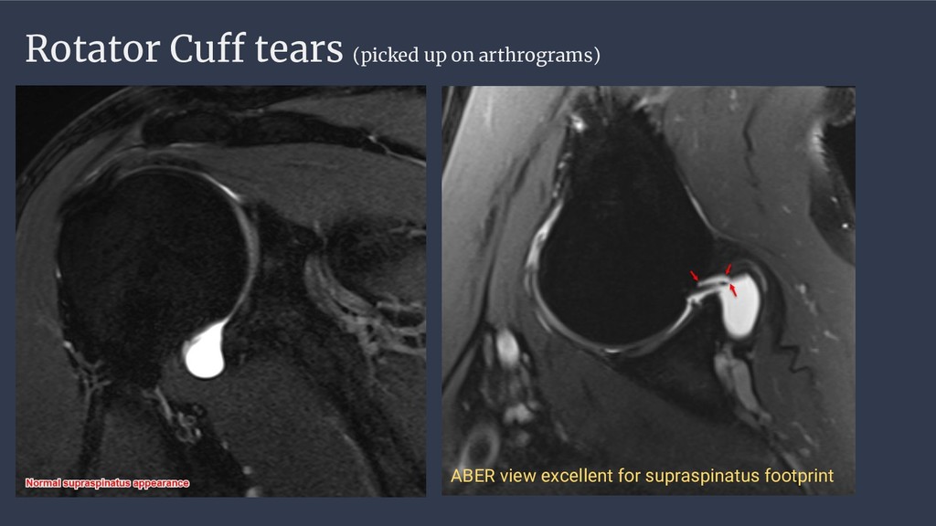
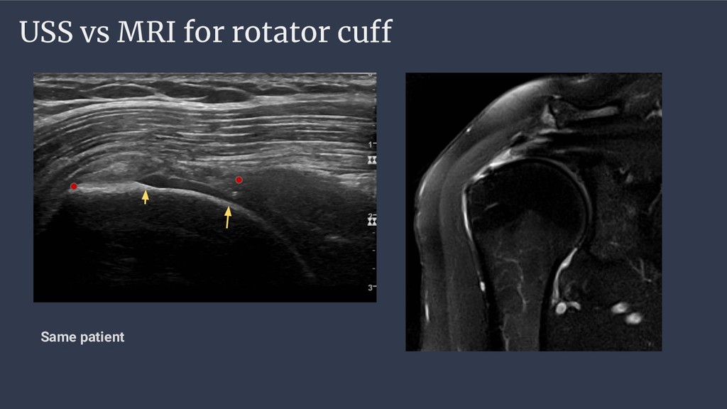
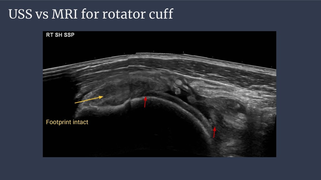
auto-arthrogram caused by blood - If it’s no good -> MR Arthrogram MR ARTHROGRAM - Delayed Imaging more likely to go direct to MR Arthrography MRI Scanning Sub Acute Questions to address with MRI/MRA • Sensitive for fracture (bone marrow oedema) • Bankart lesion (bony occult / cartilaginous) • Hill Sachs impaction fracture • Labral injuries (shopping list) • Articular damage • Rotator cuff (tear < contusion) Clinic Debate: 3T non arthrographic?