Upgrade to Pro — share decks privately, control downloads, hide ads and more …

Lateral Hip Pain - A Radiological Approach

Lateral Hip Pain - A Radiological Approach

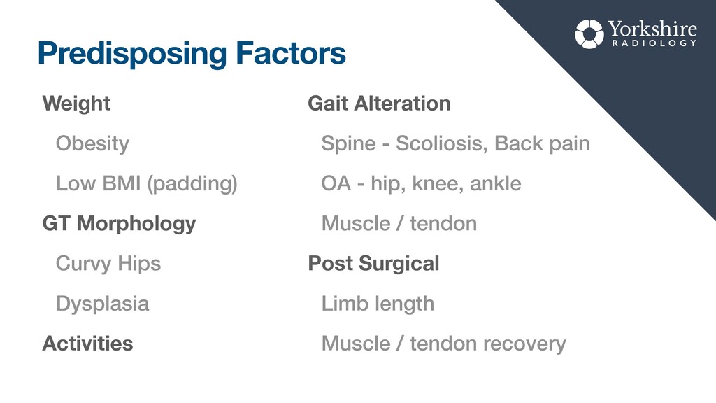
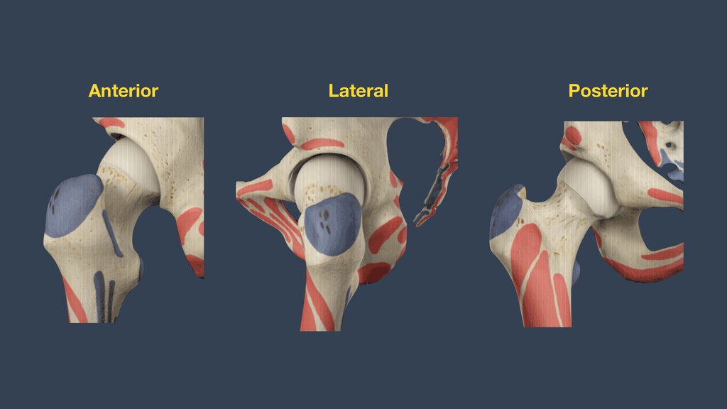
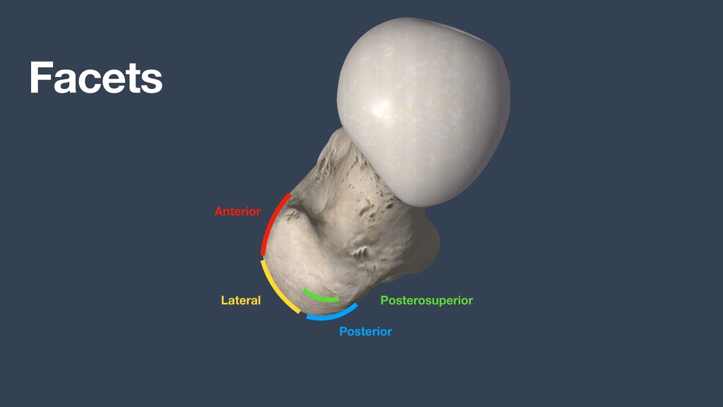
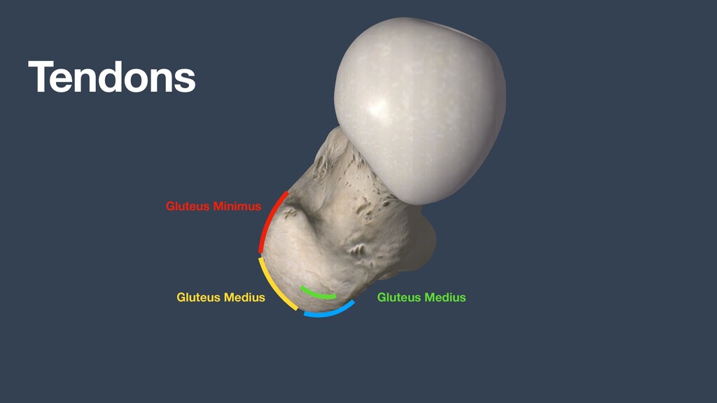
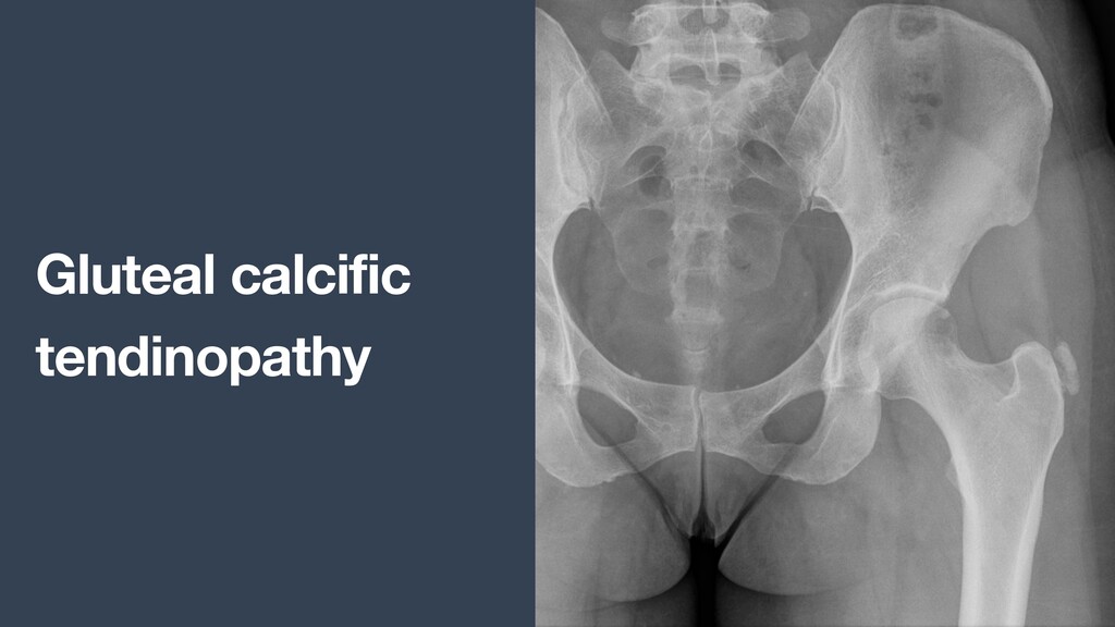
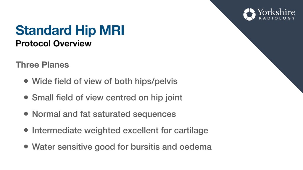
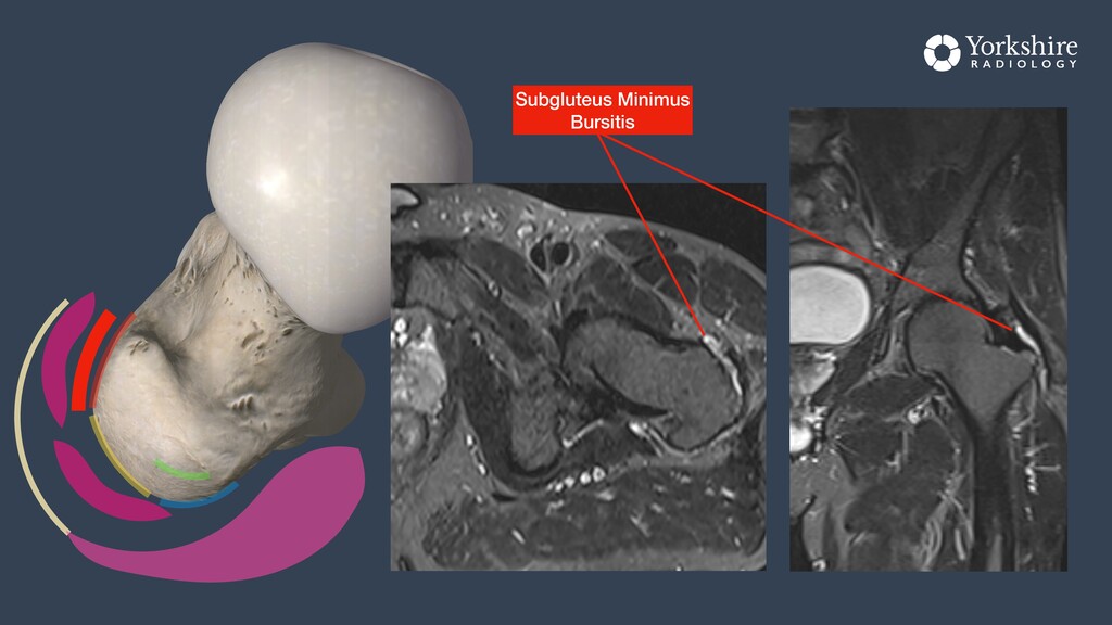
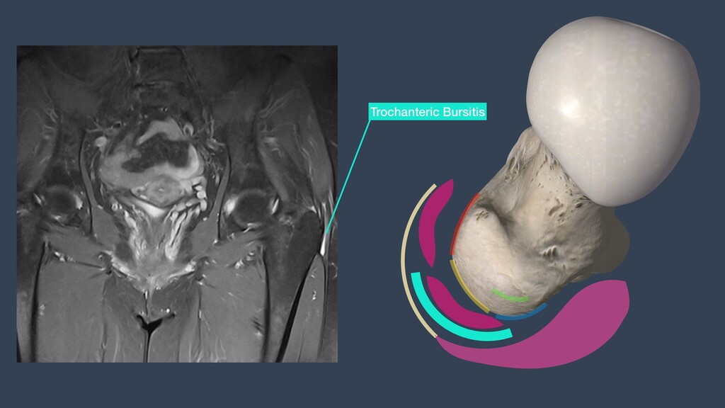
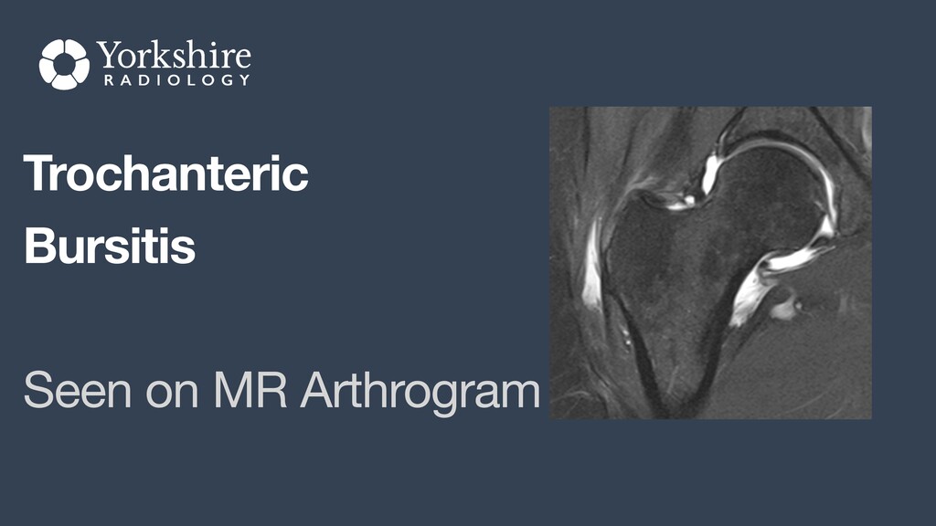
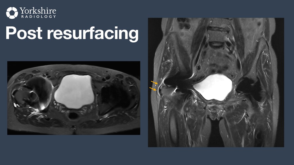
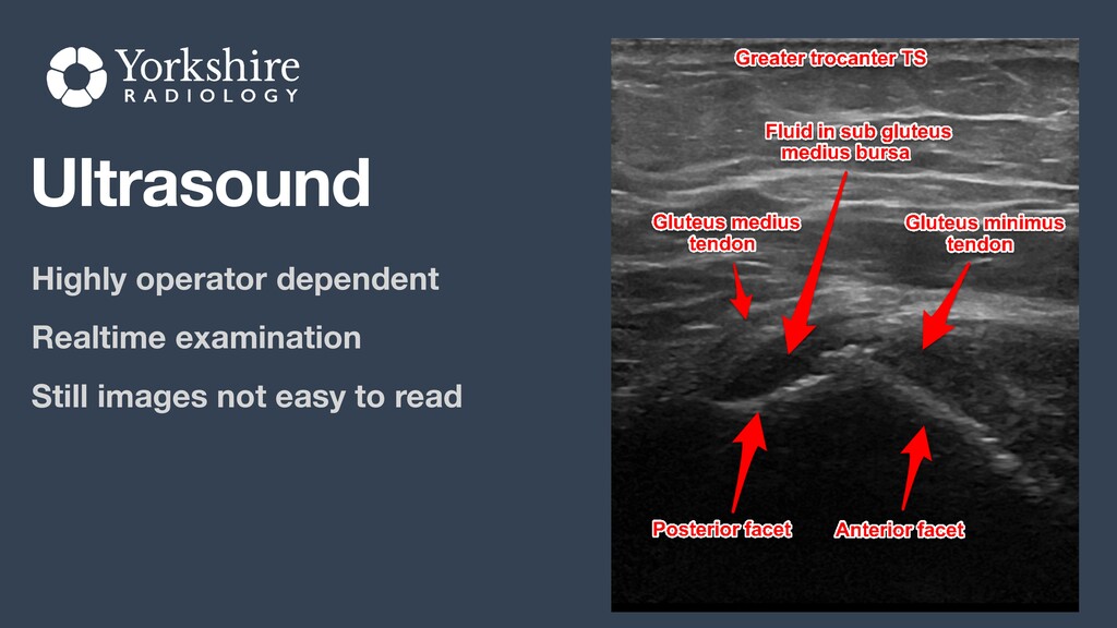
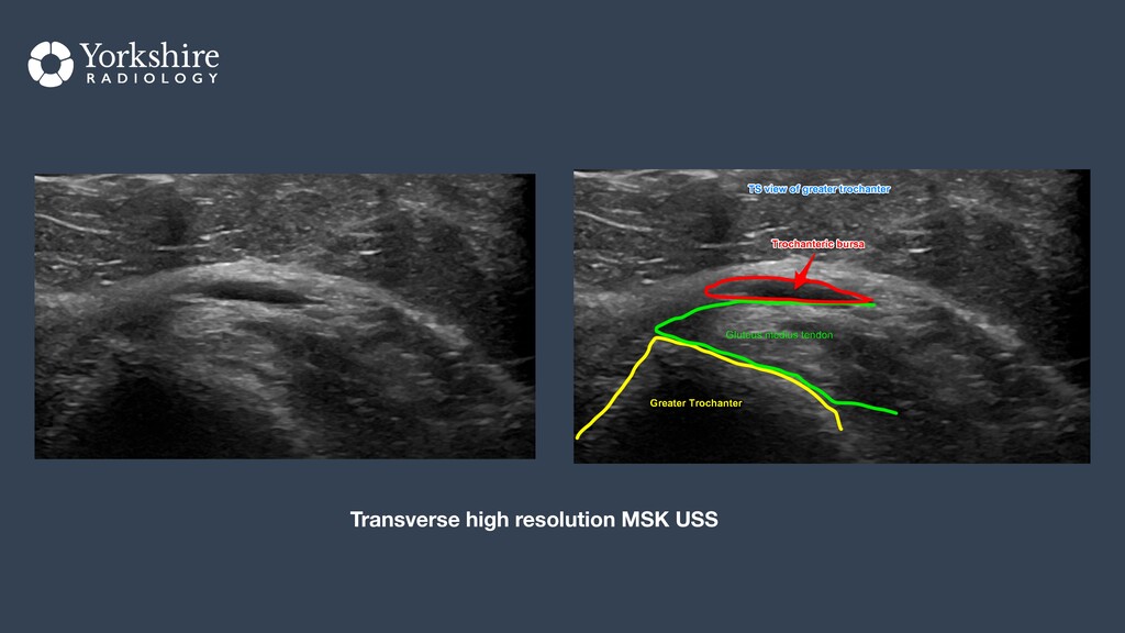
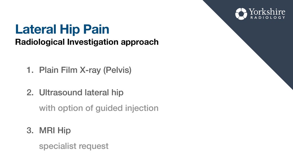
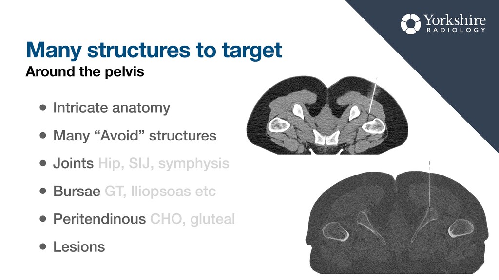
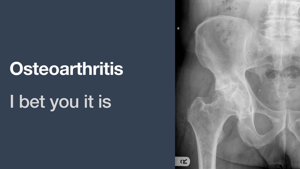
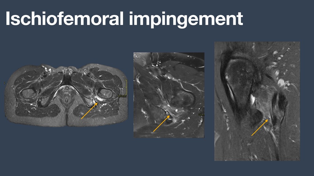
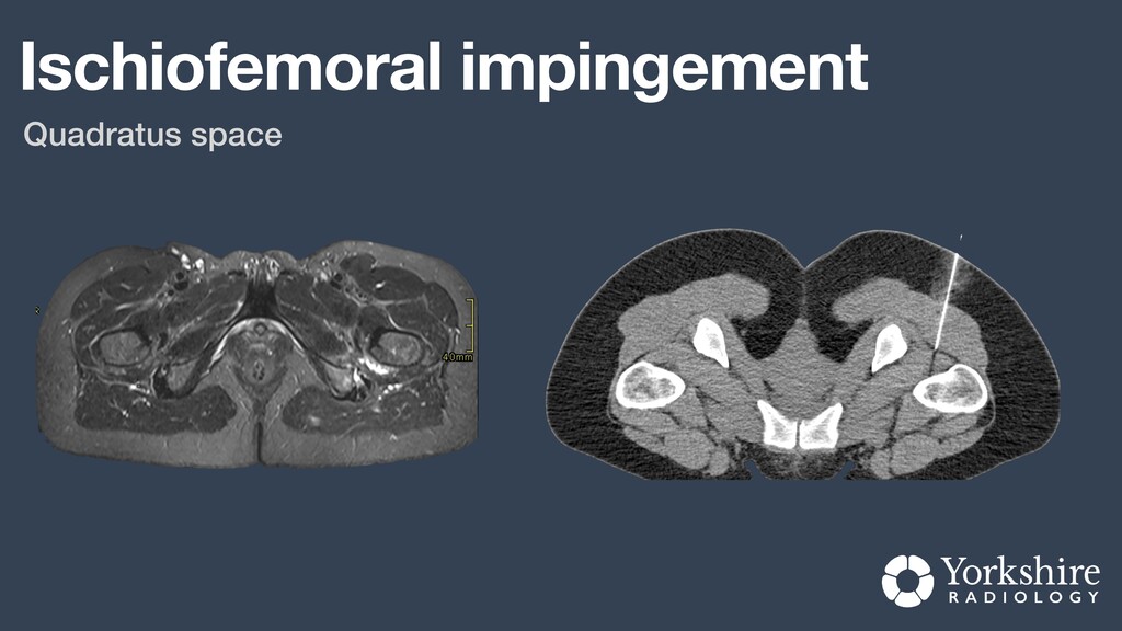
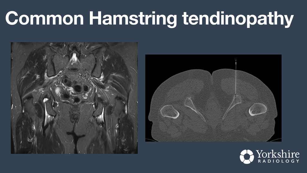
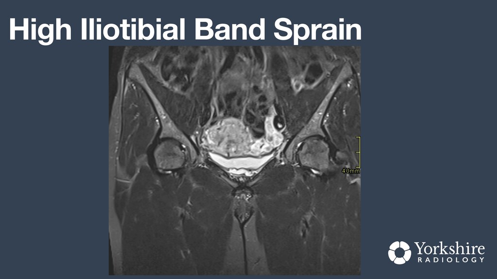
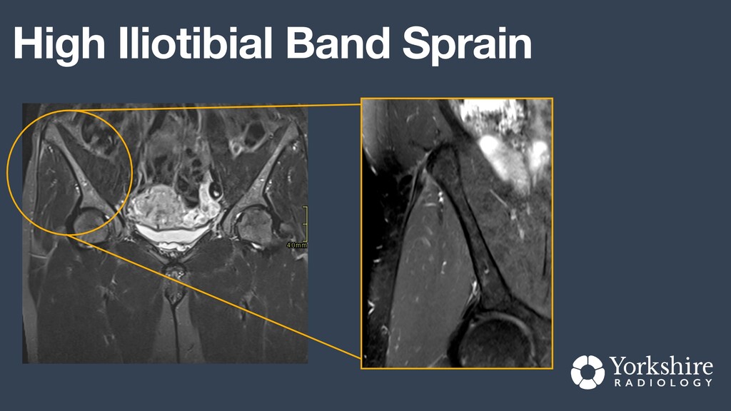
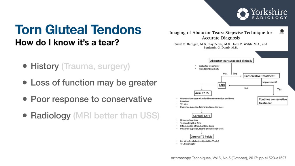
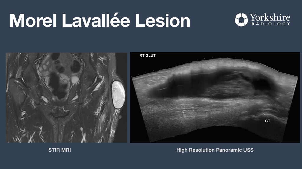
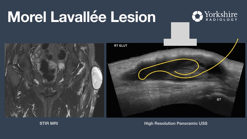
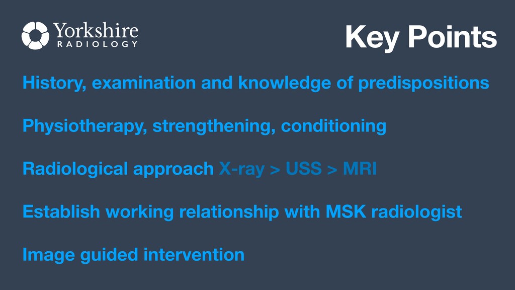
An exploration of the aetiology of common causes of lateral hip pain and some alternative swerve ball diagnoses. The radiological anatomy and approach to investigation as well as the common appearances seen on multimodality imaging (x-ray, MR, CT, ultrasound) are discussed.

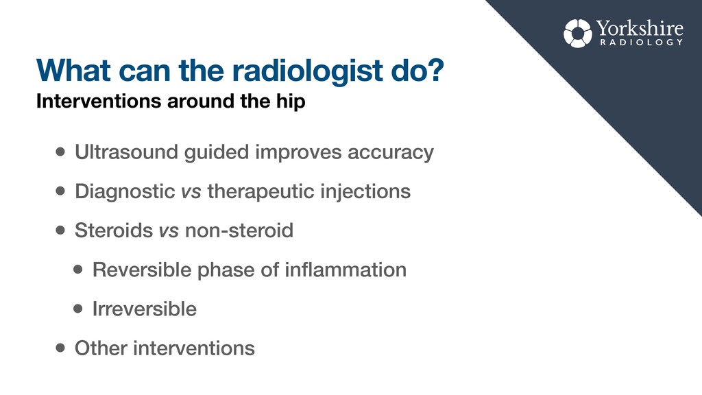
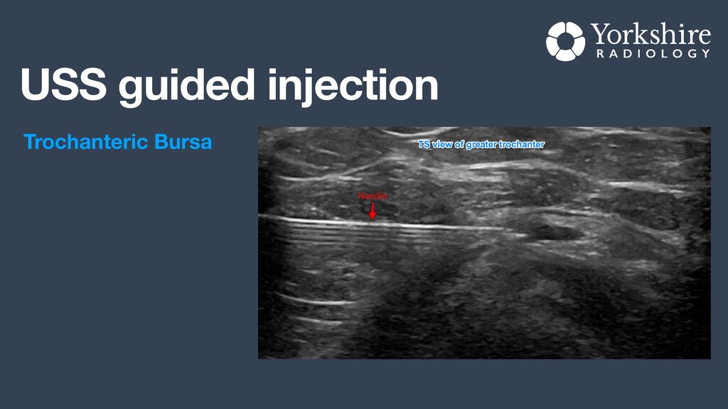
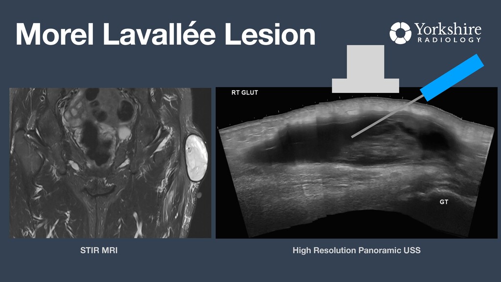
We also touch on interventional techniques for greater trochanteric pain syndrome and some of the other conditions around the hip.

Dr Daniel Fascia

August 08, 2021
Tweet

More Decks by Dr Daniel Fascia

Other Decks in Science

Transcript

  1. About me Trained in Imperial College London and Perth WA

    Consultant MSK Radiogist in Harrogate (2014) Regional Clinical Lead (YIC) Director of Medical Imaging - NHS Nightingale NHS and Private Practice Illiya Vjestica via unsplash
  2. What I’m going to cover Diagnostic Imaging A recommended imaging

    algorithm How to request imaging The role for interventional radiology Swerve balls Key points
  3. Trochanteric Pain Syndrome Con fl icting terminology often used •

    Trochanteric Bursitis • Gluteal tendinopathy • The rotator cuff syndrome of the hip? What exactly is it?
  4. Predisposing Factors Weight Obesity Low BMI (padding) GT Morphology Curvy

    Hips Dysplasia Activities Gait Alteration Spine - Scoliosis, Back pain OA - hip, knee, ankle Muscle / tendon Post Surgical Limb length Muscle / tendon recovery
  5. Plain Film Radiography Pelvic x-ray • Osteoarthritis? • Morphological features?

    • Enthesopathy? • Another cause of hip pain? X-Rays
  6. Enthesopathy Are Radiographic Trochanteric Surface Irregularities Associated with Abductor Tendon

    Abnormalities? Lenca Steinert, Marco Zanetti, Juerg Hodler, Christian W. A. P fi rrmann, Claudio Dora, and Nadja Saupe Radiology 2010 257:3, 754-763
  7. Magnetic Resonance (MR) Not usually fi rst line imaging Useful

    when the diagnosis is not clear More expensive Less accessible Time consuming Pros and Cons
  8. Magnetic Resonance (MR) Is GTPS the primary reason for the

    MRI? YES - Standard hip MRI NO - Depends on indication Which Protocol?
  9. Magnetic Resonance (MR) Is GTPS the primary reason for the

    MRI? YES - Standard hip MRI NO - Depends on indication Which Protocol? Just write a good concise history and di ff erential diagnosis. Let the radiologist choose
  10. Standard Hip MRI Three Planes • Wide fi eld of

    view of both hips/pelvis • Small fi eld of view centred on hip joint • Normal and fat saturated sequences • Intermediate weighted excellent for cartilage • Water sensitive good for bursitis and oedema Protocol Overview
  11. Lateral Hip Pain 1. Plain Film X-ray (Pelvis) 2. Ultrasound

    lateral hip 
 with option of guided injection 3. MRI Hip 
 specialist request Radiological Investigation approach
  12. What makes a great request? • Build a relationship with

    a friendly MSK radiologist • Liberally communicate and discuss cases • Establish preferences: terminology, pathways, grading • Write concise, clear referrals • Always include differential diagnosis / clinical question • Let the radiologist choose the protocol Top tips to getting the most out of radiology
  13. What makes a great request? IMAGING REQUEST: MRI Left Hip

    CLINICAL DETAILS: Left sided lateral hip pain radiating to buttock. Started 4-months ago after increase in running activities during lockdown. Limited response to rest, NSAIDs and physiotherapy. 
 Had CDH as child treated with osteotomy. Already had x-ray at Churchill Hospital. ? GTPS ? OA ? Impingement Top tips to getting the most out of radiology
  14. What can the radiologist do? • Ultrasound guided improves accuracy

    • Diagnostic vs therapeutic injections • Steroids vs non-steroid • Reversible phase of in fl ammation • Irreversible • Other interventions Interventions around the hip
  15. Many structures to target • Intricate anatomy • Many “Avoid”

    structures • Joints Hip, S IJ , symphysis • Bursae GT, Iliopsoas etc • Peritendinous CHO, gluteal • Lesions Around the pelvis
  16. Torn Gluteal Tendons • History (Trauma, surgery) • Loss of

    function may be greater • Poor response to conservative • Radiology (MRI better than USS) How do I know it’s a tear? Arthroscopy Techniques, Vol 6, No 5 (October), 2017: pp e1523-e1527
  17. Key Points History, examination and knowledge of predispositions Physiotherapy, strengthening,

    conditioning Radiological approach X-ray > USS > MRI Establish working relationship with MSK radiologist Image guided intervention