Upgrade to Pro — share decks privately, control downloads, hide ads and more …

First Contact Physio: Musculoskeletal Radiology Primer

First Contact Physio: Musculoskeletal Radiology Primer

Synopsis
MSK physiotherapists are taking on a new role as primary consulting practitioners across the land. This is both exciting and daunting at the same time and there is currently a gap in rapid education to equip physios for this role.

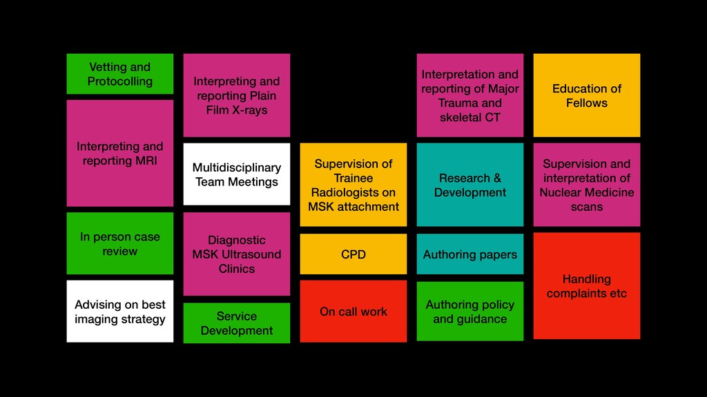
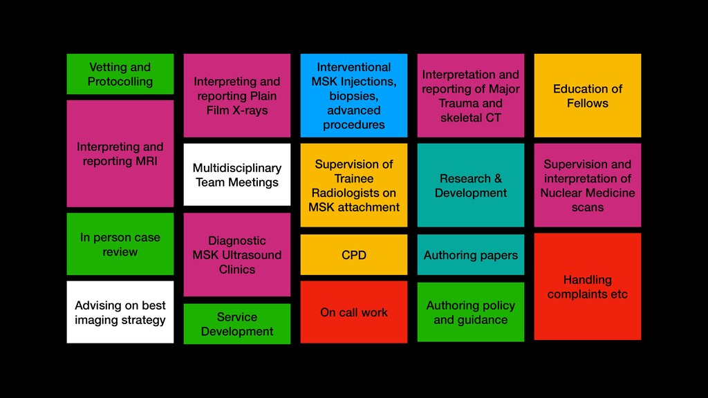
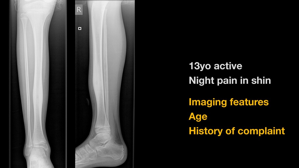
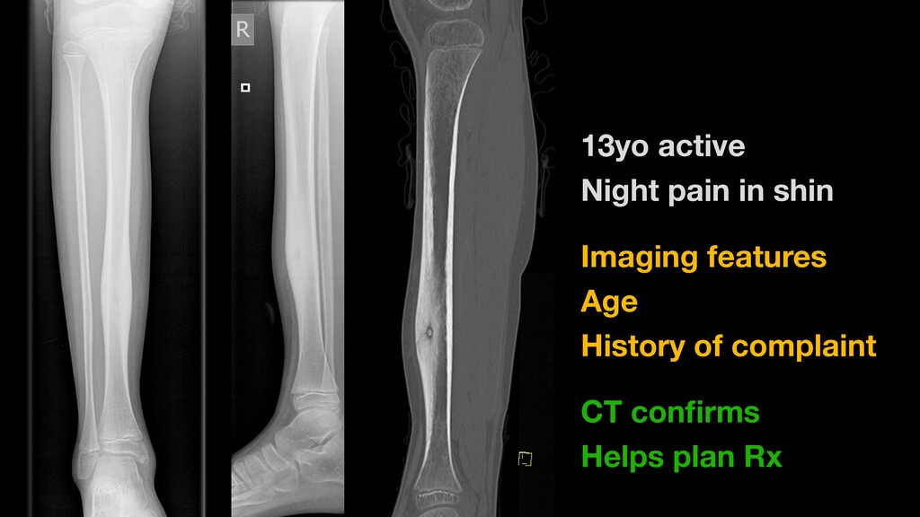
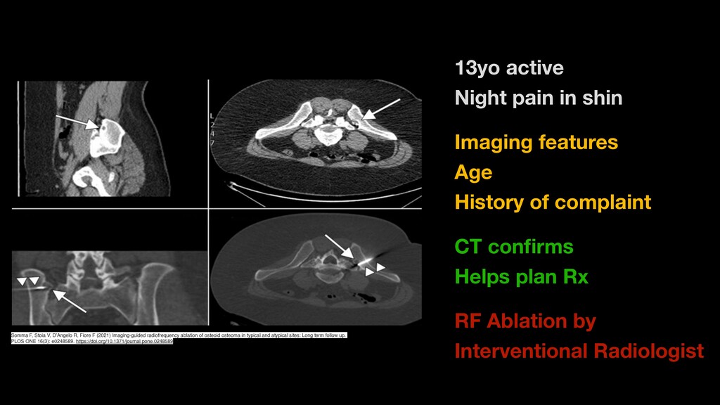
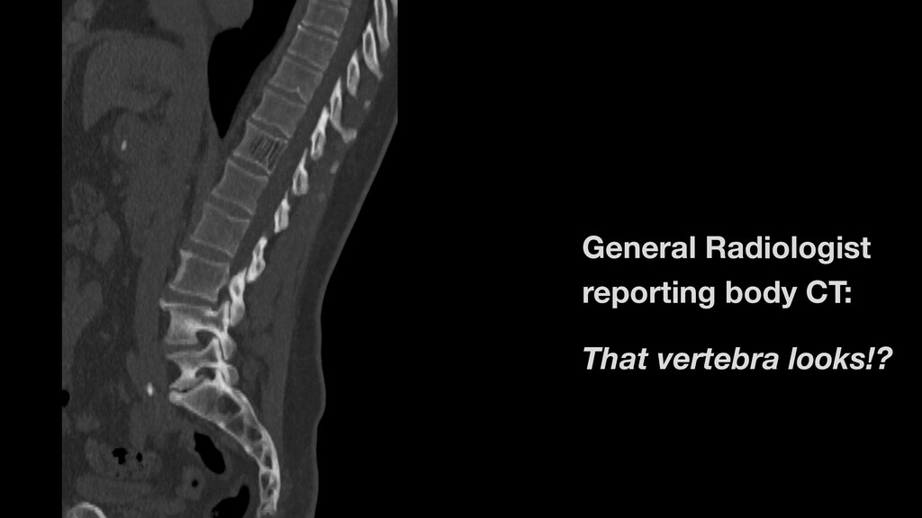
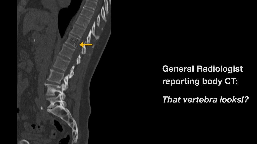
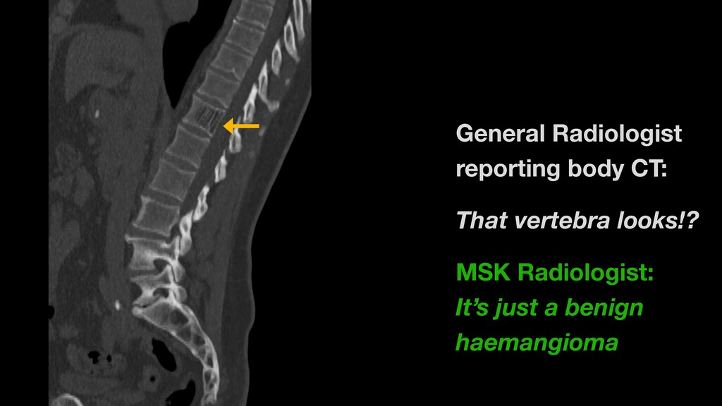
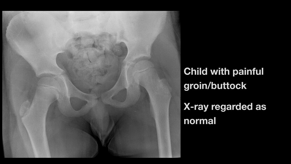
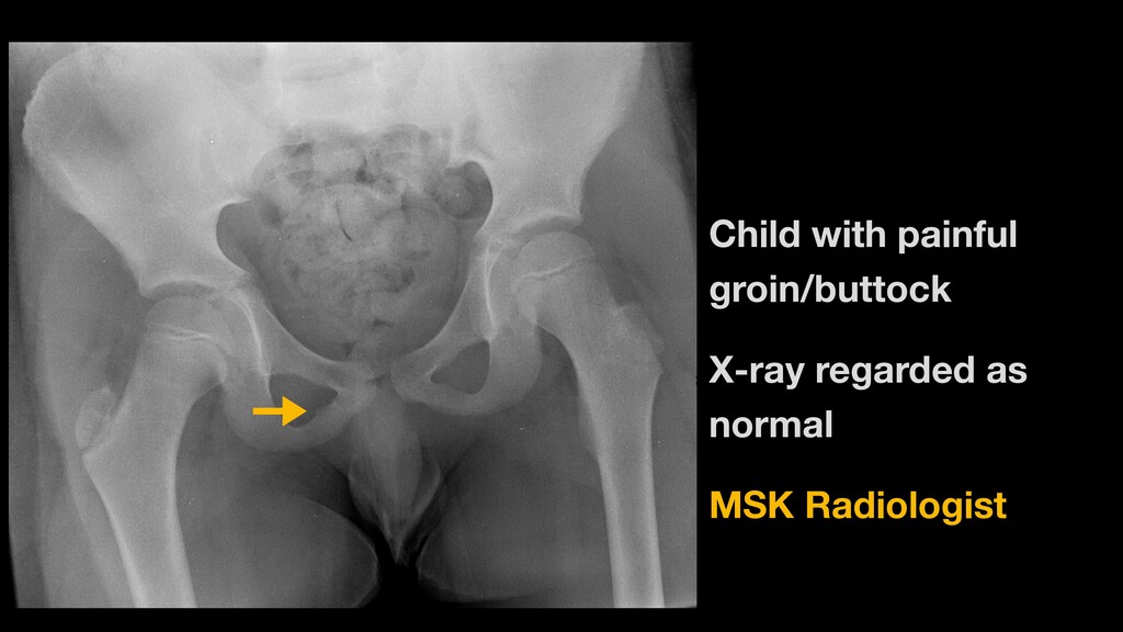
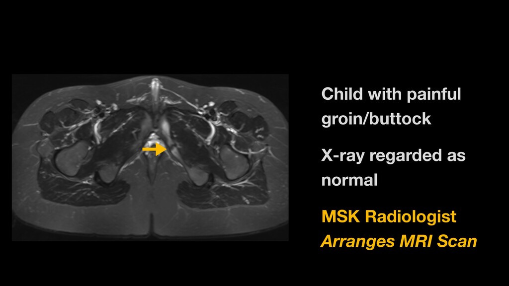
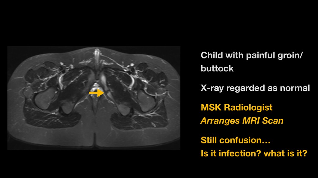
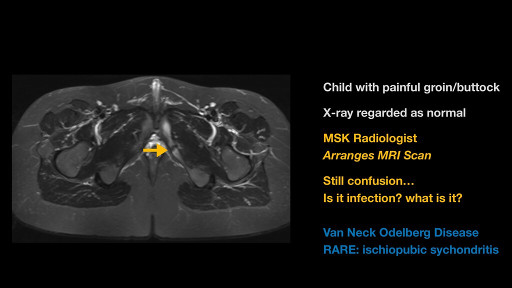
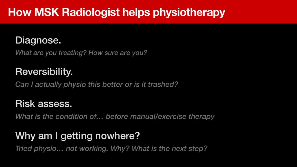
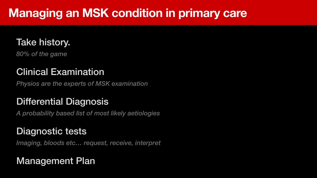
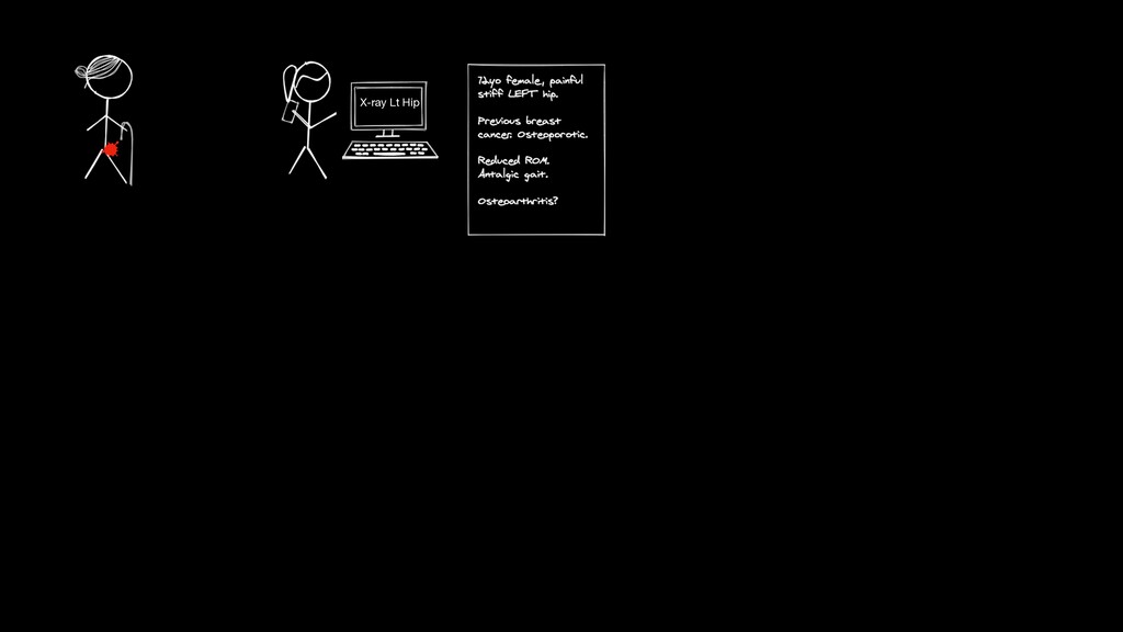
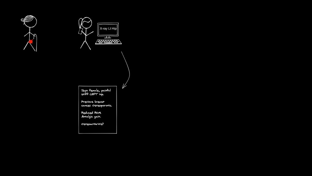
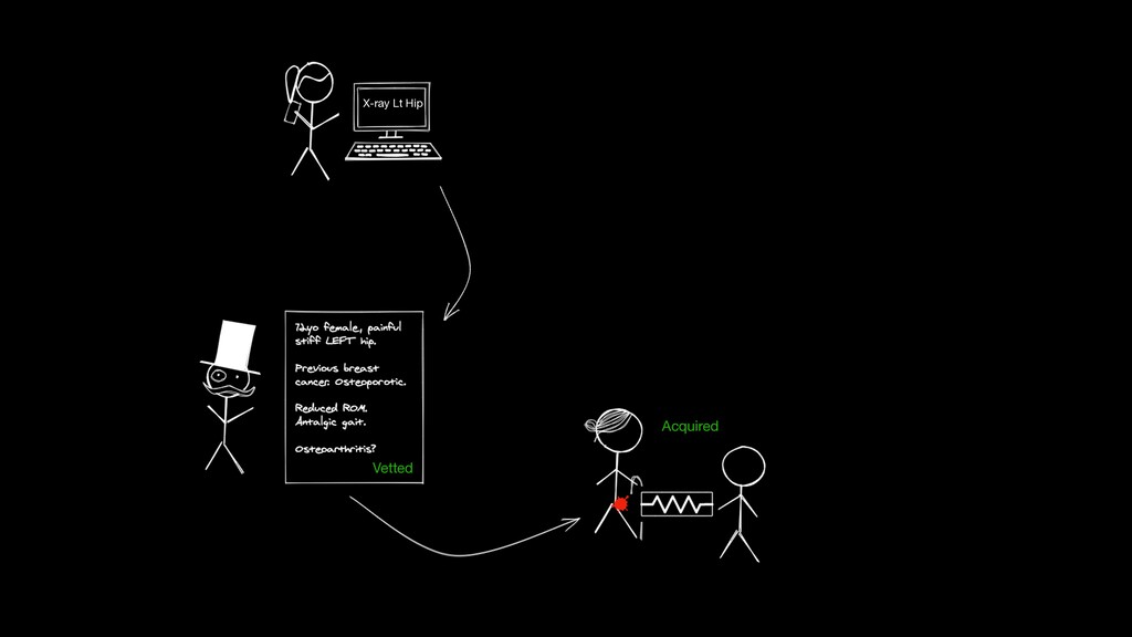
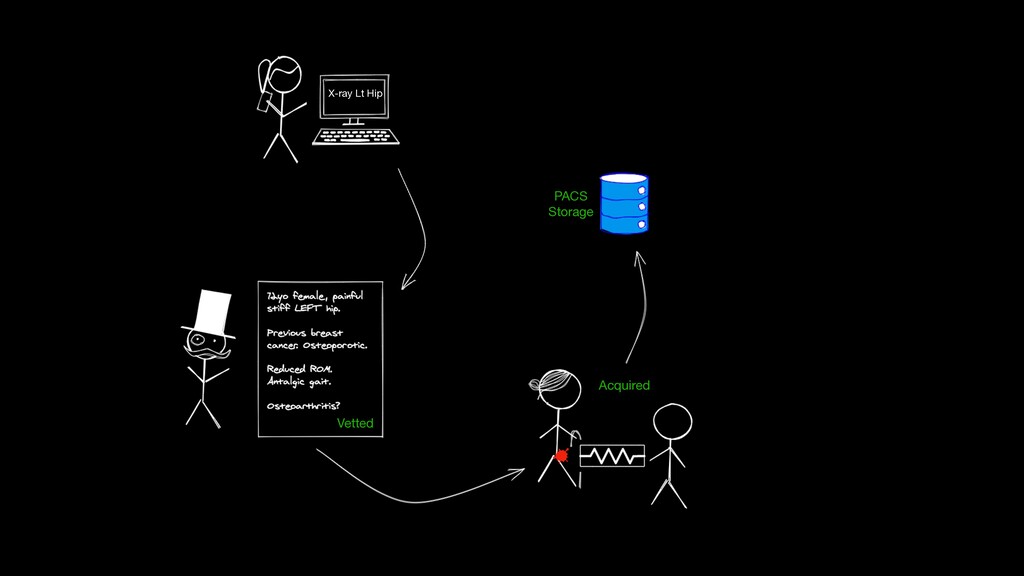
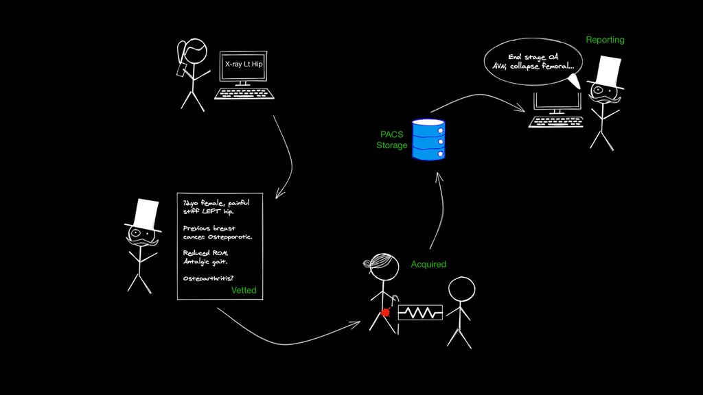
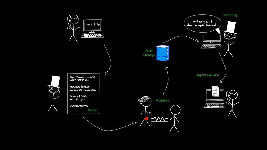
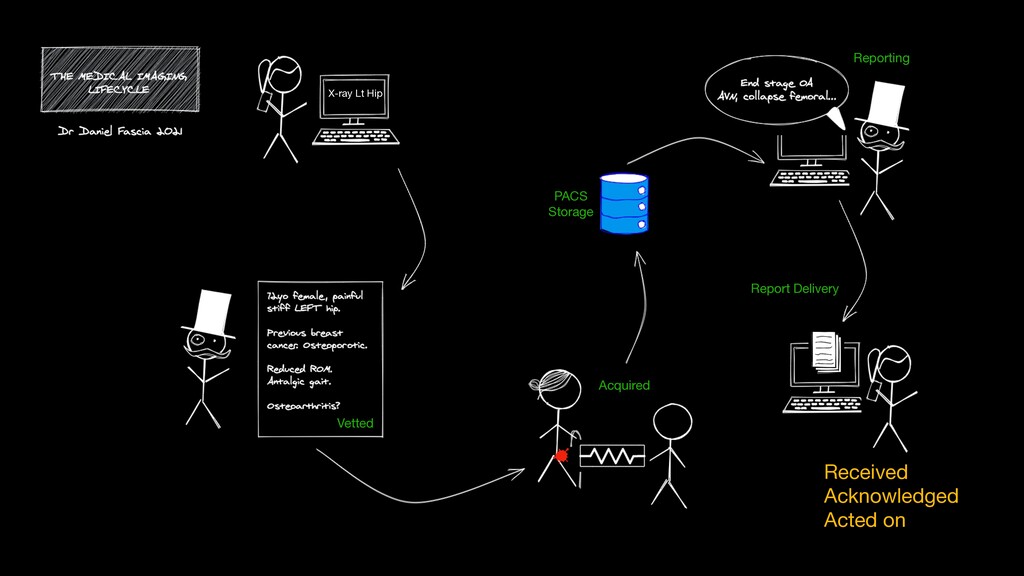
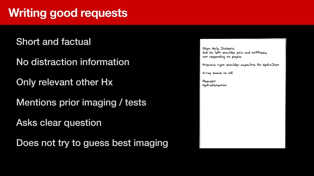
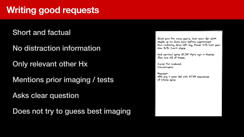
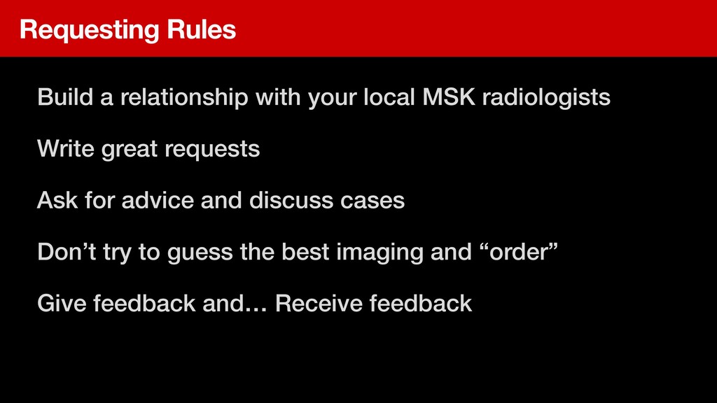
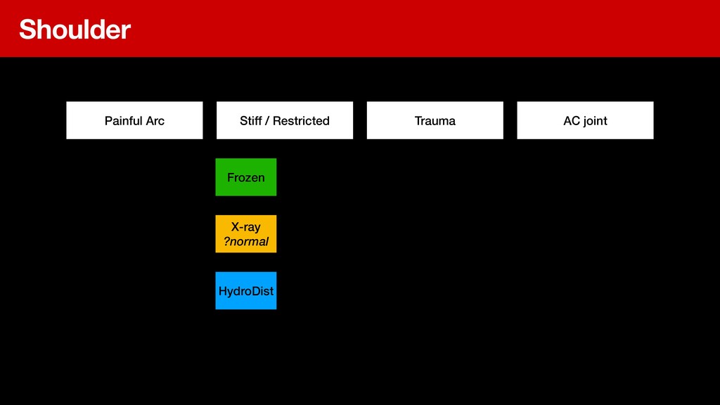
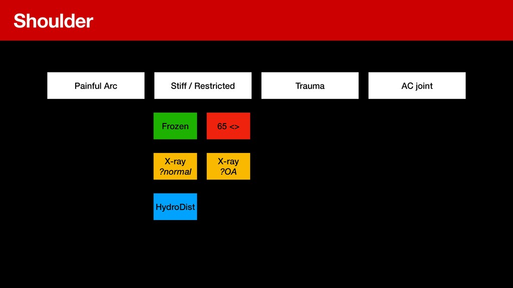
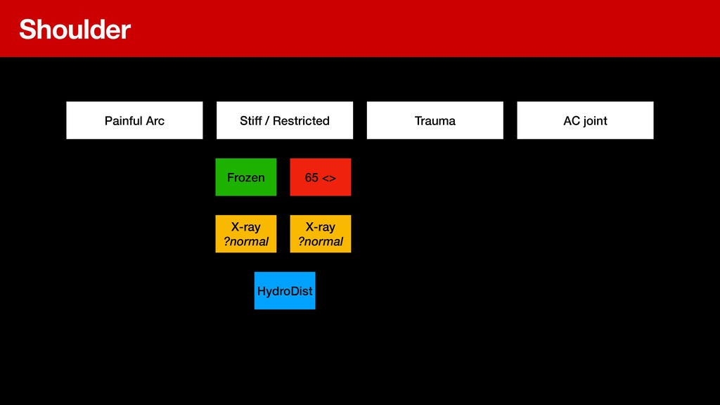
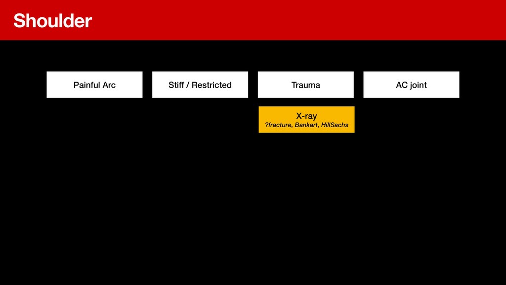
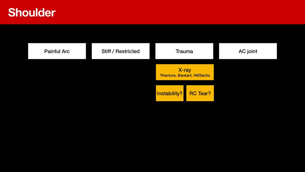
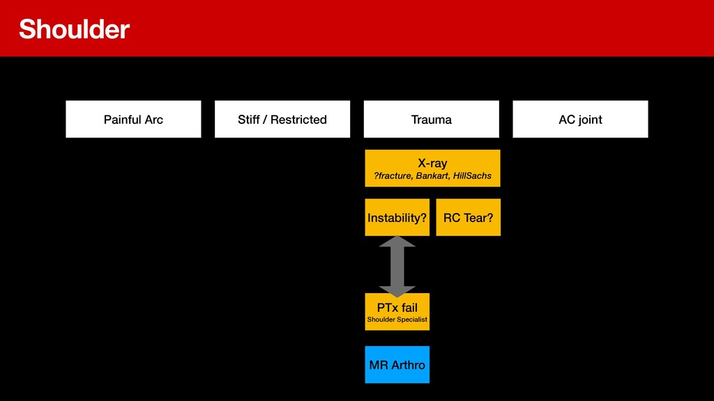
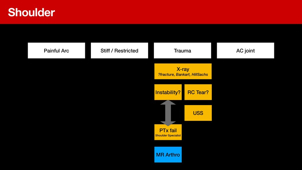
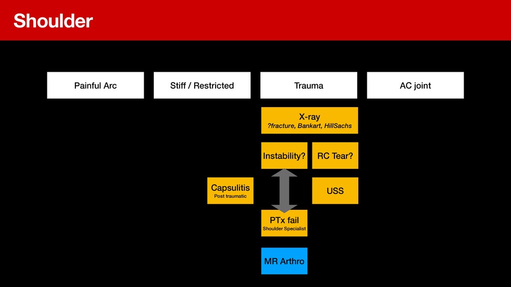
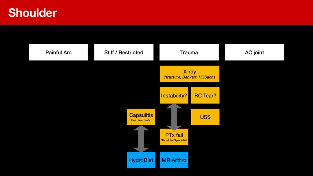
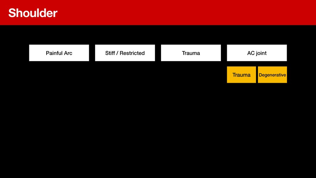
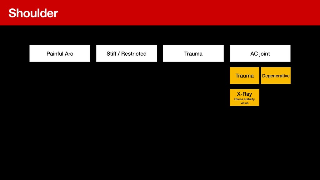
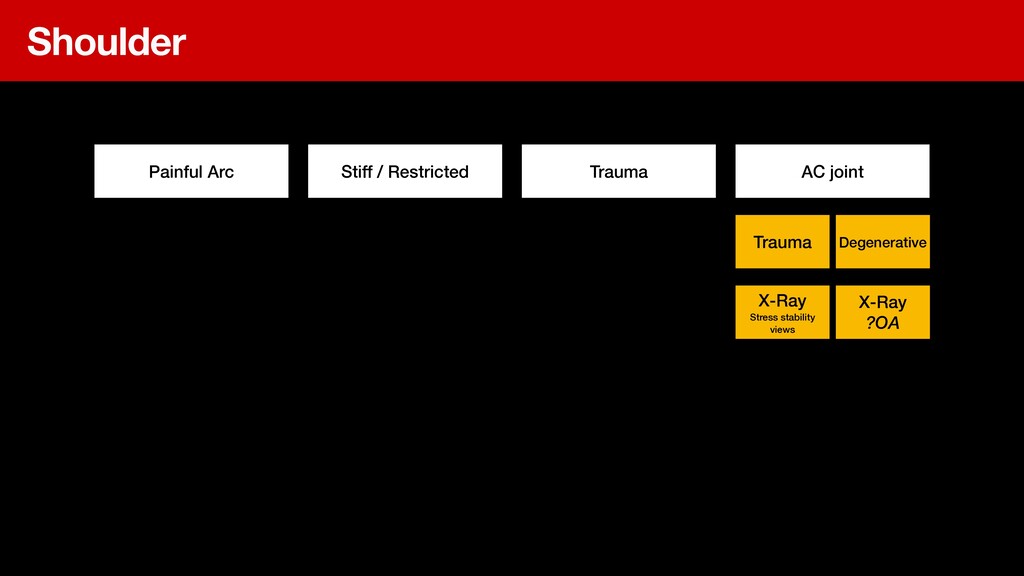
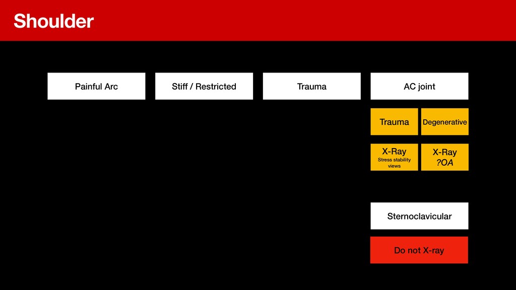
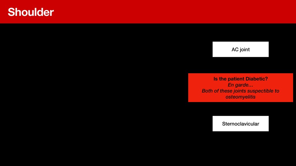
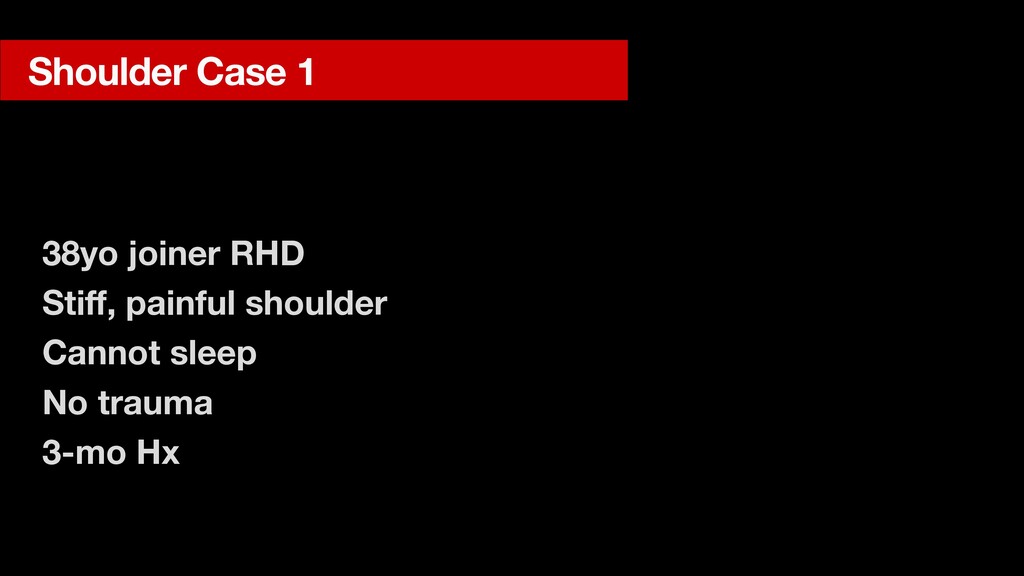
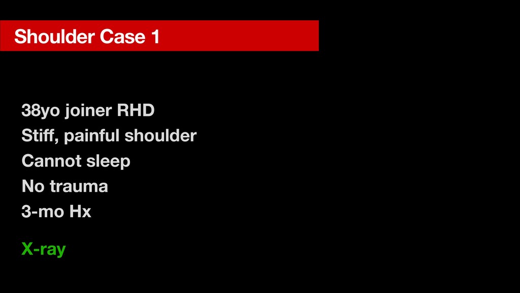
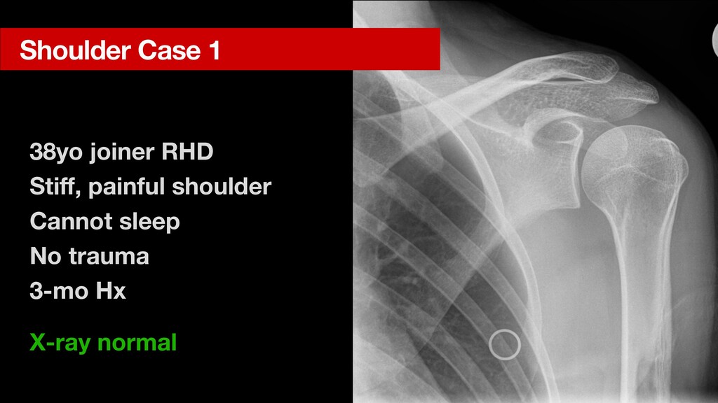
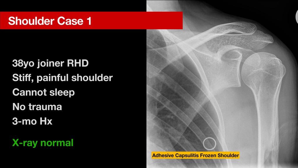
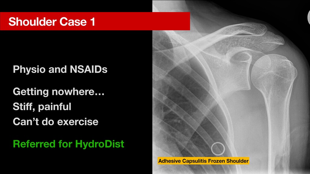
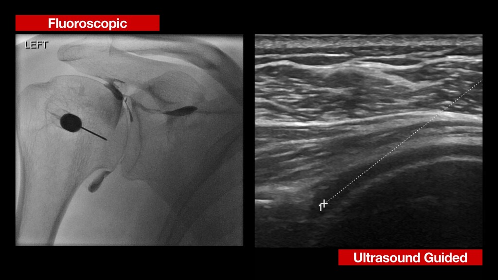
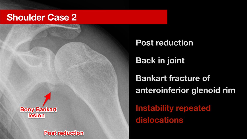
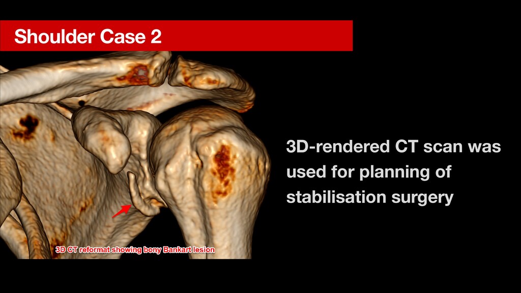
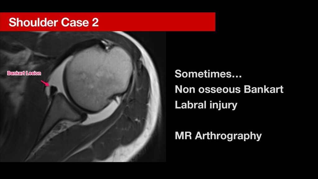
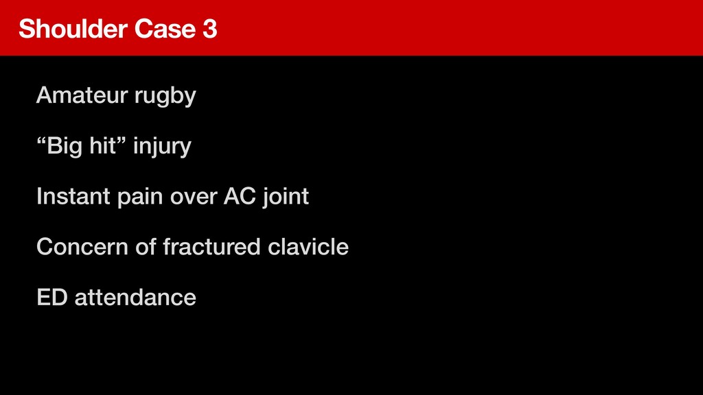
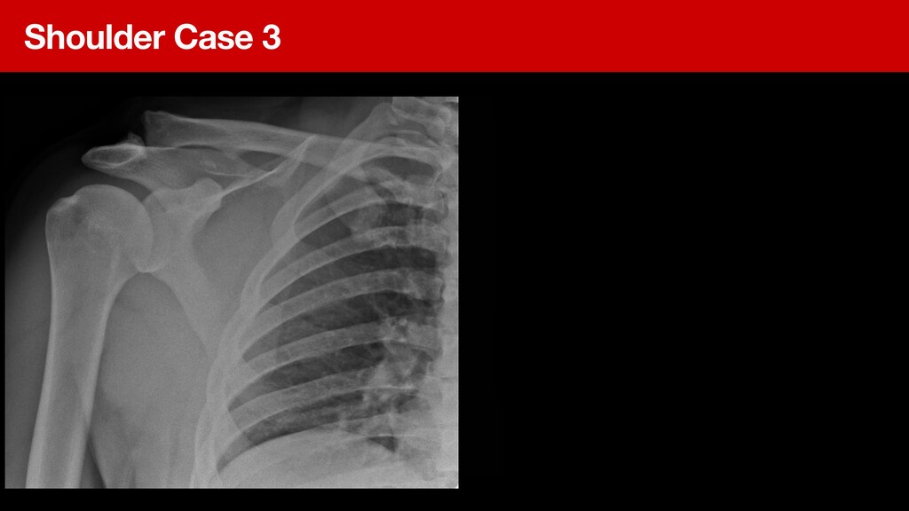
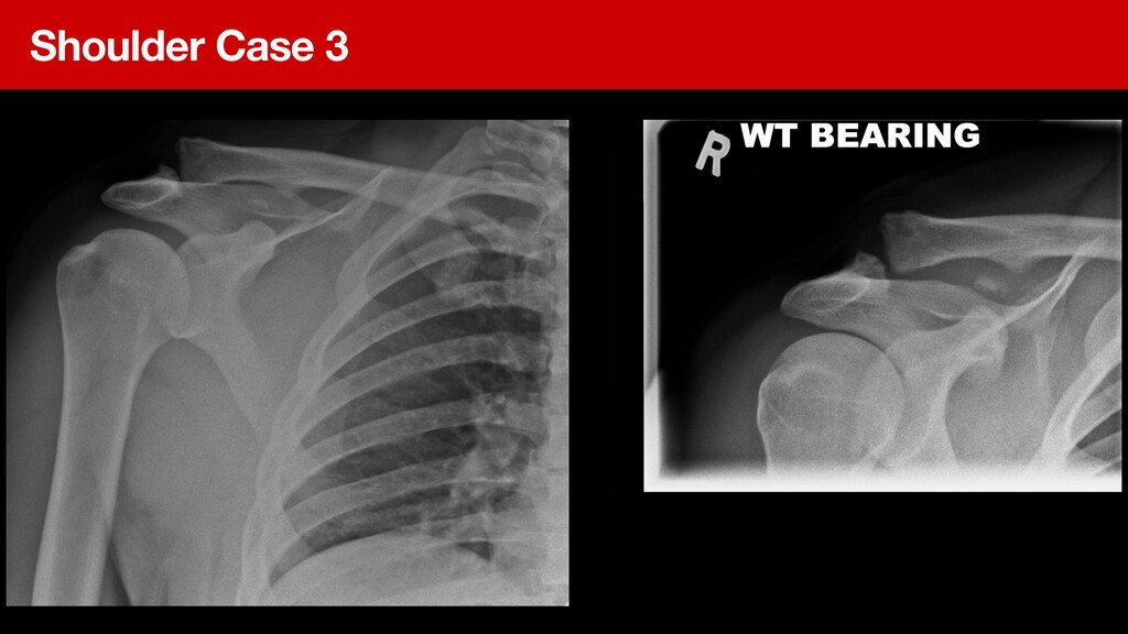
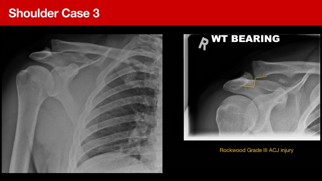
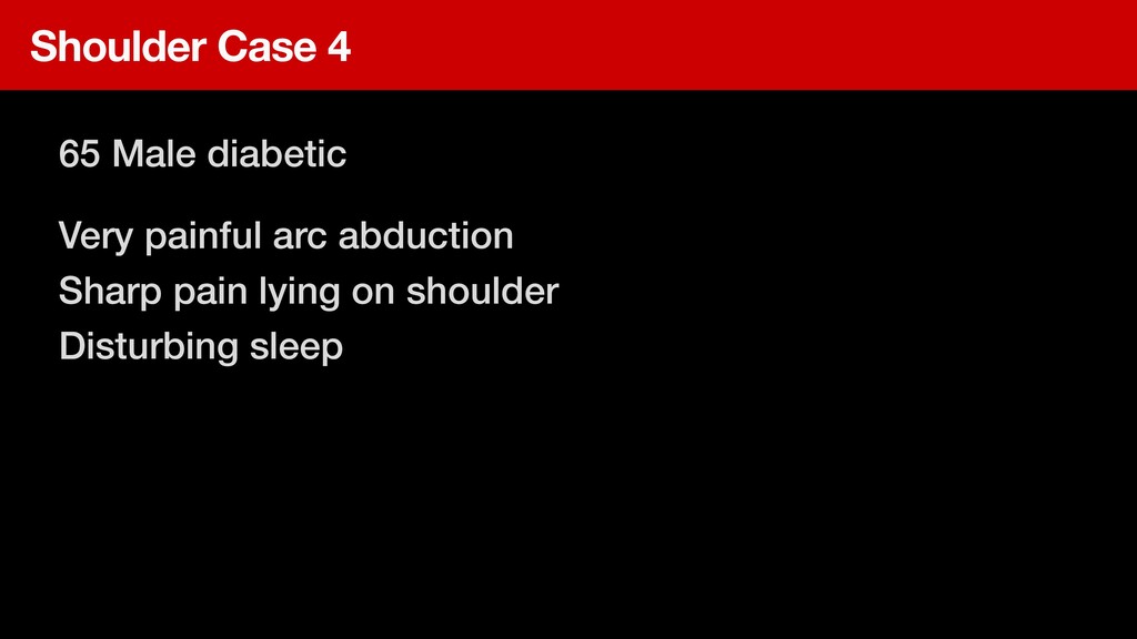
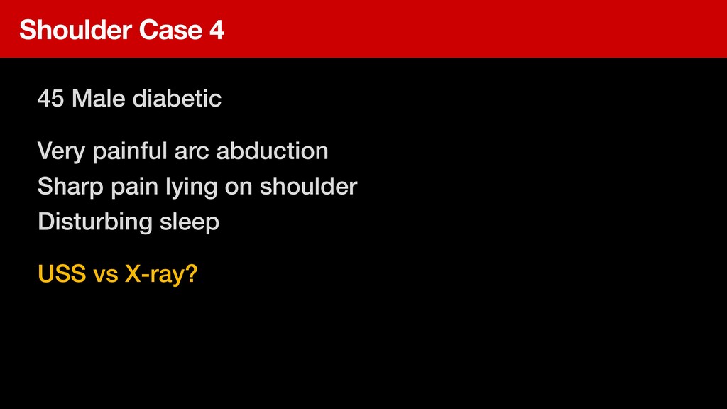
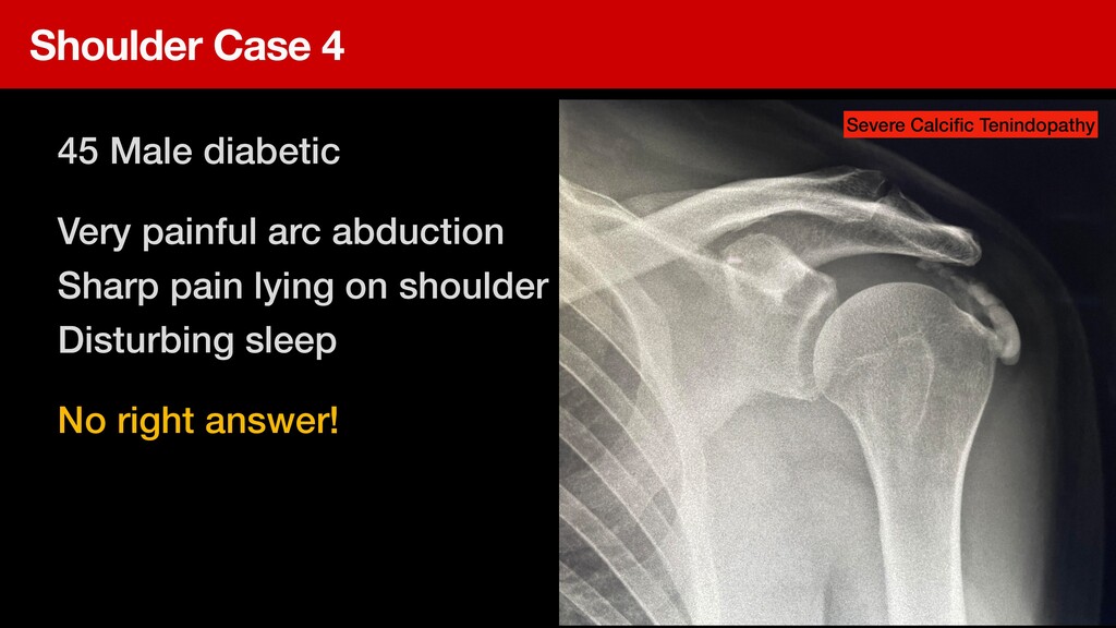
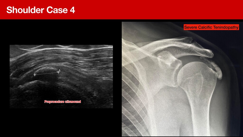
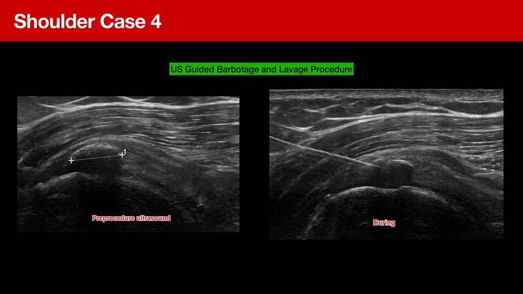
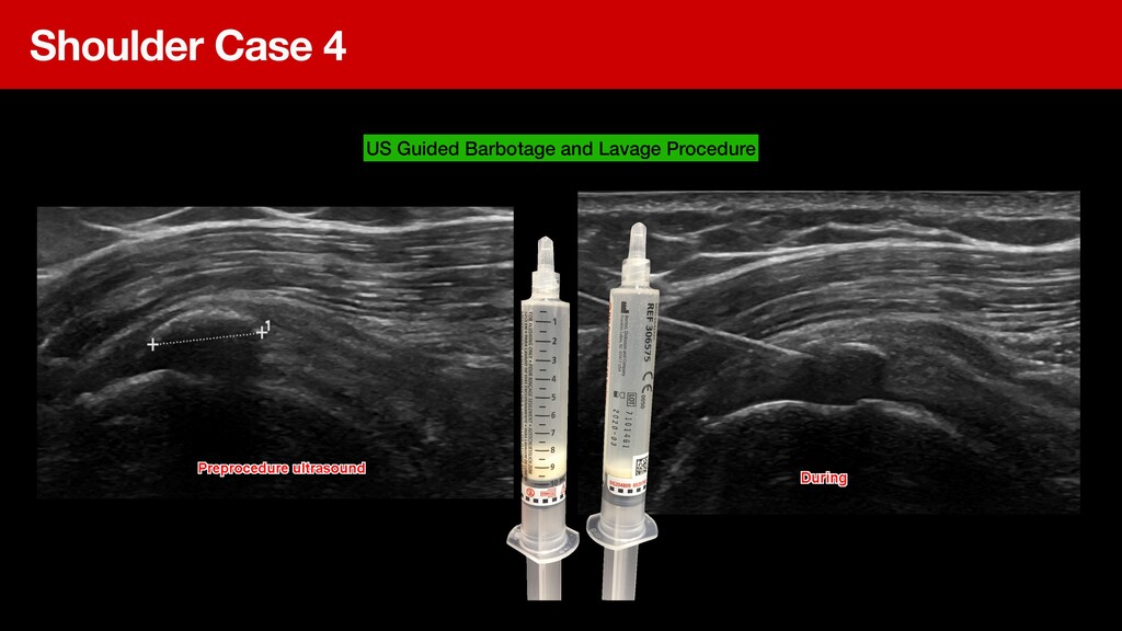
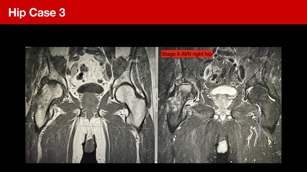
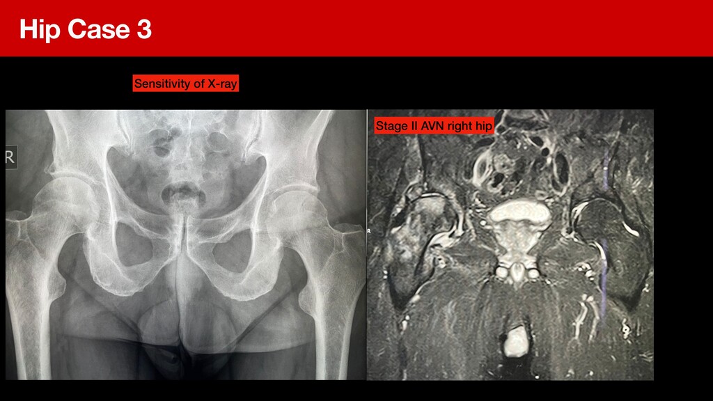
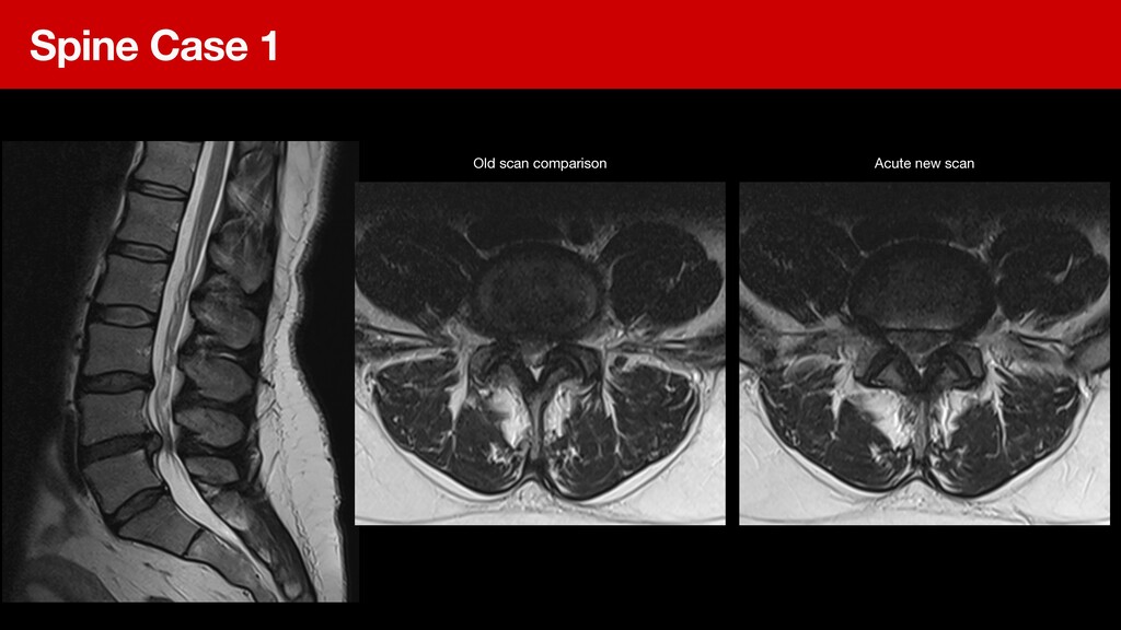
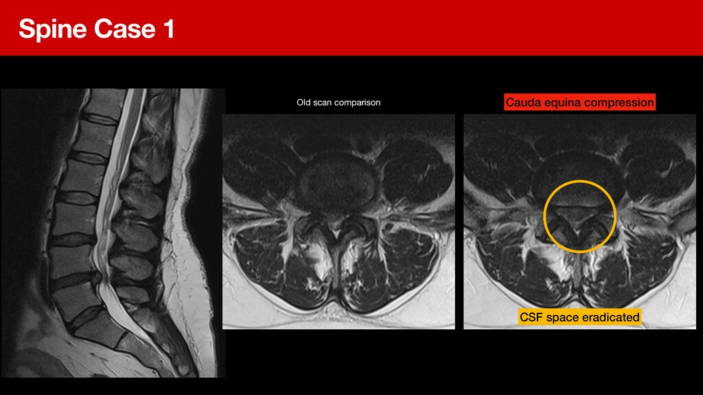
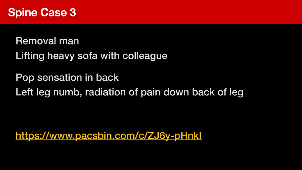
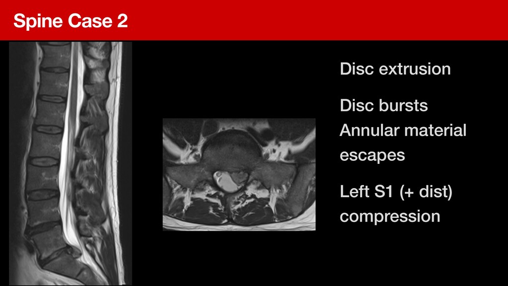
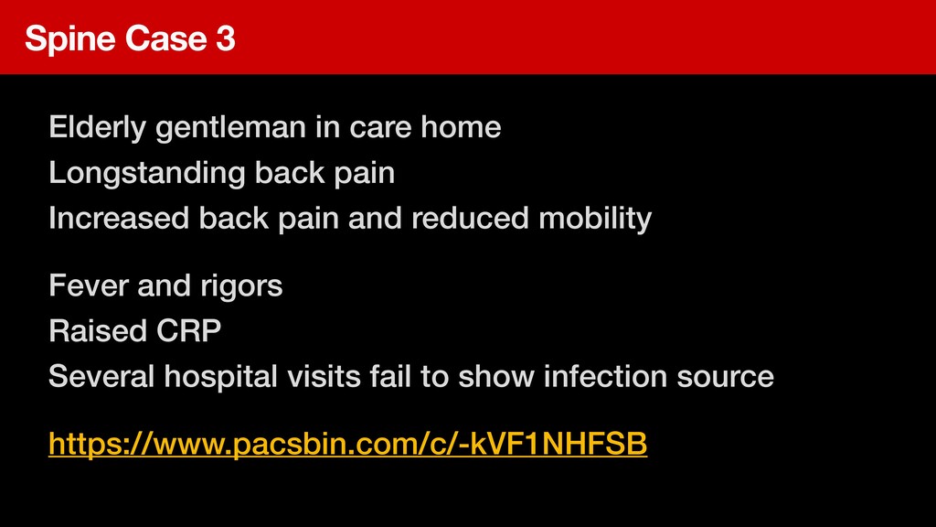
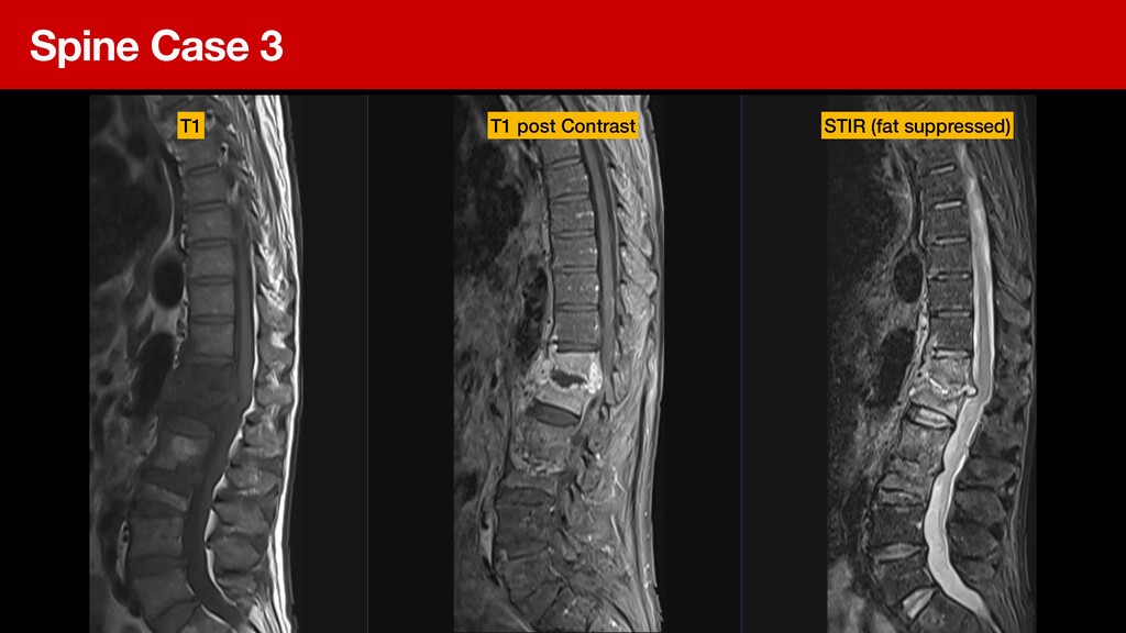
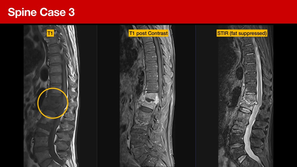
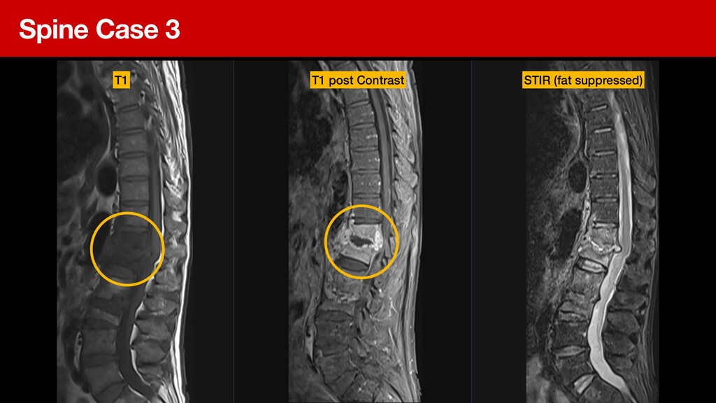
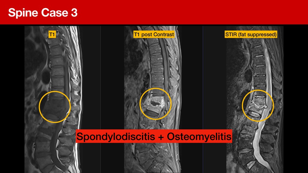
This lecture is a primer with case discussions on musculoskeletal radiology for the FCP including how radiology works and how to write a great referral.

About the author

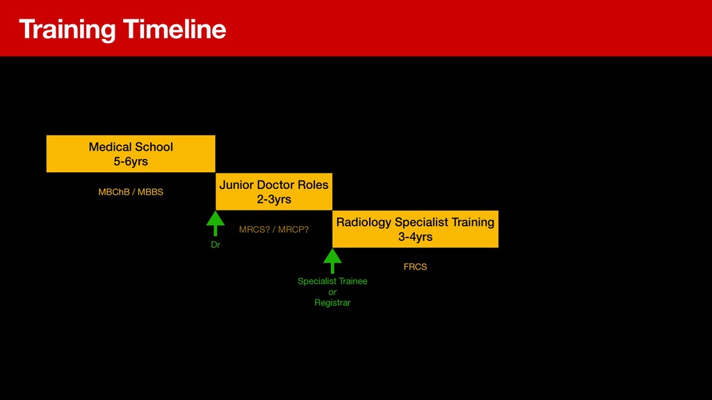
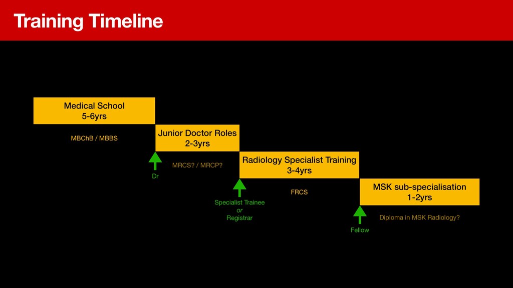
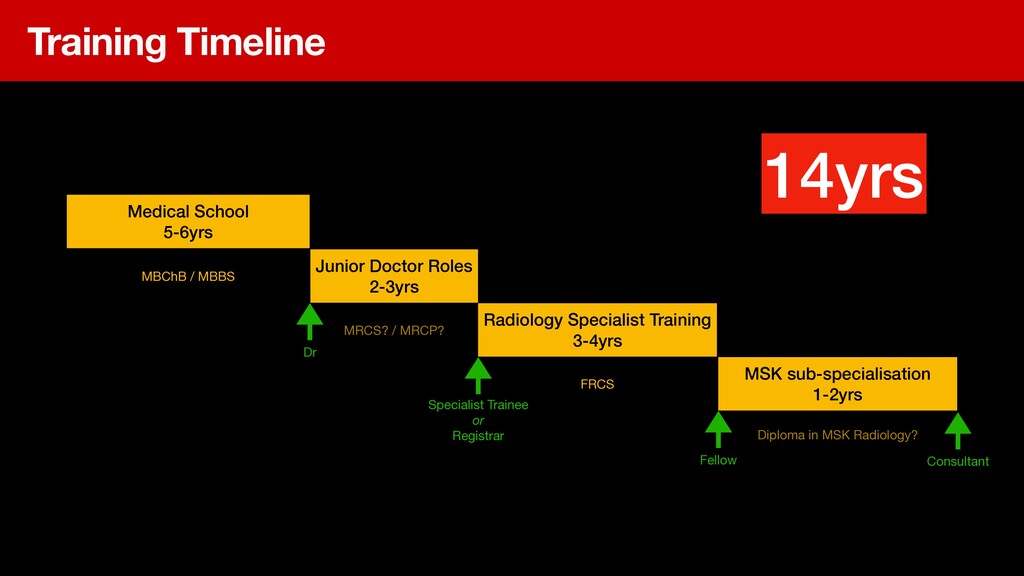
Daniel is an MSK Radiologist specialising in diagnostic and interventional radiology for orthopaedic, sports injury and rheumatological conditions. He trained at Imperial College London and undertook a senior fellowship in MSK/Sports Imaging in Perth, Western Australia before returning to take a consultant post in Harrogate, Yorkshire.

Daniel plays an active role in education as a BSSR Executive board member, an educator in ultrasound guided injections and the founder of the Radiology Masters online learning platform.

He is also regional clinical lead of the Yorkshire Imaging Collaborative and was Director of Imaging for NHS Nightingale. Artificial Intelligence and Radiology Informatics form a major interest as a Royal College of Radiologists Informatics Committee Officer.

Dr Daniel Fascia

June 02, 2021
Tweet

More Decks by Dr Daniel Fascia

Other Decks in Science

Transcript

  1. About the presenter Radiology SpR (Imperial College, London) Senior Fellowship

    (Perth, WA) Consultant Musculoskeletal Radiologist Regional Clinical Lead 
 Yorkshire Imaging Collaborative
  2. Special Interests MSK diagnostics Novel image guided intervention Spinal intervention

    Health tech and informatics Medical Education (Radiology Masters)
  3. Today’s schedule - Part 1 What is an MSK Radiologist

    and what do they do? How we interoperate with other clinical staff The Imaging Lifecycle: from thought to report Q&A Break
  4. Training Timeline Medical School 
 5-6yrs Junior Doctor Roles 


    2-3yrs Radiology Specialist Training 
 3-4yrs MBChB / MBBS MRCS? / MRCP? FRCS Dr Specialist Trainee or Registrar
  5. Training Timeline Medical School 
 5-6yrs Junior Doctor Roles 


    2-3yrs Radiology Specialist Training 
 3-4yrs MSK sub-specialisation 
 1-2yrs MBChB / MBBS MRCS? / MRCP? FRCS Diploma in MSK Radiology? Dr Specialist Trainee or Registrar Fellow
  6. Training Timeline Medical School 
 5-6yrs Junior Doctor Roles 


    2-3yrs Radiology Specialist Training 
 3-4yrs MSK sub-specialisation 
 1-2yrs MBChB / MBBS MRCS? / MRCP? FRCS Diploma in MSK Radiology? Dr Consultant Specialist Trainee or Registrar Fellow 14yrs
  7. Interpreting and reporting MRI Interpreting and reporting Plain Film X-rays

    Diagnostic 
 MSK Ultrasound 
 Clinics Interpretation and reporting of Major Trauma and skeletal CT Supervision and interpretation of Nuclear Medicine scans
  8. Interpreting and reporting MRI Vetting and Protocolling In person case

    review Interpreting and reporting Plain Film X-rays Diagnostic 
 MSK Ultrasound 
 Clinics Service Development Interpretation and reporting of Major Trauma and skeletal CT Authoring policy and guidance Supervision and interpretation of Nuclear Medicine scans
  9. Interpreting and reporting MRI Vetting and Protocolling In person case

    review Advising on best imaging strategy Interpreting and reporting Plain Film X-rays Multidisciplinary Team Meetings Diagnostic 
 MSK Ultrasound 
 Clinics Service Development Interpretation and reporting of Major Trauma and skeletal CT Authoring policy and guidance Supervision and interpretation of Nuclear Medicine scans
  10. Interpreting and reporting MRI Vetting and Protocolling In person case

    review Advising on best imaging strategy Interpreting and reporting Plain Film X-rays Multidisciplinary Team Meetings Diagnostic 
 MSK Ultrasound 
 Clinics Service Development Supervision of Trainee Radiologists on MSK attachment CPD Interpretation and reporting of Major Trauma and skeletal CT Authoring policy and guidance Education of Fellows Supervision and interpretation of Nuclear Medicine scans
  11. Interpreting and reporting MRI Vetting and Protocolling In person case

    review Advising on best imaging strategy Interpreting and reporting Plain Film X-rays Multidisciplinary Team Meetings Diagnostic 
 MSK Ultrasound 
 Clinics Service Development Supervision of Trainee Radiologists on MSK attachment CPD Interpretation and reporting of Major Trauma and skeletal CT Research & Development Authoring papers Authoring policy and guidance Education of Fellows Supervision and interpretation of Nuclear Medicine scans
  12. Interpreting and reporting MRI Vetting and Protocolling In person case

    review Advising on best imaging strategy Interpreting and reporting Plain Film X-rays Multidisciplinary Team Meetings Diagnostic 
 MSK Ultrasound 
 Clinics Service Development Supervision of Trainee Radiologists on MSK attachment On call work CPD Interpretation and reporting of Major Trauma and skeletal CT Research & Development Authoring papers Authoring policy and guidance Education of Fellows Supervision and interpretation of Nuclear Medicine scans Handling complaints etc
  13. Interpreting and reporting MRI Vetting and Protocolling In person case

    review Advising on best imaging strategy Interpreting and reporting Plain Film X-rays Multidisciplinary Team Meetings Diagnostic 
 MSK Ultrasound 
 Clinics Service Development Interventional 
 MSK Injections, biopsies, advanced procedures Supervision of Trainee Radiologists on MSK attachment On call work CPD Interpretation and reporting of Major Trauma and skeletal CT Research & Development Authoring papers Authoring policy and guidance Education of Fellows Supervision and interpretation of Nuclear Medicine scans Handling complaints etc
  14. Common clinical team links Orthopaedic Surgeons Rheumatologists Emergency Department Sports

    and Exercise Medicine Physiotherapists Neurosurgery, spine, oncology, physicians, other AHPs
  15. Underwriters of medicine Advice on diagnostic plan Interpretation of results

    Part of treatment plan How we help other medical teams
  16. 13yo active
 Night pain in shin Imaging features
 Age
 History

    of complaint CT con fi rms
 Helps plan Rx
  17. 13yo active
 Night pain in shin Imaging features
 Age
 History

    of complaint CT con fi rms
 Helps plan Rx RF Ablation by Interventional Radiologist Somma F, Stoia V, D’Angelo R, Fiore F (2021) Imaging-guided radiofrequency ablation of osteoid osteoma in typical and atypical sites: Long term follow up. 
 PLOS ONE 16(3): e0248589. https://doi.org/10.1371/journal.pone.0248589
  18. Child with painful groin/ buttock X-ray regarded as normal MSK

    Radiologist
 Arranges MRI Scan Still confusion…
 Is it infection? what is it?
  19. Child with painful groin/buttock X-ray regarded as normal MSK Radiologist


    Arranges MRI Scan Still confusion…
 Is it infection? what is it?
 
 Van Neck Odelberg Disease
 RARE: ischiopubic sychondritis
  20. How MSK Radiologist may help you Advise on diagnostic strategy

    Correlate fi ndings with extensive medical knowledge Recognise rare diseases and variants of common Actuary of medicine: probability + evidence Inform treatment approach Deliver intervention
  21. How MSK Radiologist helps physiotherapy Diagnose. 
 What are you

    treating? How sure are you? Reversibility. 
 Can I actually physio this better or is it trashed? Risk assess. 
 What is the condition of… before manual/exercise therapy Why am I getting nowhere? 
 Tried physio… not working. Why? What is the next step?
  22. Managing an MSK condition in primary care Take history. 


    80% of the game Clinical Examination 
 Physios are the experts of MSK examination Differential Diagnosis 
 A probability based list of most likely aetiologies Diagnostic tests 
 Imaging, bloods etc… request, receive, interpret Management Plan
  23. Writing good requests Short and factual No distraction information Only

    relevant other Hx Mentions prior imaging / tests Asks clear question Does not try to guess best imaging
  24. Writing good requests Short and factual No distraction information Only

    relevant other Hx Mentions prior imaging / tests Asks clear question Does not try to guess best imaging
  25. Tips Sort out your IT Passwords and permissions to request

    electronically Make sure you can receive and act on results Understand your responsibility in using specialist imaging
  26. Requesting Rules Build a relationship with your local MSK radiologists

    Write great requests Ask for advice and discuss cases Don’t try to guess the best imaging and “order” Give feedback and… Receive feedback
  27. Shoulder Painful Arc Stiff / Restricted Trauma AC joint Trauma

    X-ray 
 ?fracture USS 
 ?RC tear No X-ray >65 
 ?OA USS (inj) failed PTx
  28. Shoulder Painful Arc Stiff / Restricted Trauma AC joint Frozen

    X-ray 
 ?normal HydroDist 65 <> X-ray 
 ?OA
  29. Shoulder Painful Arc Stiff / Restricted Trauma AC joint Frozen

    X-ray 
 ?normal HydroDist 65 <> X-ray 
 ?normal
  30. Shoulder Painful Arc Stiff / Restricted Trauma AC joint X-ray

    
 ?fracture, Bankart, HillSachs Instability? RC Tear?
  31. Shoulder Painful Arc Stiff / Restricted Trauma AC joint X-ray

    
 ?fracture, Bankart, HillSachs Instability? RC Tear? PTx fail 
 Shoulder Specialist MR Arthro
  32. Shoulder Painful Arc Stiff / Restricted Trauma AC joint X-ray

    
 ?fracture, Bankart, HillSachs Instability? RC Tear? USS PTx fail 
 Shoulder Specialist MR Arthro
  33. Shoulder Painful Arc Stiff / Restricted Trauma AC joint X-ray

    
 ?fracture, Bankart, HillSachs Instability? RC Tear? USS PTx fail 
 Shoulder Specialist MR Arthro Capsulitis 
 Post traumatic
  34. Shoulder Painful Arc Stiff / Restricted Trauma AC joint X-ray

    
 ?fracture, Bankart, HillSachs Instability? RC Tear? USS PTx fail 
 Shoulder Specialist MR Arthro Capsulitis 
 Post traumatic HydroDist
  35. Shoulder Painful Arc Stiff / Restricted Trauma AC joint Trauma

    Degenerative X-Ray 
 Stress stability views
  36. Shoulder Painful Arc Stiff / Restricted Trauma AC joint Trauma

    Degenerative X-Ray 
 Stress stability views X-Ray 
 ?OA
  37. Shoulder Painful Arc Stiff / Restricted Trauma AC joint Trauma

    Degenerative X-Ray 
 Stress stability views X-Ray 
 ?OA Sternoclavicular Do not X-ray
  38. Shoulder AC joint Sternoclavicular Is the patient Diabetic? 
 En

    garde… 
 Both of these joints suspectible to osteomyelitis
  39. 38yo joiner RHD
 Sti ff , painful shoulder
 Cannot sleep


    No trauma
 3-mo Hx X-ray Shoulder Case 1
  40. 38yo joiner RHD
 Sti ff , painful shoulder
 Cannot sleep


    No trauma
 3-mo Hx X-ray normal Shoulder Case 1
  41. 38yo joiner RHD
 Sti ff , painful shoulder
 Cannot sleep


    No trauma
 3-mo Hx X-ray normal Shoulder Case 1 Adhesive Capsulitis Frozen Shoulder
  42. Physio and NSAIDs Getting nowhere…
 Sti ff , painful
 Can’t

    do exercise Referred for HydroDist Shoulder Case 1 Adhesive Capsulitis Frozen Shoulder
  43. Post reduction Back in joint Bankart fracture of anteroinferior glenoid

    rim Instability repeated dislocations Shoulder Case 2
  44. Shoulder Case 3 Amateur rugby “Big hit” injury Instant pain

    over AC joint Concern of fractured clavicle ED attendance
  45. Shoulder Case 4 65 Male diabetic Very painful arc abduction

    
 Sharp pain lying on shoulder 
 Disturbing sleep
  46. Shoulder Case 4 45 Male diabetic Very painful arc abduction

    
 Sharp pain lying on shoulder 
 Disturbing sleep USS vs X-ray?
  47. Shoulder Case 4 45 Male diabetic Very painful arc abduction

    
 Sharp pain lying on shoulder 
 Disturbing sleep No right answer! Severe Calci fi c Tenindopathy
  48. Spine Case 1 Attends primary care Long Hx of back

    pain with prior MR showing disc hernia Worsening Symptoms Dif fi culty passing urine. 
 Change in sensation around groin. https://www.pacsbin.com/c/WkIPkiEkZD
  49. Spine Case 1 Old scan comparison Acute new scan Cauda

    equina compression CSF space eradicated
  50. Spine Case 3 Removal man 
 Lifting heavy sofa with

    colleague Pop sensation in back 
 Left leg numb, radiation of pain down back of leg https://www.pacsbin.com/c/ZJ6y-pHnkI
  51. Spine Case 2 Disc extrusion Disc bursts 
 Annular material

    escapes Left S1 (+ dist) 
 compression
  52. Spine Case 3 Elderly gentleman in care home 
 Longstanding

    back pain 
 Increased back pain and reduced mobility Fever and rigors 
 Raised CRP 
 Several hospital visits fail to show infection source https://www.pacsbin.com/c/-kVF1NHFSB
  53. MRI the problem solver Fantastic problem solving algorithm Design scan

    sequences to suit problem with radiologist 
 Provide them good information! Anatomical information Biological information 
 Inflammation, blood flow etc