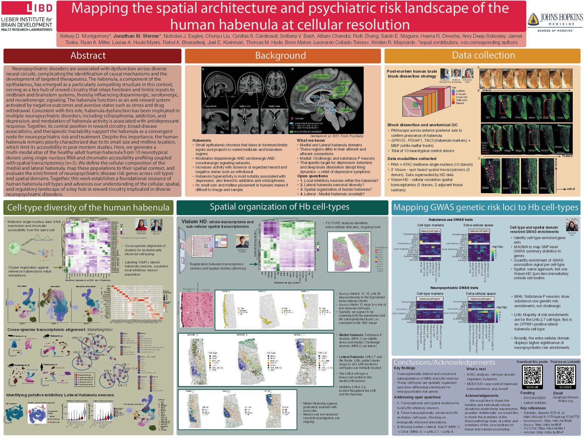
human habenula at cellular resolution Abstract Neuropsychiatric disorders are associated with dysfunction across diverse neural circuits, complicating the identi cation of causal mechanisms and the development of targeted therapeutics. The habenula, a component of the epithalamus, has emerged as a particularly compelling structure in this context, serving as a key hub of reward circuitry that relays forebrain and limbic inputs to midbrain and brainstem systems, thereby in uencing dopaminergic, serotonergic, and noradrenergic signaling. The habenula functions as an anti-reward system activated by negative outcomes and aversive states such as stress and drug withdrawal. Consistent with this role, habenula dysfunction has been implicated in multiple neuropsychiatric disorders, including schizophrenia, addiction, and depression; and modulation of habenula activity is associated with antidepressant response. Together, its central position in reward circuitry, broad disease associations, and therapeutic tractability support the habenula as a convergent node for neuropsychiatric risk and treatment. Despite this importance, the human habenula remains poorly characterized due to its small size and midline location, which limit its accessibility in post-mortem studies. Here, we generate a multi-modal atlas of the healthy adult human habenula from 10 neurotypical donors using single-nucleus RNA and chromatin accessibility pro ling coupled with spatial transcriptomics (n=5). We de ne the cellular composition of the medial and lateral habenula, map these populations to their spatial context, and evaluate the enrichment of neuropsychiatric disease risk genes across cell types and spatial domains. Together, this work establishes a foundational resource of human habenula cell types and advances our understanding of the cellular, spatial, and regulatory landscape of a key hub in reward circuitry implicated in diverse neuropsychiatric disorders. Background Data collection Cell-type diversity of the human habenula Spatial organization of Hb cell-types Mapping GWAS genetic risk loci to Hb cell-types Conclusions/Acknowledgements A B C 1000 μm Anterior Posterior Lateral Ventral TAC3 MBP GPR151 POU4F1 1 cm 5 mm 0μm 220μm 440μm 660μm 880μm 1100μm 1320μm 1540μm 1760μm 1980μm 2200μm 2420μm 2640μm d c a b 220μm 880μm 1540μm 2200μm 1500μm 220μm 880μm 1540μm 2200μm Kelsey D. Montgomery*, Jonathan M. Werner*, Nicholas J. Eagles, Chunyu Liu, Cynthia S. Cardinault, Svitlana V. Bach, Atharv Chandra, Ruth Zhang, Sarah E. Maguire, Heena R. Divecha, Amy Deep-Soboslay, James Tooke, Ryan A. Miller, Louise A. Huuki-Myers, Rahul A. Bharadwaj, Joel E. Kleinman, Thomas M. Hyde, Brion Maher, Leonardo Collado-Torres+, Kristen R. Maynard+, *equal contributors, +co-corresponding authors UMAP 1 UMAP 2 RNA UMAP O LIG 2 PD G FR A M O G S100B C LD N 5 PEC AM 1 P2RY12 C X3C R 1 SLC 1A2 G FAP FO XJ1 PIFO G AP43 SN AP25 SLC 17A6 SLC 17A7 LYPD 6B ADAR B2 D R D 2 SO X14 RO R B G AD 1 G AD 2 SLC 32A1 LYN X1 C H R M 3 G ABR A1 PC D H 10 H TR 2C G PR 151 PO U 4F1 C H R N B4 C H AT SLC 5A7 SLC 18A3 TAC 1 TAC 3 Percent Expressing 0 25 50 75 Mean Exp. z−score −1 0 1 2 3 OPC Oligo Endo Microglia Astrocyte Ependymal Excit.Thal Inhib.Thal LHb.4 LHb.1.3.4 LHb.1.3 LHb.1 LHb.2.7 MHb.3 MHb.2 MHb.1.2 MHb.1 ATAC UMAP UMAP 1 UMAP 2 WNN (RNA+ATAC) UMAP UMAP 1 UMAP 2 X X X X X X X X X X X X X X X X X X X X X X X X X * X X X X X X X X X X X X X X X X X X X X X X X X C.05.LHb.2.7 C.18.LHb.1.3.4 C.23.LHb.1 C.33.LHb.1.3 C.08.LHb.4 C.13.LHb.4 C.09.LHb.4 C.06.LHb.4 C.04.LHb.4 C.16.MHb.1.2 C.07.MHb.2 C.11.MHb.1.2 C.10.MHb.1 C.14.MHb.1 C.36.MHb.3 C.24.LHb.4 C.30.LHb.7 C.40.LHb.4 C.02.Oligo C.22.Oligo C.29.Endo C.26.OPC C.21.Astrocyte C.20.Astrocyte C.27.Microglia C.41.Microglia C.39.Inhib.Thal C.31.LHb.4 C.25.Excit.Thal C.32.Excit.Thal C.17.Excit.Thal C.12.Excit.Thal C.35.Excit.Thal C.15.Excit.Thal C.03.Excit.Thal C.37.Thal C.38.Inhib.Thal C.19.Inhib.Thal C.01.Inhib.Thal C.28.Inhib.Thal Inhib.Thal Excit.Thal LHb.4 LHb.3 LHb.1 LHb.5 OPC LHb.7 LHb.2 MHb.2 MHb.1 LHb.6 MHb.3 Endo Astrocyte Microglia Oligo Cor −1 0 1 * * * * * *Doublet-enriched cluster; dropped Wallace.Mouse Hashikawa.Mouse Yalcinbas.Human multiome.Human Pandey.Zebrafish 1 LHb.1 LHb.1.3 LHb.1.3.4 LHb.2.7 LHb.4 MHb.1 MHb.1.2 MHb.2 MHb.3 2 3 4 5 6 7 1 2 3 4 5 1 2 3 4 5 6 7 8 9 10 11 12 1 2 3 4 5 6 7 8 9 10 Hashikawa et. al. Non-neurons Lateral Habenula Medial Habenula UMAP 1 UMAP 2 UMAP 1 UMAP 2 Yalcinbas et. al Thalamus Medial Habenula Lateral Habenula Non-neurons LHb-6 MHb subP-cholinergic MHb subp-cholinergic MHb.1.2 9 5 3 MHb.1 MHb-1 MHb-subP MHb substance P Dorsolateral-left-subP 1 6 2 4 MHb.2 2 1 1 1 2 3 2 3 MHb-2 MHb-cholinergic 1 MHb-cholinergic 2 MHb-cholinergic 3 MHb -cholinergic Dorsomedial-right-cholinergic-GAT1 Dorsomedial-right-cholinergic Dorsomedial neuron LHb.1, .1.3, .1.3.4 LHb-1 LHb-5 LHb-1-2 LHb-1-4 LHb-1 Ventral Ventral immediate early 4 8 4 5 6 8 10 LHb.2.7 LHb-2 LHb-7 LHb-1-5 LHb-2 LHb-2 5 11 5 10 12 LHb.4 LHb-3 LHb-4 LHb-1-1 LHb-1-3 LHb-1 Inhibitory GAP43 6 7 4 7 7 9 Co-expression Both Neither SLC17A6 only SLC32A1 only SLC32A1 and SLC17A6 Co−expression UMAP 2 UMAP 1 UMAP 1 UMAP 2 LHb 4 neurons 0 1000 2000 M H b N on−neurons Excit_Thal LH b Inhib_LH b_4.2 Inhib_LH b_4.1 Inhib_Thal Top 25 markers (Avg. CPM) Inhibitory Thalamus markers 0 250 500 750 N on−neurons Inhib_Thal M H b Excit_Thal Inhib_LH b_4.2 Inhib_LH b_4.1 LH b Top 25 markers (Avg. CPM) Non−neurons Inhib_Thal MHb Excit_Thal Inhib_LHb_4.2 Inhib_LHb_4.1 LHb Lateral Habenula markers Yalcinbas, Ajanaku et. al 2025. Am J Psychiatry Cross-species transcriptomic alignment: MetaNeighbor Identifying putative inhibitory Lateral Habenula neurons • Cross-species alignment of clusters for evolutionarily informed cell-typing • Labeling VGAT+ lateral habenula neurons, a putative local inhibitory neuron population Open questions • 1. Local inhibitory neurons within the habenula? • 2. Lateral habenula neuronal diversity? • 3. Spatial organization of human habenula? • 4. Lateral - Medial Habenula crosstalk? What we know • Medial and Lateral habenula domains • These regions differ in their afferent and efferent connections • Medial: Cholinergic and substance P neurons • Therapeutic target for depression: ketamine and deep-brain stimulation disrupt firing dynamics -> relief of depressive symptoms Habenula • Small epithalamic structure that takes in forebrain/limbic inputs and projects to varied midbrain and brainstem regions • Modulates dopaminergic AND serotonergic AND noradrenergic signaling networks • Increases activity with absence of expected reward and negative states such as withdrawal • Habenula hyperactivity is most notably associated with depression; also linked to addiction and schizophrenia • Its small size and midline placement in humans makes it difficult to image and sample Post-mortem human brain block dissection strategy Challenges of the Habenula Project • Anatomy • RNAscope QC 1-8 times • Dissections of over 180 blocks to collect the 60 donors used Best Worst Svitlana Bach Rahul Bharadwaj Ruth Zhang Atharv Chandra Kelsey Montgomery Challenges of the Habenula Project • Anatomy • RNAscope QC 1-8 times • Dissections of over 180 blocks to collect the 60 donors used Best Worst Svitlana Bach Rahul Bharadwaj Ruth Zhang Atharv Chandra Kelsey Montgomery Cynthia S. Cardinault Jonathan M. Werner Nicholas J. Eagles Kelsey D. Montgomery Rahul A. Bharadwaj Svitlana V. Bach Chunyu Liu • Registration between transcriptomic clusters and spatial clusters (Banksy) • Banksy clusters 12, 16, and 24 map exclusively to the Ependymal transcriptional cluster • Banksy cluster 13 maps to a mix of non-neuronal cell-types • Spatially, we appear to be capturing both the ependymal and the sub-ependymal layers, as annotated in the H&E image • Lateral Habenula: LHb 2.7 and the Excite. LHb. spatial cluster (maps to all 3 LHb excitatory cell-types) are medially located. • Identify cell-type-enriched gene sets • MAGMA to map SNP-level GWAS summary statistics to genes • Quantify enrichment of GWAS association signal per cell-type • Spatial: same approach, but use Visium HD 2µm bins immediately outside cell bodies Cell-type and spatial domain resolved GWAS enrichments • MHb: Substance P neurons show substance use genetic risk enrichments, not cholinergic • RNAscope across anterior-posterior axis to confirm prescence of habenula • GPR151, POU4F1, TAC3 (Habenula markers) + MBP (white matter tracks) • Total of 10 neurotypical control donors • RNA + ATAC multiome single-nucleus (10 donors) • 3’ Visium - spot based spatial transcriptomics (3 donors). Data superseded by HD data • Visium HD - cellular resolution spatial transcriptomics (5 donors, 2 adjacent tissue sections) Data modalities collected Block dissection and anatomical QC Germann et. al. 2021. Front. Psychiatry • 1. Transcriptional and spatial evidence for local LHb inhibitory neurons • 2. Three transcriptionally conserved LHb excitatory cell-types. Working on biologically informed annotations. • 3. Moving medial-> lateral: Sub.P (MHb.1) -> Chol. (MHb.2) -> LHb.2.7 -> LHb.4. Addressing open questions What’s next • ATAC analysis: cell type-specific regulatory footprints • MDD/OUD case-control habenula transcriptomics; stay tuned! Download this poster Find me on LinkedIn Funding Key references • Yalcinbas, Ajanaku 2025 et. al: https://doi.org/10.1176/appi.ajp.20240776 • MetaNeighbor: https://rdcu.be/ff6qN • Banksy: https://rdcu.be/ff6rP • FICTURE: https://rdcu.be/ff6sT • MAGMA: https://rdcu.be/ff6vF • R01DA055823 • Lieber Institute Dorsal Habenula Non-neurons Pandey et. al. UMAP 1 UMAP 2 Ventral Habenula Wallace et. al. Lateral Habenula Medial Habenula Non-neurons UMAP 1 UMAP 2 Cell type OPC Oligo Microglia MHb.3 MHb.2 MHb.1.2 MHb.1 LHb.4 LHb.2.7 LHb.1.3.4 Inhib.Thal Inhib_LHb_4.2 Inhib_LHb_4.1 Excit.Thal Ependymal Endo Astrocyte Factor 0 Factor 1 Factor 2 Factor 3 Factor 4 Factor 5 Factor 6 Factor 7 Factor 8 Factor 9 FICTURE factor Email Jonathan.Werner @libd.org • Multiome single-nucleus data: RNA expression and chromatin accessibility from the same cell • Cluster registration against reference habenula for initial annotations X X X X X X X X X X X X X X X X X X X X X X X X X X X X X X X X X X X X X 19 15 8 23 7 20 25 5 9 11 6 16 12 24 14 1 26 21 17 22 18 2 13 27 4 3 10 OPC Endo Microglia Astrocyte Ependymal Oligo MHb.3 MHb.2 MHb.1 MHb.1.2 LHb.2.7 Inhib.Thal Inhib_LHb_4.1 Excit.Thal Inhib_LHb_4.2 LHb.4 LHb.1.3.4 Cor −1 0 1 Multiome cell-type clusters Visium HD Banksy clusters Visium HD: whole-transcriptome and sub-cellular spatial transcriptomics • FICTURE analysis identifies extra-cellular domains, ongoing work We would like to thank the families and individuals whose donations made these experiments possible. Additionally, we would like to thank the members of the Neuropathology team at Lieber and members of the core facilities for tissue and sample processing. Acknowledgements Key findings • Transcriptionally distinct and conserved subpopulations of MHb and LHb neurons • These cell-types are spatially organized and show differential enrichment for neuropsychiatric risk genes ? * *? * * ? ? * * ? ? ? * * ? ? * ? * ? Habenula cell-types AUD_Zhou CUD_Johnson CUD_Pasman OUD_Deak SUD_Hotoum SUD_F5_Grotzinger SUD_Polimanti Astrocyte Endo Endo/microglia Ependymal Excit_LHb Excit.Thal Excit.Thal/Inhib_LHb_4.2 LHb.2.7 LHb.4 LHb.4/Inhib_LHb_4.2 MHb.1 MHb.2 Oligo OPC Subependymal Cell-type markers −log10(p) 1 2 3 4 * ? * ? * ? * * * ? * * *? * ? ? Habenula cell-types AUD_Zhou CUD_Johnson CUD_Pasman OUD_Deak SUD_Hotoum SUD_F5_Grotzinger SUD_Polimanti Astrocyte Endo Endo/microglia Ependymal Excit_LHb Excit.Thal Excit.Thal/Inhib_LHb_4.2 LHb.2.7 LHb.4 LHb.4/Inhib_LHb_4.2 MHb.1 MHb.2 Oligo OPC Subependymal Substance use GWAS traits Extra-cellular space ? * ? ? * ? * ? * ? ? ? * ? * ? * ? * ? * Habenula cell-types compulsive_F1_Grotzinger externalizing_Linnér internalizing_F4_Grotzinger MDD_Howard neurodev_F3_Grotzinger panic_Forster SCZ_Trubetskoy SCZ/BPD_F2_Grotzinger Astrocyte Endo Endo/microglia Ependymal Excit_LHb Excit.Thal Excit.Thal/Inhib_LHb_4.2 LHb.2.7 LHb.4 LHb.4/Inhib_LHb_4.2 MHb.1 MHb.2 Oligo OPC Subependymal −log10(p) 1 2 3 4 * ? * ? ? * ? * * ? * * ? * ? * ? compulsive_F1_Grotzinger externalizing_Linnér internalizing_F4_Grotzinger MDD_Howard neurodev_F3_Grotzinger panic_Forster SCZ_Trubetskoy SCZ/BPD_F2_Grotzinger Astrocyte Endo Endo/microglia Ependymal Excit_LHb Excit.Thal Excit.Thal/Inhib_LHb_4.2 LHb.2.7 LHb.4 LHb.4/Inhib_LHb_4.2 MHb.1 MHb.2 Oligo OPC Subependymal Habenula cell-types Cell-type markers Neuropsychiatric GWAS traits Extra-cellular space • LHb: Majority of risk enrichments are for the LHb.2.7 cell-type, this is an OPRM1-positive lateral habenula cell-type • Broadly, the extra-cellular domain displays higher significance of neuropsychiatric risk enrichments Final cell-type annotations Choroid Plexus/ ependymal cells? Subependym al layer Habenula M yelin layer Ependymal layer Transit amplifying cells? Sub-ependymal layer Myelin layer Habenula Ependymal layer • The LHb.4 cell-type is dorsal and ventral to this medial LHb domain. • Inhibitory LHb.4.2 is located throughout the LHb and the thalamus • Medial Habenula: Substance P neurons (MHb.1) are slightly dorsal and medial. Cholinergic neurons (MHb.2) are lateral. • Medial Habenula appears particularly enriched with astrocytes. • Neuron and non-neuronal crosstalk investigations are ongoing...


 jonathanmwerner
 codingconduct
 nikkihalliwell
 honnibal
 reverentgeek
 axbom
 davetheseo
 malarkey
 aleyda
 palkan
 stephaniewalter
 bkeepers
 baasie