Upgrade to Pro — share decks privately, control downloads, hide ads and more …

montgomery2022

 montgomery2022

Leonardo Collado-Torres

July 22, 2022
Tweet

More Decks by Leonardo Collado-Torres

Other Decks in Science

Transcript

  1. 29
    Applications, limitations, and future
    directions of spatial transcriptomics
    technology in the human brain
    Leonardo Collado-Torres, Ph.D.
    Lieber Institute for Brain Development
    RNAseqWorkshop
    Montgomery College
    July 22, 2022
    Keri Martinowich Stephanie C Hicks
    Lieber Institute Johns Hopkins
    @lcolladotor
    #spatialLIBD
    Kristen R Maynard
    Lieber Institute

    View Slide

  2. The spatial architecture of the brain is
    fundamentally connected to its function
    2
    chartdiagram.com slideshare.net

    View Slide

  3. Laminar position of a cell influences its gene
    expression, morphology, physiology, and function
    3
    Kwan et al., 2012, Development

    View Slide

  4. 4
    Image Credit: Bo Xia, https://twitter.com/boxia7/status/1261464021322137600?s=12
    Studying gene expression in human brain
    Bulk RNA-seq Single cell/nucleus RNA-seq Spatial transcriptomics

    View Slide

  5. Visium & Single nucleus RNA-sequencing technologies
    (Commercial platform 10x Genomics)
    5
    Single Cell Gene Expression
    Spatial Gene Expression

    View Slide

  6. Overview
    6
    1. Identification of layer-enriched genes in human cortex using Visium.
    2. Spatial registration of single-nucleus RNA-seq data.
    3. Resources and tools for analysis of spatial transcriptomics data.
    4. Using spatial transcriptomics to better understand brain disorders.

    View Slide

  7. Study design for Visium experiments in
    dorsolateral prefrontal cortex (DLPFC)
    7
    Maynard, Collado-Torres, et al, Nat Neuro, 2021

    View Slide

  8. Visualizing gene expression in a histological context
    8
    logcounts logcounts logcounts
    Maynard, Collado-Torres, et al, Nat Neuro, 2021

    View Slide

  9. 2 pairs spatial adjacent replicates x subject = 12 sections
    9
    Subject 1
    Subject 2
    Subject 3
    Adjacent spatial replicates (0µm) Adjacent spatial replicates (300µm)
    PCP4
    Maynard, Collado-Torres, et al, Nat Neuro, 2021

    View Slide

  10. “Pseudo-bulking” collapses data: spot to layer level
    10
    Maynard, Collado-Torres, et al, Nat Neuro, 2021

    View Slide

  11. Three statistical models to assess laminar enrichment
    “ANOVA”
    model
    11
    “Enrichment”
    model
    “Pairwise”
    model
    Is any layer different? Is one layer > the rest? Is layer X > layer Y?
    Maynard, Collado-Torres, et al, Nat Neuro, 2021

    View Slide

  12. 12
    Identification of laminar enriched genes
    “Enrichment” model
    Is one layer > the rest?
    Group FDR<0.05
    Layer1 3033
    Layer2 1562
    Layer3 183
    Layer4 740
    Layer5 643
    Layer6 379
    WM 9124
    Only a subset of previous layer
    marker genes in mouse and human
    showed laminar association
    Maynard, Collado-Torres, et al, Nat Neuro, 2021

    View Slide

  13. 13 ISH images courtesy of Allen Human Brain Atlas: http://human.brain-map.org/ (Hawrylycz et al., 2012)
    Visium replicates layer-enrichment of previously
    identified layer marker genes
    L4>rest, p=1.74e-09
    L6>WM, p=4.48e-19
    logcounts
    logcounts
    Maynard, Collado-Torres, et al, Nat Neuro, 2021

    View Slide

  14. Identification & validation of novel layer-enriched genes
    14
    L5>rest, p=4.33e-12
    L6>rest, p=5.05e-12
    L1>rest, p=1.47e-10
    L2>rest, p=9.73e-11
    ”dotdotdot” for smFISH analysis
    Maynard et al, Nucleic Acids Research, 2020

    View Slide

  15. 15
    Segmentation of histology data identifies spots
    containing single cell bodies and neuropil
    50um
    Gray matter White matter
    Neuron
    Neuropil
    Glial cell
    Mouse Brain Tissue Postmortem Human DLPFC
    Madhavi Tippani
    @MadhaviTippani
    Joseph L Catallini II

    View Slide

  16. http://spatial.libd.org/spatialLIBD/
    Maynard, Collado-Torres, et al, Nature Neuroscience, 2021

    View Slide

  17. Overview
    17
    1. Identification of layer-enriched genes in human cortex using Visium.
    2. Spatial registration of single-nucleus RNA-seq data.
    3. Resources and tools for analysis of spatial transcriptomics data.
    4. Using spatial transcriptomics to better understand brain disorders.

    View Slide

  18. L4
    L3
    L2
    L1
    0.0
    0.2
    0.4
    0.6
    0.8
    (A) (B)
    (C)
    Maynard, Collado-Torres, et al, Nature Neuroscience, 2021
    Spatial registration of sc/snRNA-seq data
    snRNA-seq data from Allen Institute:
    manual dissection of cortical layers
    from middle temporal gyrus
    (Hodge et al, Nature, 2019)
    Visium

    View Slide

  19. 19
    Matthew N Tran
    @mattntran
    Generation of snRNA-seq data in DLPFC
    n= 5,231 total nuclei
    n= 2 neurotypical donors
    n=6 broad cell classes
    n= 30 preliminary clusters (20 neuronal)

    View Slide

  20. Maynard, Collado-Torres, et al, Nature Neuroscience, 2021
    WM
    Layer6
    Layer5
    Layer4
    Layer3
    Layer2
    Layer1
    22 (Oligo)
    3 (Oligo)
    23 (Oligo)
    17 (Oligo)
    21 (Oligo)
    7 (Astro)
    5 (Astro)
    9 (OPC)
    26 (OPC)
    1 (Micro)
    24 (Drop)
    13 (Excit)
    10 (Excit)
    27 (Excit)
    29 (Inhib)
    14 (Inhib)
    15 (Inhib)
    18 (Inhib)
    2 (Excit)
    31 (Excit)
    8 (Excit)
    16 (Inhib)
    28 (Inhib)
    30 (Inhib)
    20 (Inhib)
    11 (Inhib)
    25 (Inhib)
    4 (Excit)
    12 (Excit)
    6 (Excit)
    19 (Excit)
    −0.8
    −0.6
    −0.4
    −0.2
    0.0
    0.2
    0.4
    0.6
    0.8
    Spatial registration of snRNA-seq data in DLPFC
    DLPFC snRNA-seq clusters
    Visium Data

    View Slide

  21. Spatial registration of sc/snRNA-seq data
    in Alzheimer’s Disease
    snRNAseq data from Mathys et al, Nature, 2019

    View Slide

  22. 22 Matthew N Tran
    @mattntran
    https://libd.shinyapps.io/tran2021_AMY/

    View Slide

  23. 23

    View Slide

  24. 24
    Image Credit
    @bayraktar_lab

    View Slide

  25. Overview
    25
    1. Identification of layer-enriched genes in human cortex using Visium.
    2. Spatial registration of single-nucleus RNA-seq data.
    3. Resources and tools for analysis of spatial transcriptomics data.
    4. Using spatial transcriptomics to better understand brain disorders.

    View Slide

  26. bioconductor.org/packages/spatialLIBD
    Pardo et al, BMC Genomics, 2022,
    https://doi.org/10.1186/s12864-022-08601-w
    Maynard, Collado-Torres, Nat Neuro, 2021
    Brenda Pardo Abby Spangler
    @PardoBree @abspangler

    View Slide

  27. 27
    SpatialExperiment: infrastructure for spatially resolved
    transcriptomics data in R using Bioconductor
    Righelli, Weber, Crowell, et al, Bioinformatics, 2022
    DOI https://doi.org/10.1093/bioinformatics/btac299
    Dario Righellli Helena L Crowell
    @drighelli @CrowellHL
    Lukas M Weber
    @lmwebr

    View Slide

  28. 28
    Madhavi Tippani
    @MadhaviTippani
    bioRxiv, doi: https://doi.org/10.1101/2021.08.04.452489

    View Slide

  29. 29

    View Slide

  30. 30 Zhao et al, Nature Biotechnology, 2021

    View Slide

  31. 31 Zhao et al, Nature Biotechnology, 2021

    View Slide

  32. Openly sharing data accelerates science:
    share and you will reap the benefits too!
    32
    Us: 346 days Them: 271 days
    Total sequential (fictional): 617 days
    Reality (preprint to BayesSpace pub): 461 days
    Difference saved: 156 days
    Preprints: 190 days

    View Slide

  33. 33
    What helps also: provide a ground truth and a path
    towards benchmarking
    • Fully unsupervised was initially very far from the
    ground truth
    • Truth has caveats and should be considered a
    guideline
    • Ultimately, the goal is not to fully reproduce the
    ground truth, but learn what helps and what doesn’t
    • Ground truth will evolve ;)

    View Slide

  34. 34
    High accessions, citations,
    AltMetric, …
    This data is way more
    challenging than the mouse:
    mouse you are looking at
    different brain regions

    View Slide

  35. Unsupervised clustering across all samples
    35
    0.0
    0.2
    0.4
    0.6
    Graph−Based Graph−based(BC) BayesSpace BayesSpace(BC) SpaGCN
    Clustering Method
    Adjusted Rand Index
    You want to do this if
    you want cluster 1 from
    sample 1 to mean the
    same thing as cluster 1
    from sample 2
    Batch correction (BC)
    helps
    BayesSpace + BC was
    the best option we
    checked
    Abby Spangler
    @abspangler
    @Nick-Eagles (GH)
    Nicholas J Eagles

    View Slide

  36. The Development Process
    - Making a module
    - New, experimental software can change dramatically (function
    and syntax) between versions
    - Promotes collaboration by allowing two researchers to share
    exact code and instantly run software without special set-up
    SpatialExperiment release 3.14
    SpatialExperiment devel 3.15
    module load tangram/1.0.2
    module load cell2location/0.8a0
    module load spagcn/1.2.0
    @Nick-Eagles (GH)
    Nicholas J Eagles
    https://github.com/LieberInstitute/jhpce_mod_source
    https://github.com/LieberInstitute/jhpce_module_config

    View Slide

  37. The Development Process
    - Regular interaction with software
    authors to clarify functionality and
    report bugs
    - Documentation for code and author
    responsiveness on GitHub can be
    critical in successfully applying
    software to our data
    @Nick-Eagles (GH)
    Nicholas J Eagles

    View Slide

  38. Documentation + wrapper functions + tests (GitHub Actions +
    Bioconductor)
    38
    http://bioconductor.org/packages/spatialLIBD
    http://bioconductor.org/packages/release/data/experiment/vignettes/spatialLIBD/
    inst/doc/TenX_data_download.html

    View Slide

  39. Overview
    39
    1. Identification of layer-enriched genes in human cortex using Visium.
    2. Spatial registration of single-nucleus RNA-seq data.
    3. Resources and tools for analysis of spatial transcriptomics data.
    4. Using spatial transcriptomics to better understand brain disorders.

    View Slide

  40. Gandal et al, Science, 2018
    SFARI GENE; 2.0 by Abrahams et al, Mol Autism, 2013
    Jaffe et al, Nature Neuroscience, 2020
    - Curated lists
    - GWAS/TWAS
    hits
    - Differential
    expression
    - …
    Layer-enriched gene expression profiling

    View Slide

  41. 0
    2
    4
    6
    8
    10
    12
    WM
    L6
    L5
    L4
    L3
    L2
    L1
    SFAR
    I
    ASC
    102
    ASD
    53
    D
    D
    ID
    49
    D
    E.U
    p
    D
    E.D
    ow
    n
    2.7
    2.1
    2.7
    4
    3.6
    4.9
    4.5
    2.5
    5
    2.8
    5
    6.4
    2.8
    ASD
    0
    2
    4
    6
    8
    10
    12
    WM
    L6
    L5
    L4
    L3
    L2
    L1
    PE.U
    p
    PE.D
    ow
    n
    BS2.U
    p
    BS2.D
    ow
    n
    BS2.U
    p
    BS2.D
    ow
    n
    PE.U
    p
    PE.D
    ow
    n
    2.1
    2
    3.1
    1.8
    2.2
    1.8
    8.8
    5
    2.7
    2.6
    4.6
    SCZD−DE SCZD−TWAS
    (A) (B)
    DIY at
    http://spatial.libd.org/spatialLIBD/
    Laminar-enrichment of clinical gene sets
    Autism Spectrum Disorder (ASD)
    • SFARI: Abrahams et al, Mol Autism,
    2013
    • ASC102: Satterstrom et al, Cell,
    2020
    Break up into:
    • ASD53: ASD dominant traits
    • DDID49: neurodevelopmental
    delay
    COLOR is significance (-log10[p])
    NUMBER is enrichment (odds ratio)
    41
    Maynard, Collado-Torres, et al, Nat Neuro, 2021

    View Slide

  42. 0
    2
    4
    6
    8
    10
    12
    WM
    L6
    L5
    L4
    L3
    L2
    L1
    SFAR
    I
    ASC
    102
    ASD
    53
    D
    D
    ID
    49
    D
    E.U
    p
    D
    E.D
    ow
    n
    2.7
    2.1
    2.7
    4
    3.6
    4.9
    4.5
    2.5
    5
    2.8
    5
    6.4
    2.8
    ASD
    0
    2
    4
    6
    8
    10
    12
    WM
    L6
    L5
    L4
    L3
    L2
    L1
    PE.U
    p
    PE.D
    ow
    n
    BS2.U
    p
    BS2.D
    ow
    n
    BS2.U
    p
    BS2.D
    ow
    n
    PE.U
    p
    PE.D
    ow
    n
    2.1
    2
    3.1
    1.8
    2.2
    1.8
    8.8
    5
    2.7
    2.6
    4.6
    SCZD−DE SCZD−TWAS
    (A) (B)
    DIY at
    http://spatial.libd.org/spatialLIBD/
    Layer-enriched gene expression profiling
    Gandal et al, Science, 2018
    Collado-Torres et al, Neuron, 2019
    Maynard, Collado-Torres, Nat Neuro, 2021

    View Slide

  43. 43
    Adopted and modified from B Wang (2018) and the Brain from the Top to Bottom in McGill University
    Progressive neurodegeneration
    in Alzheimer’s disease

    View Slide

  44. Integration of proteomic and transcriptomic data
    with Visium-Immunofluorescence (Visium-IF)
    44
    Can we define pathology-associated changes in gene
    expression in Alzheimer’s Disease in human brain?

    View Slide

  45. 45
    Visium-IF AD Study Design (Inferior Temporal Cortex)
    Sang Ho Kwon
    @sanghokwon17
    (Kwon et al., in preparation)

    View Slide

  46. 46
    Case AgeDeath Race RIN Braak CERAD
    Neurotypical
    Br3874
    73 EUR/CAUC 7.2 B2 C0
    AD #1
    Br3854
    65 EUR/CAUC 7.0 B3 C3
    AD #2
    Br3873
    88 EUR/CAUC 7.2 B3 C3
    AD #3
    Br3880
    90 EUR/CAUC 7.1 B3 C3
    Study design
    Whole genome +
    Targeted sequencing

    View Slide

  47. 10X Genomics Targeted Gene Expression
    47
    1.Target-specific enrichment 2. Lower sequencing cost (~90%)

    View Slide

  48. Human Neuroscience Panel
    48
    Layer Gene Ensemble ID
    L1 RELN ENSG00000189056
    L2 & L3 CALB1 ENSG00000104327
    L4 PVALB ENSG00000100362
    L5 HTR2C ENSG00000147246
    L6 NR4A2 ENSG00000153234
    WM NKX6-2 ENSG00000148826
    Maynard et al, Nature Neuroscience, 2021
    http://spatial.libd.org/spatialLIBD
    *Layer-specific/associated genes
    *AD-associated genes

    View Slide

  49. Visium
    * ~5k spots in honeycomb
    * gene expression per spot
    * tissue (H&E staining)
    Immunofluorescence (IF)
    * multi-channel (6) images
    * identifies morphological features of interest
    * large: might be broken in tiles
    Channel 1
    * triangle feature
    Channel 2
    * cloud feature
    Channel 6
    * xyz feature
    Tissue (bright field image)
    Visium spot
    Channel 1 feature
    Channel 2 feature
    +
    Visium-IF raw data: 2 types

    View Slide

  50. Spot ID # Triangle # Cloud % triangle % cloud
    spot0001 0 12 0 17
    spot0002 4 0 27 0
    Merge Visium & IF
    IF
    Spot ID Gene 1 Gene 2 Gene X In
    Tissue
    # cells
    spot0001 0 12 39 true 3
    spot0002 4 0 27 false 0
    Visium
    downstream
    * QC
    * analyses

    View Slide

  51. 51
    Registering pathology maps with gene expression spots
    Madhavi Tippani
    @MadhaviTippani
    (Kwon et al., in preparation)
    Prop IF/Spot
    VistoSeg now supports Visium-IF

    View Slide

  52. Annotating and pseudo-bulking spots by pathology
    for differential expression analyses
    52 Sowmya Parthiban
    @sowmyapartybun (Kwon et al., in preparation)

    View Slide

  53. 53
    0.0
    2.5
    5.0
    7.5
    10.0
    V10A27106_A1_Br3874 MOBP
    0
    1
    2
    3
    4
    5
    V10T31036_A1_Br3874 MOBP
    0
    1
    2
    3
    V10A27004_A1_Br3874 MOBP
    0
    50
    100
    150
    0
    10
    20
    30
    V10A27106_A1_Br3874 SNAP25
    0
    10
    20
    30
    10T31036_A1_Br3874 SNAP25 10A27004_A1_Br3874 SNAP25
    1
    2
    V10A27106_A1_Br3874
    1
    2
    V10T31036_A1_Br3874
    1
    2
    V10A27004_A1_Br3874

    View Slide

  54. 54
    1
    2
    V10A27106_B1_Br3854
    1
    2
    V10A27106_C1_Br3873
    1
    2
    V10A27106_D1_Br3880
    1
    2
    V10T31036_B1_Br3854
    1
    2
    V10T31036_C1_Br3873
    1
    2
    V10T31036_D1_Br3880
    1
    2
    V10A27004_D1_Br3880

    View Slide

  55. 55
    none
    Ab+
    next_Ab+
    pT+
    next_pT+
    both
    next_both
    V10A27106_B1_Br3854
    none
    Ab+
    next_Ab+
    pT+
    next_pT+
    both
    next_both
    V10A27106_C1_Br3873
    none
    Ab+
    next_Ab+
    pT+
    next_pT+
    both
    next_both
    V10A27106_D1_Br3880
    none
    Ab+
    next_Ab+
    pT+
    next_pT+
    both
    next_both
    V10T31036_B1_Br3854
    none
    Ab+
    next_Ab+
    pT+
    next_pT+
    both
    next_both
    V10T31036_C1_Br3873
    none
    Ab+
    next_Ab+
    pT+
    next_pT+
    both
    next_both
    V10T31036_D1_Br3880
    none
    Ab+
    next_Ab+
    pT+
    next_pT+
    both
    next_both
    V10A27004_D1_Br3880

    View Slide

  56. Pathology is less common in the white matter
    56
    640
    155
    49
    6
    55
    17
    709
    15
    438
    43
    156
    309
    7
    709 104
    581
    11
    1193
    49
    2
    3 1
    12
    277
    631
    73
    73
    57
    283
    1776
    21
    666
    68
    148
    3
    301
    20
    376 46
    30
    10
    44
    14
    1757
    37
    831
    67
    132
    4
    137
    19
    1448
    102
    279
    21
    883
    73
    4
    11
    6
    219
    524
    15
    36
    17
    141
    2450
    54
    712
    136
    117
    1
    121 31
    205
    899
    45
    39
    35
    141
    2112
    22
    556
    101
    173
    1
    216
    13
    S1_B1_3854 S1_C1_3873 S1_D1_3880 S2_B1_3854 S2_C1_3873 S2_D1_3880 S3_D1_3880
    0.5 1.0 1.5 2.0 2.5
    0.5 1.0 1.5 2.0 2.5
    0.5 1.0 1.5 2.0 2.5
    0.5 1.0 1.5 2.0 2.5
    0.5 1.0 1.5 2.0 2.5
    0.5 1.0 1.5 2.0 2.5
    0.5 1.0 1.5 2.0 2.5
    0%
    25%
    50%
    75%
    100%
    Percentage

    View Slide

  57. Gray matter only pseudo-bulk analysis
    57
    P1
    P7
    Pathology
    Pathology Groups

    View Slide

  58. 58
    Whole genome
    Targeted sequencing
    −20
    −10
    0
    10
    20
    −20 0 20 40 60 80
    runPCA 01 (42%)
    runPCA 02 (10%)
    path_groups
    none
    Ab+
    next_Ab+
    pT+
    next_pT+
    both
    next_both
    −20
    −10
    0
    10
    20
    −20 0 20 40 60 80
    runPCA 01 (42%)
    runPCA 02 (10%)
    sample_id
    V10A27004_D1_Br3880
    V10A27106_B1_Br3854
    V10A27106_C1_Br3873
    V10A27106_D1_Br3880
    V10T31036_B1_Br3854
    V10T31036_C1_Br3873
    V10T31036_D1_Br3880
    −20
    0
    20
    40
    −25 0 25 50
    runPCA 01 (33%)
    runPCA 02 (17%)
    path_groups
    none
    Ab+
    next_Ab+
    pT+
    next_pT+
    both
    next_both
    −20
    0
    20
    40
    −25 0 25 50
    runPCA 01 (33%)
    runPCA 02 (17%)
    sample_id
    V10A27004_D1_Br3880
    V10A27106_B1_Br3854
    V10A27106_C1_Br3873
    V10A27106_D1_Br3880
    V10T31036_B1_Br3854
    V10T31036_C1_Br3873
    V10T31036_D1_Br3880

    View Slide

  59. Identification of genes associated with AD pathology
    59
    (Kwon et al., in preparation)
    p<0.05 in
    targeted
    sequencing
    panel

    View Slide

  60. Working with Visium
    • It’s very powerful
    • Open source 
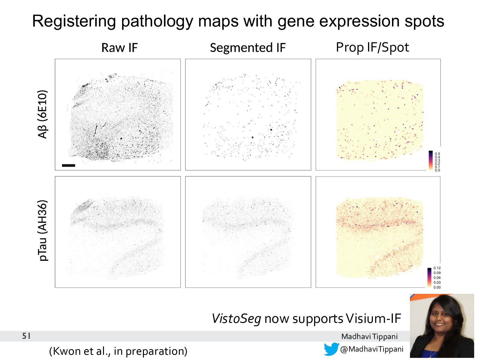
friendly
    • 6.5 mm2 too restrictive? Opportunity for creativity
    • Visium and Visium-IF have required the development of software
    • It’s fun to work on something where there are no answers on Google =)
    but also a challenge
    60

    View Slide

  61. 61
    @HeenaDivecha
    Heena R Divecha

    View Slide

  62. 62
    B1
    A1
    D1
    C1
    C1
    B1
    A1
    D1
    @CerceoPage
    Stephanie C Page

    View Slide

  63. Summary
    63
    • Identification of layer-enriched genes in human dorsolateral
    prefrontal cortex using Visium technology.
    • Spatial registration of single-nucleus RNA-seq data to determine
    enrichment of cell populations in specific cortical layers.
    • Single nucleus and spatial transcriptomics approaches can be used
    to better understand molecular associations with brain disorders,
    including neurodevelopmental and neurodegenerative disorders.
    • Development of tools and resources to analyze spatial
    transcriptomics data.

    View Slide

  64. Future Directions
    • Integration of proteomic and transcriptomic data
    • Visium-IF AD proof-of-concept
    • Integration of snRNA-seq and Visium data
    • Visium + snRNA-seq on LC
    • Increasing resolution (# spots) and area (array size)
    • Visium HD
    • Leveraging rich histology/imaging data
    • Clustering (SpaGCN), spot deconvolution, etc.
    • Building educational resources
    • Completing Orchestrating Spatially Resolved
    Transcriptomics Analysis with Bioconductor (OSTA)
    64

    View Slide

  65. #spatialLIBD is a supportive LIBD & JHU team
    65
    Check for your yourself at
    https://twitter.com/lcolladotor/status/1516587531369811971
    https://lcolladotor.github.io/team_surveys/

    View Slide

  66. We are hiring! https://www.libd.org/careers/
    66

    View Slide

  67. Acknowledgements
    Lieber Institute
    Sang Ho-Kwon
    MadhaviTippani
    Abby Spanger
    Brenda Pardo
    Joseph L. Catallini II
    Matthew N. Tran
    Vijay Sadashivaiah
    Heena Divecha
    Kelsey Montgomery
    Nick Eagles
    Josh Stolz
    Louise Huuki
    Rahul Bharadwaj
    Stephanie Page
    Leonardo Collado-Torres
    Keri Martinowich
    Andrew Jaffe
    Joel E. Kleinman
    Thomas M. Hyde
    Daniel R. Weinberger
    JHU Biostatistics Dept
    Stephanie Hicks
    Lukas Weber
    Sowmya Parthiban
    10x Genomics
    Courtney Anderson
    Cedric Uytingco
    Stephen R. Williams
    Charles Bruce
    Jennifer Chew
    YifengYin
    Nikhil Rao
    Michelle Mak
    Guixia Yu
    Julianna Avalos-Gracia
    JHU Oncology Tissue Services (Kristen Lecksell)
    JHU SKCCC Flow Core (Jessica Gucwa)
    JHU Transcriptomics & Deep Sequencing Core (Linda Orzolek)
    JHU Tumor Microenvironment Core (Liz Engle)
    We are hiring! https://www.libd.org/careers/
    @kr_maynard
    @lcolladotor
    #spatialLIBD team

    View Slide

  68. 68
    https://twitter.com/bayraktar_lab/status/1481719801789939712

    View Slide