Upgrade to Pro — share decks privately, control downloads, hide ads and more …

Understanding the FBC in 15 mins - A quick lit review.

Simon Mark
June 02, 2019
38

Understanding the FBC in 15 mins - A quick lit review.

Simon Mark

June 02, 2019
Tweet

Transcript

  1. Understanding the Full blood count in 15 mins - a

    ‘quick-lit-review’. Simon Mark Daley (2018)
  2. Constituents of the FBC… ▪Red Blood Cells ▪ Haemoglobin, Mean

    cell volume, Reticulocyte count, Red cell count, Haematocrit / Packed cell volume, Mean corpuscular haemoglobin, Red blood cell distribution width. ▪Platelets ▪White Blood Cells ▪ Neutrophil count, Lymphocyte count, Monocyte count, Eosinophil count, Basophil count.
  3. Red blood cells… Constituents of the red blood cell. ▪

    Haemoglobin (Hb); concentration of haemoglobin in the blood. Hb in the protein that carries oxygen in the blood. Low Hb = anaemia. ▪ Mean cell volume (MVC); mean volume of the red cells (“-cytic”). This is used to classify anaemia type. ↓MVC = microcytic, ↑MVC = macrocytic. ▪ Reticulyte count; concentration of immature red blood cells – increased in blood loss and haemolytic anaemia because the bone marrow works harder to replace lost cells. ▪ Red cell count (RCC); concentration of red blood cells within the blood. ▪ Haematocrit (HCT) / packed cell volume (PCV); volume percentage of red blood cells within the blood. ▪ Mean corpuscular haemoglobin (MCH); mean haemoglobin quantity with the blood cells – affects the colour of the cells (“-chromic”). ▪ most normocytic/macrocytic anaemias rare normochromic, most microcytic anaemias are hypochromic. ▪ Mean corpuscular haemoglobin concentration (MCHC); mean haemoglobin concentration within the blood cells. ▪ Red blood cell distribution width (RDW); measure of the variation of red blood cell volumes. It is used in conjunction with MCV to determine if anaemia is due to mixed or single cause. Raised RDW = “anisocytosis”. MOST IMPORTANT LESS IMPORTANT
  4. Red blood cells… Anaemia. Anaemia = a reduced concentration of

    haemoglobin within the blood. Other relevant tests; ▪ WCC and platelet count; if both also abnormal, a bone marrow cause is likely ▪ Reticulocyte count; if raised, the cause is blood loss or haemolytic anaemia ▪ Mean cell volume (MCV); Causes of anaemia by MVC Microcytic ▪ Sideroblastic ▪ Iron deficiency ▪ Thalassaemia Normocytic ▪ Acute blood loss ▪ Haemolytic anaemia ▪ Sickle cell anaemia Macrocytic anaemia ▪ Megaloblastic ▪ ↓B12, ↓folate ▪ Non-megaloblastic ▪ Alcohol, reticulocytosis, liver disease, pregnancy Chronic disease Hypothyroidism Bone marrow failure (aplastic anaemia, myelodyplastia, leukaemia, myelofibrosis)
  5. Red blood cells… Anaemia. Tests for specific causes; ▪ Haematinics;

    B12 % folate, ferritin. ▪ Iron studies: Iron, transferritin / total iron binding capacity (TIBC), transferritin saturation, ferritin, soluble transferritin receptor. ▪ TFTs ▪ Blood film +/- bone marrow biopsy (if bone marrow cause/haemolytic anaemia/sideroblastic anaemia suspected). ▪ Hb electrophoresis (if thalassaemia / sickle cell suspected) ▪ Bilirubin (raised in haemolysis)
  6. Red blood cells… Anaemia. Commonest causes. ▪ Iron-deficiency anaemia. ▪

    Physiology: Iron is found in red meats (haem iron) and cereal (non-haem iron). In humans, 2/3 is stored as haem, 2/9 as ferritin, 1/9 as haemosiderin. Fe2+ (ferrous) is absorbed; Fe3+ (ferric) is consumed and used. ▪ Causes: ▪ Chronic blood loss (Menstrual loss, GI tract loss (↑urea in UGI bleed), urinary tract loss). ▪ ↑demand (pregnancy, growth). ▪ ↓absorption (coeliacs, gastrectomy). ▪ Poor intake. ▪ Investigations if no clear cause: upper GI endoscopy + colonoscopy, coeliacs screen, urine dipstick. ▪ Treatment: treat cause, ferrous sulphate tablets, transfusion if Hb<70. ▪ B12 -deficiency anaemia. ▪ Physiology: Vitamin B12 is found in meat and dairy products. The stomach produces intrinsic factor which binds to B12 , allowing it to be absorbed in the terminal ileum, Body stores last up to 4 years. ▪ Causes: Pernicious anaemia, malabsorption (eg gastrectomy or terminal ileum disease/resection). ▪ Investigations for pernicious anaemia: Parietal cell antibodies, intrinsic factor antibodies, Schilling’s test. ▪ Treatment: Treat cause, hydroxocobalamin (B12 ) injections 3-monthly.
  7. Red blood cells… Anaemia. ▪ Folate-deficiency anaemia. ▪ Physiology: folate

    is found in green vegetables. Body stores only last 4 months (therefore deficiency develops earlier in malabsorption/pregnancy). ▪ Causes: ▪ Dietary (alcoholism, neglect). ▪ ↑requirements (pregnancy, haematopoiesis). ▪ Malabsorption (coeliacs, pancreatic insufficiency, gastrectomy, crohns). ▪ Drugs interfere with metabolism (phenytoin, methotrexate, trimethoprim). ▪ Treatment: treat cause, oral folic acid supplements. ▪ Anaemia of chronic disease. ▪ Causes: any chronic disease. ▪ Classically: ↓iron ↓TIBC ferritin normal (vs. iron deficiency anaemia: ↓iron, ↑TIBC, ↓ferritin). ▪ Treatment: treat cause, transfuse if Hb<70.
  8. Red blood cells… Anaemia. ▪ Haemolytic anaemia. ▪ Physiology: ▪

    When red cells are destroyed extravascularly by macrophages (normal process): 1. Hb → globulin (which is broken down to amino acids) + haem (which is broken down to bilirubin). 2. Bilirubin is then conjugated by the liver (a rate limited process0 and passed to the bowel in bile, where it is converted to urobilinogen. 3. Some of this urobilinogen is passed in stool and some is reabsorbed and excreted in urine as urinary urobilinogen. ▪ When red cells are destroyed intravascularly (abnormal process) – free Hb follows 1 of 3 pathways: ▪ Some binds to haptoglobin (and is removed by liver). ▪ Some is filtered by the glomerulus and passed as haemiglobinuria or haemosiderinuria. ▪ Some is oxidised to methaemoglobulin which dissociates to globin + ferrihaem (most ferrihaem then binds to albumin → methaemalbuminaemia). ▪ Inherited causes: ▪ Haemoglobinopathies: sickle cell, thalassaemia. ▪ Membrane defects: hereditary spherocytosis, ellptocystosis. ▪ Enzyme defects: G6PD deficiency, pyruvate kinase deficiency. ▪ Acquired causes: ▪ Immune mediated: autoimmune haemolytic anaemia, drug-induced haemolytic anaemia, alloimmune haemolytic anaemia. ▪ Non-immune mediated: DIC, TTP, physical damage by eg heart valves, toxins such as lead/uraemia/drugs, malaria, paroxysmal nocturnal haemoglobulinuria.
  9. Red blood cells… Anaemia. ▪ Investigations to confirm haemolysis; ▪

    Increased Hb breakdown: ↑unconjugated bilirubin, ↑LDH (from red cells), ↑urinary urobilinogen (on urine dipstick). ▪ Increased Hb production: ↑reticulocytes. ▪ Intravascular haemolysis: ↓free haptoglobin, haemoglobinuria (on haematuric urine microscopy), ↑urinary haemosiderin, red cell fragments on blood film. ▪ Investigations to find cause: ▪ Blood film: sickle cells scistocytes (microanglopathic haemolytic anaemia), inclusion bodies (malaria), spherocytes/elliptocytes (hereditary spherocytosis/elliptocytosis), Heinz bodies (G6PD), bite/blister cells (G6PD), distorted ‘prickle’ cells (pyruvate kinase deficiency). ▪ Direct antiglobulin (Coombs’) test (for autoimmune haemolytic anaemia). ▪ Osmotic fagility testing (for membrane abnormalities). ▪ Hb electrophoresis (for haemoglobulinopathies). ▪ Enzyme assays (for enzyme defects).
  10. Red blood cells… Polycythaemia. Polycythaemia = an increased concentration of

    red blood cells within the blood. Causes: ▪ Relative polycythaemia (ie ↓plasma volume). ▪ Acute dehydration. ▪ Chronic (associated with obesity, HTN, alcohol excess, smoking). ▪ Absolute polycythaemia (ie ↑RBC mass) ▪ Primary = polycythaemia ruba vera ▪ Secondary = due to increased EPO (eg RCC) or chronic hypoxia (eg COPD, altitude, congenital heart disease). Investigations: ▪ WCC and platelet count (both raised in primary absolute polycythaemia, but not in secondary absolute polycythaemia). ▪ 51Cr Red cell mass study (normal red cell mass in relative polycythaemia; raised red cell mass in absolute polycythaemia). ▪ Erythropoietin level ▪ If polycythaemia rub vera suspected: bone marrow biopsy, JAK-2 mutation
  11. White blood cells… WCC constituents High Low Neutrophil count Bacterial

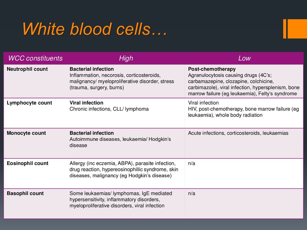
    infection Inflammation, necorosis, corticosteroids, malignancy/ myeloproliferative disorder, stress (trauma, surgery, burns) Post-chemotherapy Agranulocytosis causing drugs (4C’s; carbamazepine, clozapine, colchicine, carbimazole), viral infection, hypersplenism, bone marrow failure (eg leukaemia), Felty’s syndrome Lymphocyte count Viral infection Chronic infections, CLL/ lymphoma Viral infection HIV, post-chemotherapy, bone marrow failure (eg leukaemia), whole body radiation Monocyte count Bacterial infection Autoimmune diseases, leukaemia/ Hodgkin’s disease Acute infections, corticosteroids, leukaemias Eosinophil count Allergy (inc eczemia, ABPA), parasite infection, drug reaction, hypereosinophillic syndrome, skin diseases, malignancy (eg Hodgkin’s disease) n/a Basophil count Some leukaemias/ lymphomas, IgE mediated hypersensitivity, inflammatory disorders, myeloproliferative disorders, viral infection n/a
  12. Platelets… Thrombocytopaenia. Causes: ▪ Decreased production: bone marrow failure, aplastic

    anaemia, megaloblastic anaemia, myelosuppression. ▪ Increased destruction/ consumption: ▪ Non-immune: DIC, TTP, HUS, sequestration in hypersplenism (including portal HTN eg liver disease). ▪ Primary immune: ITP ▪ Secondary immune: SLE, CLL, viruses, drugs, alloimmune. Possible investigations: ▪ Blood film +/- bone marrow biopsy. ▪ Infection screen (eg HIV, hepatitis C). ▪ LFTs (live dysfunction can also cause thrombocytopenia). ▪ LDH (increased in haemolysis and lymphoproliferative disorders). ▪ Serum vit B12 and folate. ▪ Coagulation screen (inc fibrinogen and D-dimer (if suspect DIC). ▪ Acute phase reactants (look for evidence of infection). Treatment: ▪ Treat cause. ▪ Immunosuppressants if autoimmune eg prednisolone, azathioprine, cyslophosphamide. ▪ Plasmapheresis for TTP/ HUS. ▪ Platelet concentrate transfusion. ▪ Splenectomy.
  13. Platelets… Thrombocythaemia. Causes: ▪ Primary: essential thrombocythaemia, other myeloproliferative disorders.

    ▪ Secondary: bleeding, inflammation, infection, malignancy, post-splenectomy. Possible investigations: ▪ Blood film +/- bone marrow biopsy. ▪ Acute phase reactants (look for evidence of infection). ▪ JAK2 mutation (myloproliferative disease). Treatment: ▪ Aspirin (to prevent thromboembolic disease. ▪ Hydroxycarbamide (if primary cause). References; Mansbridge 2013 www.OSCEstop.com