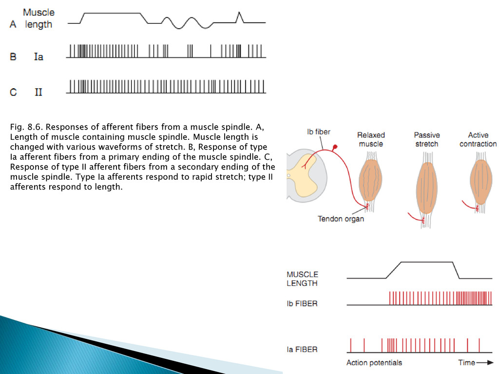
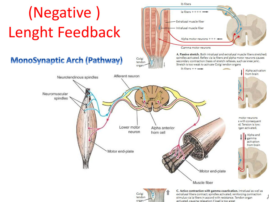
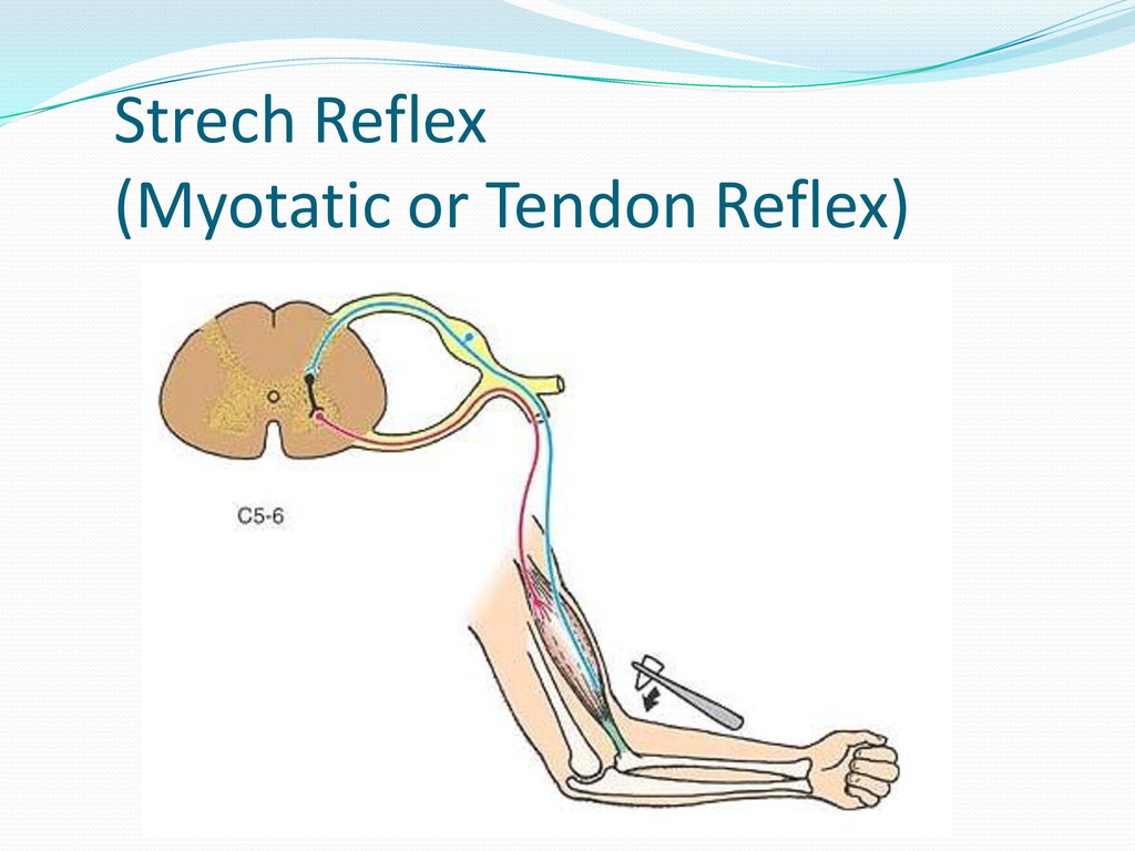
A, Length of muscle containing muscle spindle. Muscle length is changed with various waveforms of stretch. B, Response of type Ia afferent fibers from a primary ending of the muscle spindle. C, Response of type II afferent fibers from a secondary ending of the muscle spindle. Type Ia afferents respond to rapid stretch; type II afferents respond to length.
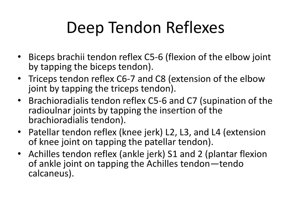
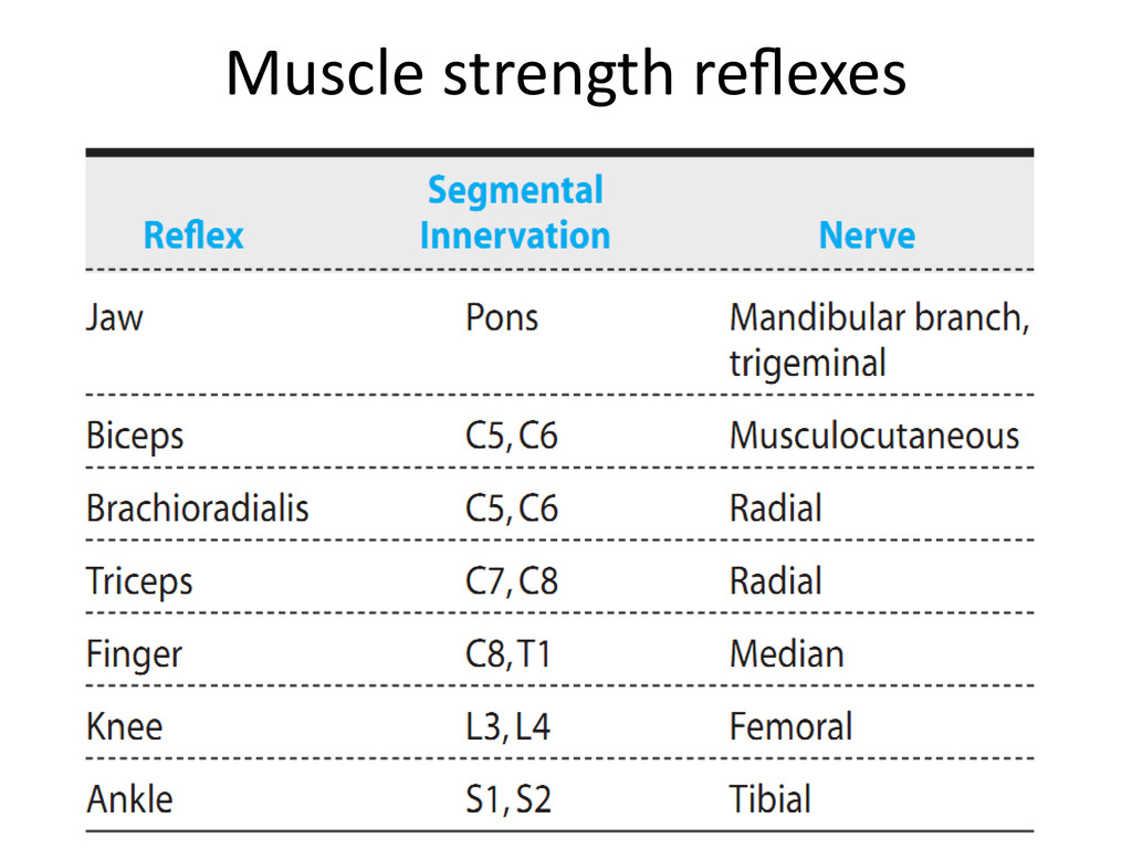
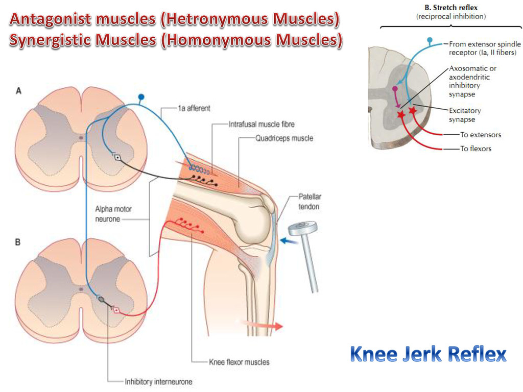
of the elbow joint by tapping the biceps tendon). • Triceps tendon reflex C6-7 and C8 (extension of the elbow joint by tapping the triceps tendon). • Brachioradialis tendon reflex C5-6 and C7 (supination of the radioulnar joints by tapping the insertion of the brachioradialis tendon). • Patellar tendon reflex (knee jerk) L2, L3, and L4 (extension of knee joint on tapping the patellar tendon). • Achilles tendon reflex (ankle jerk) S1 and 2 (plantar flexion of ankle joint on tapping the Achilles tendon—tendo calcaneus).
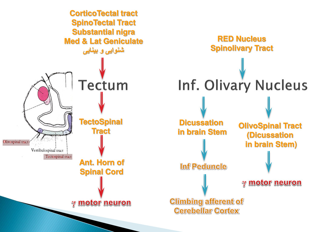
in brain Stem) CorticoTectal tract SpinoTectal Tract Substantial nigra Med & Lat Geniculate ییانیب و ییاونش RED Nucleus Spinolivary Tract Dicussation in brain Stem
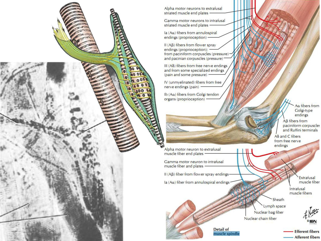
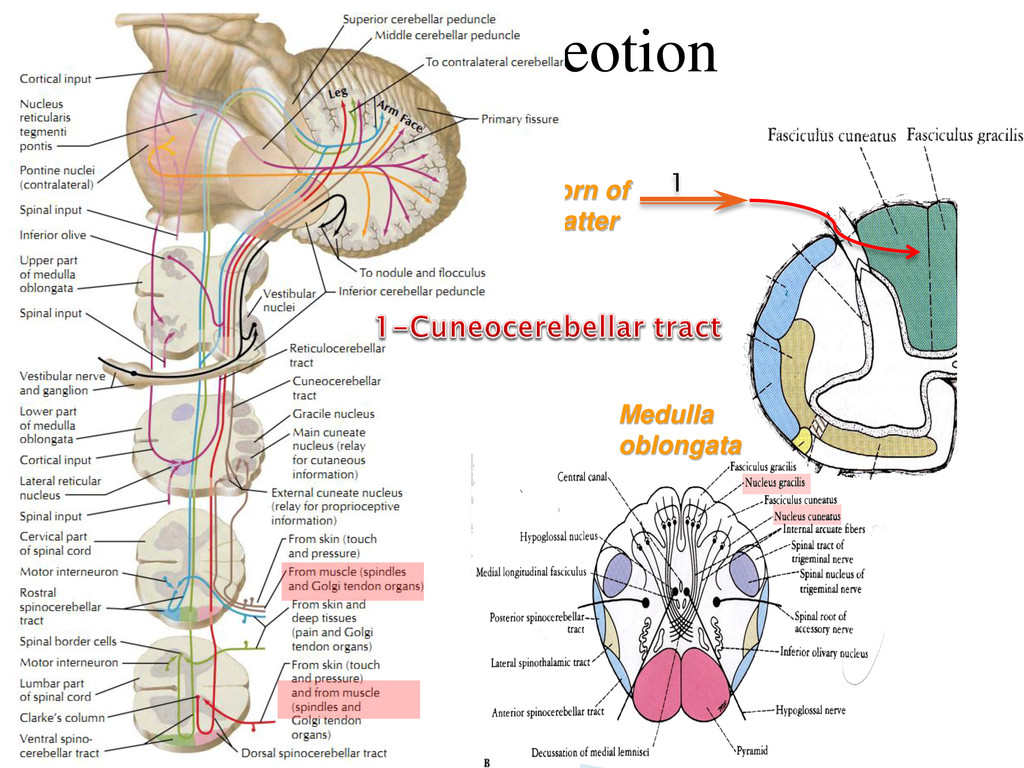
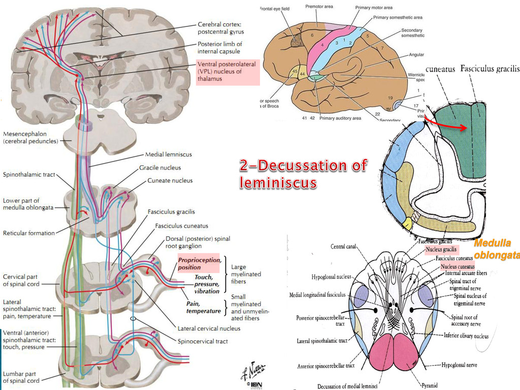
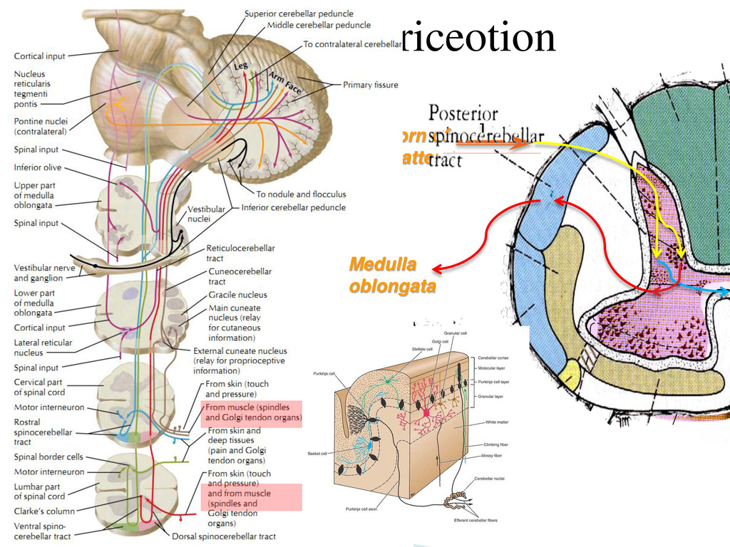
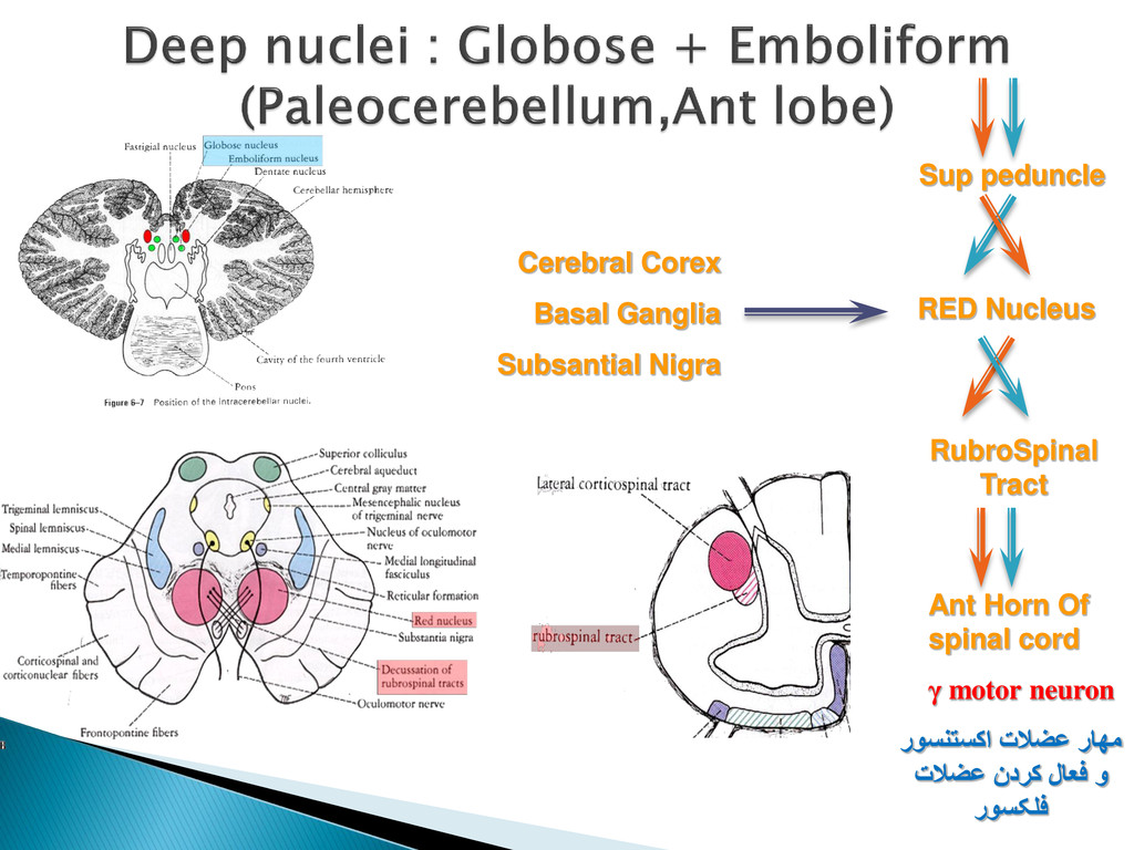
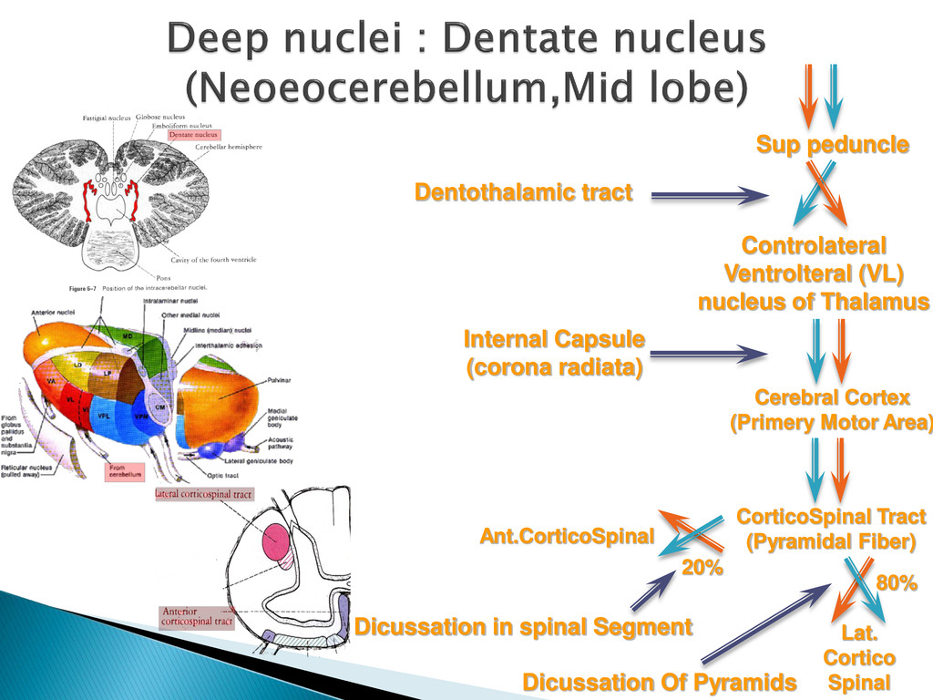
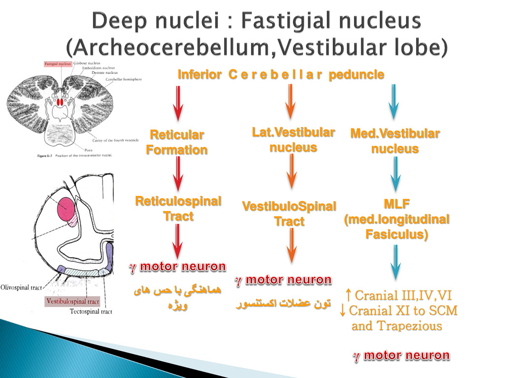
pathway of all centers involve d in moto r activity is the large anterior horn cell ( A1) and its a xon (!-motoneuron. The central regions that influence motor activity via descending pathways a re interconnecte d in many ways. The most important af fe rent pathways ste m from the cereb ellum, which receives the impulses of muscle receptors via the spinocereb ellar tracts (A2) and the stimuli of the cortex via the corticopontine tracts (A3). The cereb ellar impulses are transmitte d via the parvocellular part of the dentate nucleus ( A4) and the ventral lateral nucleus of thalamus ( A5) to the precentral corte x (area 4) (A6). The corticospinal (pyramidal) tract (A7 ) descends from area 4 to the anterior horn and gives of f collaterals in the pons ( A8) that return to the cereb ellum. Additional cereb ellar impulses are transmitte d via the emboliform nucleus ( A9 ) and the centromedian nucleus of the thalamus (A10) to the striatum ( A11) and via the magnocellular part of the dentate nucleus (A12) to the red nucleus (A13). From here f ib ers run in the central tegmental tract (A14) via the olive (A15) back to the cereb ellum and in the rubroreticulospinal tract (A16) to the anterior horn. Fib ers from the globose nucleus (A17) run to the interstitial nucleus of Cajal ( A18) and from there i n the interstitiospinal fasciculus (A19) to the anterior horn. Finally, cere b e llofugal f ib ers are relaye d in the ve stibular nuclei (A20) and in the reticular formation (A21 ) to the vestibulospinal tract (A22) and the reticulospinal tract (A23), respectively.The descending pathways can b e divide d into two groups according to their ef fect on the muscles: one group stimulates the fle xor muscles , and another group stimulates the e xtensor muscles. The corticospinal tract and the rubroret iculospinal tract activate mainly the neurons of the flexor muscles and inhibit the neurons of the extensor muscles. This cor responds to the functional importance of the corticospinal tract for delicate and precise movements, especially those of hand and f inger muscles where flexor muscles play an important role. In contrast, the f i b ers of the ve st ibulospinal tract and the f i b ers from the pontine reticular formation inhibit the flexors and activate the ex tensors. They b elong to a p hylogenetically old motor system that is directe d against the ef fect of gravity and, thus, is of special importance for body posture and balance. The peripheral f ib ers that run through the posterior ro ot into the anterior horn originate from the muscle receptors. The af ferent f i b ers of the annulospiral endings (A24 ) terminate with their collaterals directly on the !-motoneurons, while the f ib ers of the tendon organs (A25) terminate o n interneurons. Many descending pathways influence the !-neurons via the spinal reflex apparatus. They terminate on the large !neurons and on the small "-neurons ( A26). Since the "-neurons have a lower threshold of stimulation, they a re stimulate d f irst, which results i n the activation of muscle spindles. The latter send their impulses to the !-neurons. Thus, the "-neurons and muscle spindles have a starter function for voluntar y movements. A27 Accessor y olive . A28 Skeletal muscles. A29 Muscle spindle.