General Embryology 3 (Intraembryonic mesoderm , Endoderm , Embryonic folding)
This presentations shows during pregnancy what is Intraembryonic mesoderm formation & classification , Endoderm fate , and Embryonic folding steps & results
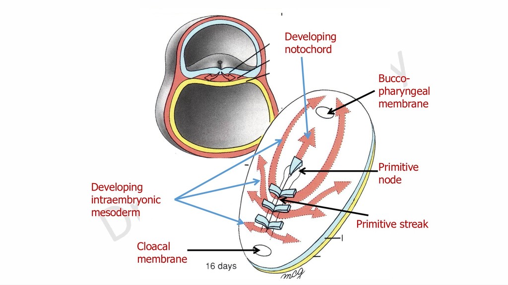
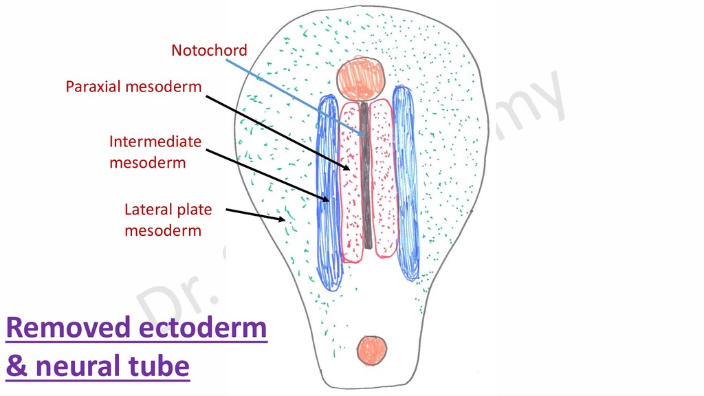
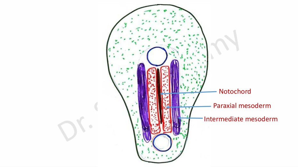
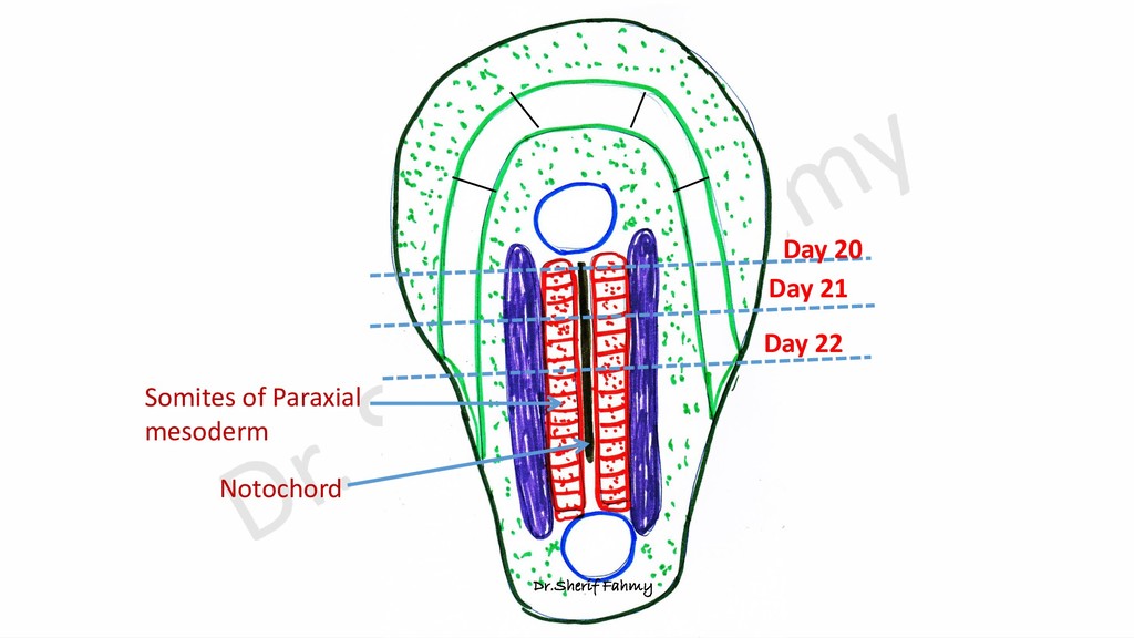
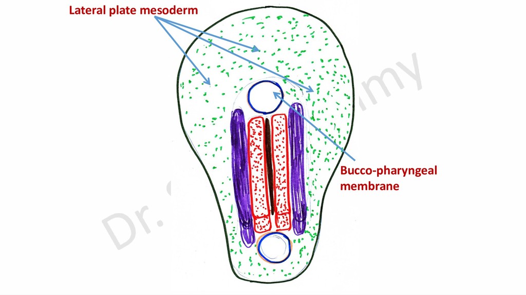
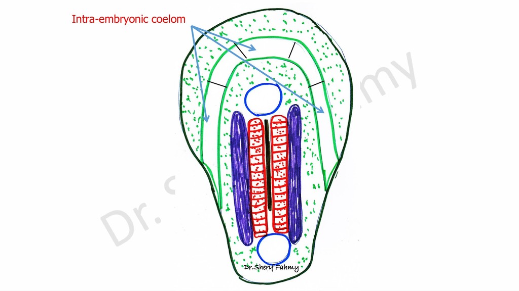
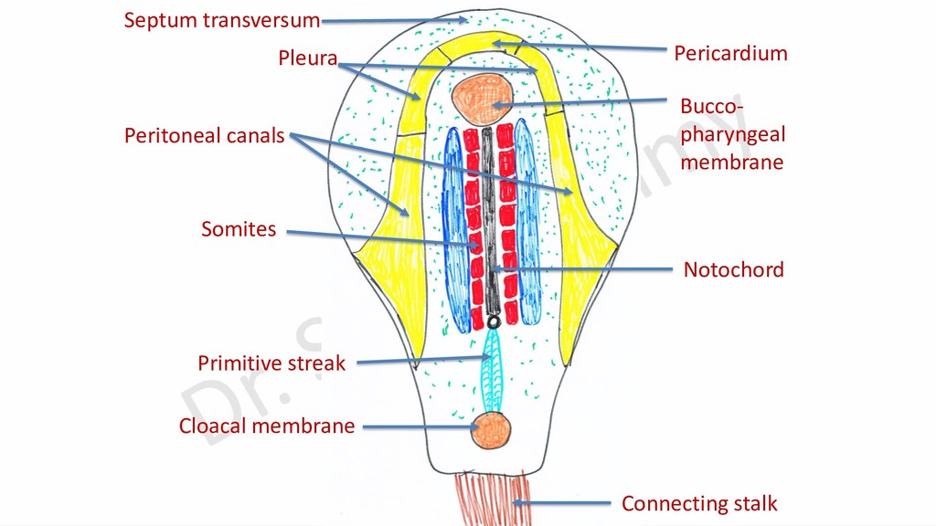
notochord, divides into: 1- Paraxial Mesoderm: on both sides of notochord. 2- Intermediate Mesoderm: Middle part of the mesoderm. 3- Lateral plate Mesoderm: Lateral part which communicates with that of the opposite side cranial to bucco-pharyngeal membrane.
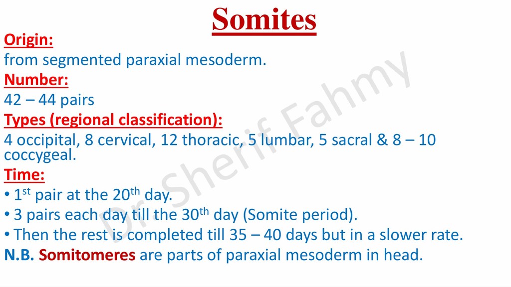
pairs Types (regional classification): 4 occipital, 8 cervical, 12 thoracic, 5 lumbar, 5 sacral & 8 – 10 coccygeal. Time: • 1st pair at the 20th day. • 3 pairs each day till the 30th day (Somite period). • Then the rest is completed till 35 – 40 days but in a slower rate. N.B. Somitomeres are parts of paraxial mesoderm in head.
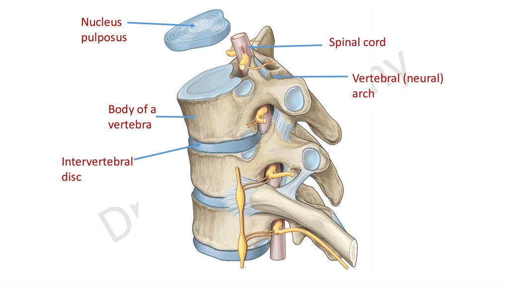
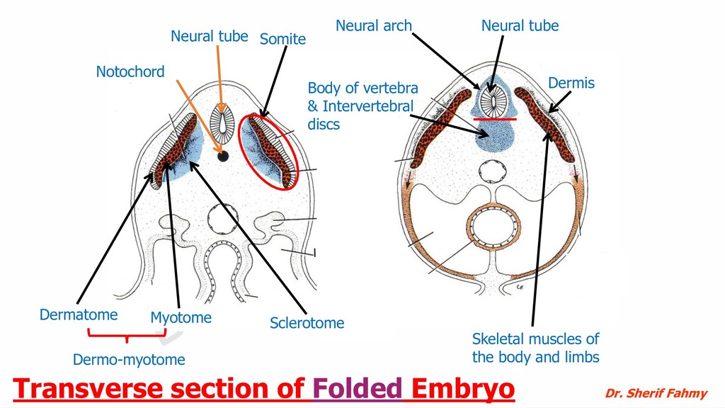
of vertebra & Intervertebral discs Skeletal muscles of the body and limbs Dermis Dermo-myotome Neural arch Transverse section of Folded Embryo Dr. Sherif Fahmy
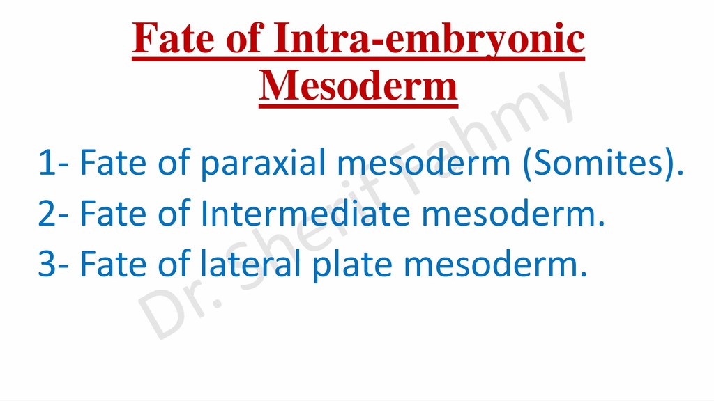
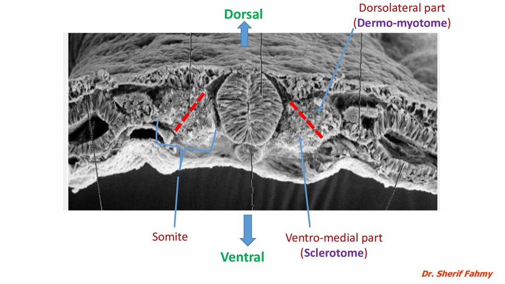
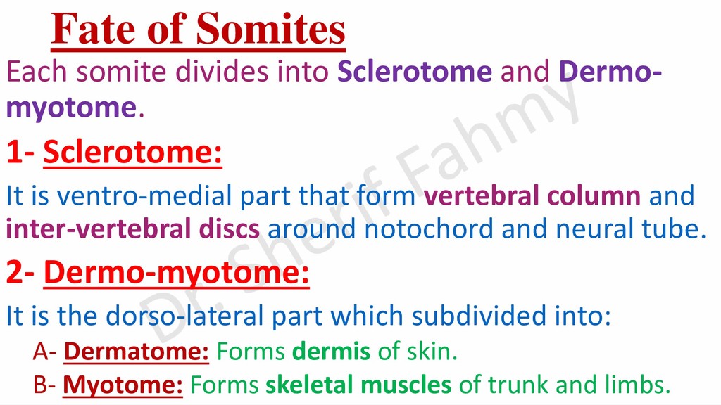
myotome. 1- Sclerotome: It is ventro-medial part that form vertebral column and inter-vertebral discs around notochord and neural tube. 2- Dermo-myotome: It is the dorso-lateral part which subdivided into: A- Dermatome: Forms dermis of skin. B- Myotome: Forms skeletal muscles of trunk and limbs.
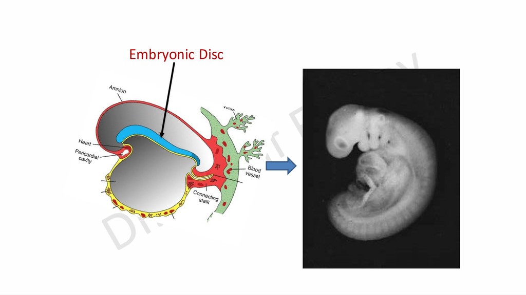
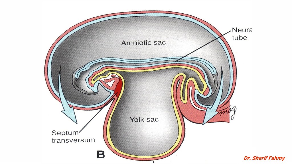
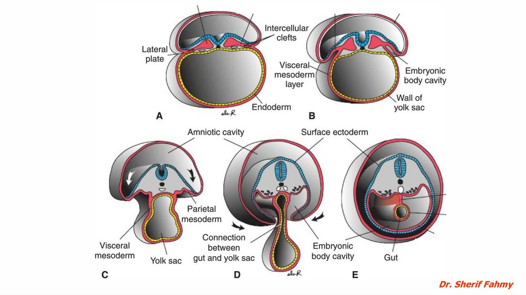
folded upon itself. Time of folding: • From the end of 3rd week to the end of 4th week. Causes of folding: • Rapid increase of cranio-caudal length due to rapid growth of neural tube and somites. • Rapid expansion of amniotic cavity. Types of folding: • Head and tail folds are folding of cranial and caudal parts of the disc. Folds are limited by relatively firm notochord and primitive streak. • Lateral folds are folding of the sides of the embryonic disc in the transverse direction.
- Ventral shift of the amnio-ectodermal junction (towards endoderm) with dorsal bulge into amniotic cavity. B-Cranio-caudal elongation of embryonic disc leads to: -Formation of head & tail folds.
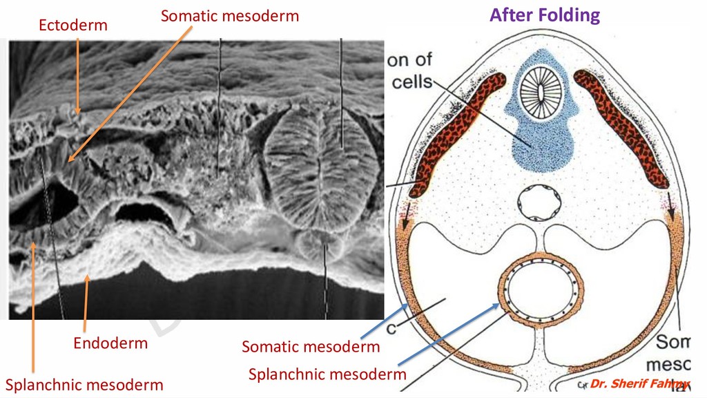
➢ Ectoderm on the outer surface. ➢ Mesoderm deep (internal) to ectoderm. ➢ Endoderm is innermost tube that forms the gut (Foregut, Midgut and Hindgut). ➢ Amniotic cavity surrounds the fetus and the umbilical cord.
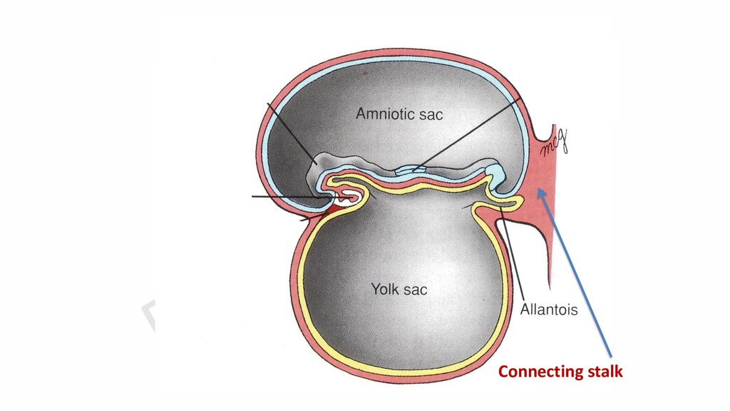
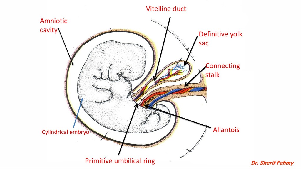
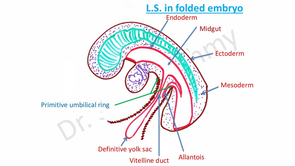
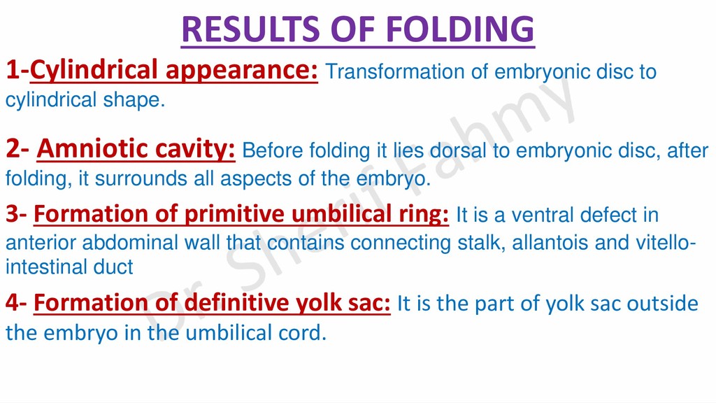
cylindrical shape. 2- Amniotic cavity: Before folding it lies dorsal to embryonic disc, after folding, it surrounds all aspects of the embryo. 3- Formation of primitive umbilical ring: It is a ventral defect in anterior abdominal wall that contains connecting stalk, allantois and vitello- intestinal duct 4- Formation of definitive yolk sac: It is the part of yolk sac outside the embryo in the umbilical cord.
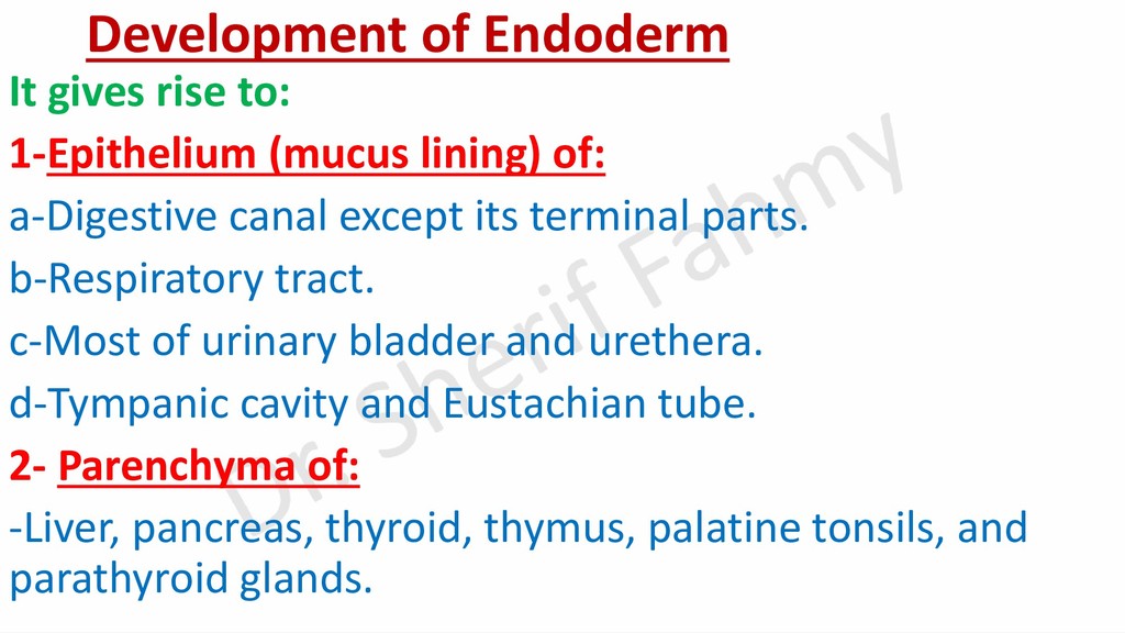
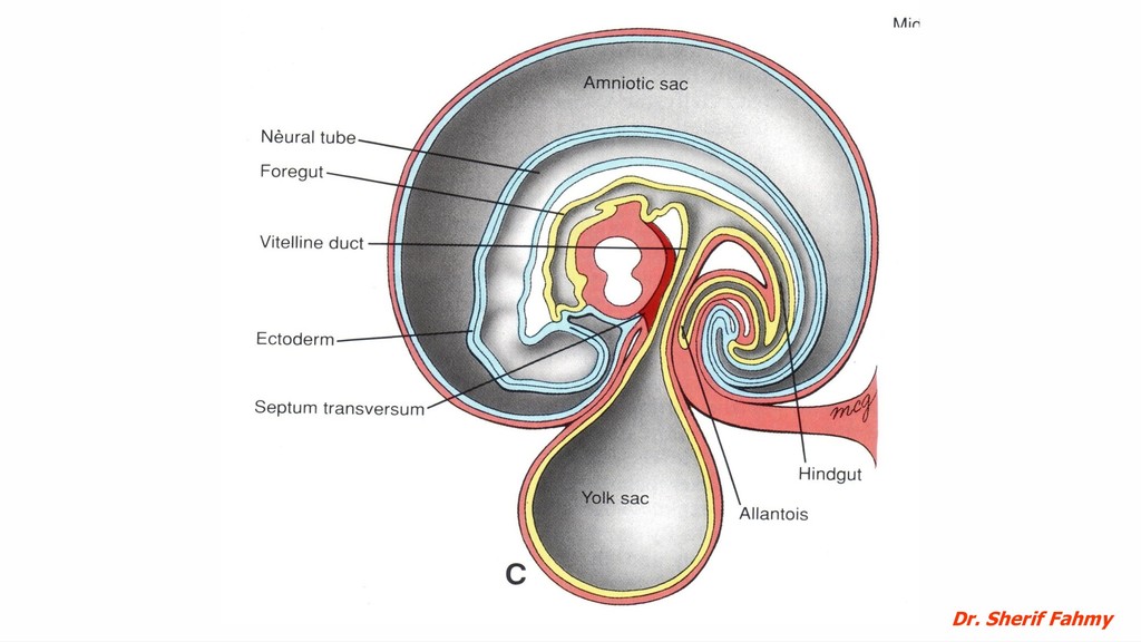
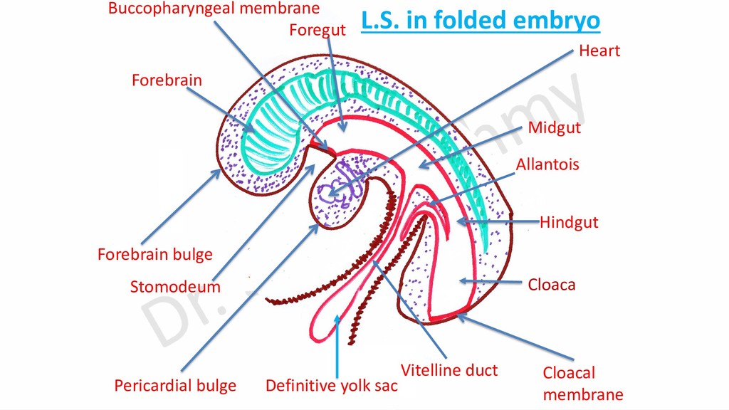
layer together with part of yolk sac. Foregut is formed in head fold with bucco-pharyngeal membrane closing its cranial end. Hindgut: is formed in tail fold and closed caudally by cloacal membrane. The caudal part is dilated and called cloaca with allantois connected to it. Midgut: is formed by lateral folds and present between foregut and hindgut. It is connected with defenitive yolk sac by vitelline duct.
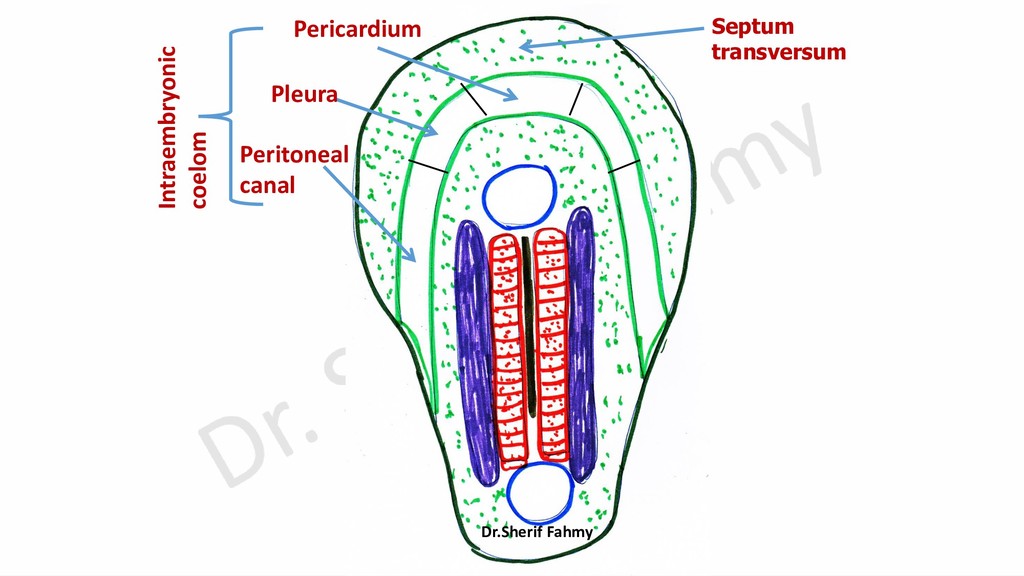
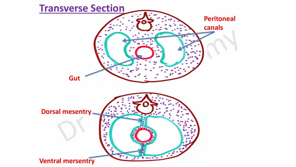
form pericardial bulge while growing heart will form pericardial bulge. 7- Formation of stomodeum: It is ectodermal depression between forebrain and pericardial bulges. It is separated from foregut by bucco-pharyngeal membrane. 8- Formation of mesenteries: When peritoneal canals surrounds the gut, ventral and dorsal mesenteries are formed. 9- Reversal of positions: -Buccopharyngeal membrane becomes the most cranial, heart and pericardium become cranial to septum transversum (before folding septum transversum is most cranial). -Connecting stalk becomes ventral and more cranial in spite of being most