Upgrade to Pro — share decks privately, control downloads, hide ads and more …

acls bradycardia algorithm and heart block

acls bradycardia algorithm and heart block

based on 2010 ACLS guidelines for bradycardia. these slides also includes ECG strips and answers.

malek ahmad

May 14, 2015
Tweet

More Decks by malek ahmad

Other Decks in Education

Transcript

  1. May 6, 2015 Learning Objectives ❖ able to recognise signs

    and symptoms ❖ able to state the treatments for symptomatic bradycardia ❖ able to determine whether signs and symptoms are caused by bradycardia or another condition ❖ able to identify AV blocks ❖ able to list out the indication for TCP and drugs used
  2. Bradycardia vs Symptomatic Bradycardia ❖ Bradycardia is defined as any

    rhythm disorder with a heart rate less than 60 beats per minute. (Typically it will be <50/min) • This could also be called asymptomatic bradycardia • Bradycardia can be a normal non-emergent rhythm • For instance, well trained athletes may have a normal heart rate that is less than 60 bpm ❖ Symptomatic bradycardia however is defined as a heart rate less than 60/min that elicits signs and symptoms, but the heart rate will usually be less than 50/min • Symptomatic bradycardia exists when the following 3 criteria are present: 1.) The heart rate is slow 2.) The patient has symptoms 3.) The symptoms are due to the slow heart rate
  3. Functional or relative bradycardia occurs when a patient may have

    a heart rate within normal sinus range, but the heart rate is insufficient for the patients condition. An example would be a patient with an heart rate of 80 bpm when they are experiencing septic shock.
  4. Bradycardia algorithm ❖ The decision point for ACLS intervention in

    the bradycardia algorithm is determination of adequate perfusion ❖ For the patient with adequate perfusion, you should observe and monitor ❖ If the patient has poor perfusion, preparation for transcutaneous pacing should be initiated, and an assessment of contributing causes (H’s and T’s) should be carried out
  5. Bradycardia pharmacology ❖ There are 3 medications that are used

    in the Bradycardia ACLS Algorithm ❖ They are : 1. Atropine: • The first drug of choice for symptomatic bradycardia • Dose is 0.5mg IV push and may repeat up to a total dose of 3mg 2. Dopamine: • Second-line drug for symptomatic bradycardia when atropine is not effective • Dosage is 2-10 micrograms/kg/min infusion 3. Epinephrine: • Can be used as an equal alternative to dopamine when atropine is not effective • Dosage is 2-10 micrograms/min infusion Smith I, Monk TG, White PF. Comparison of transesophageal atrial pacing with anticholinergic drugs for the treatment of intraoperative bradycardia. Anesth Analg. 1994;78:245–252. Brady WJ, Swart G, DeBehnke DJ, Ma OJ, Aufderheide TP. The efficacy of atropine in the treatment of hemodynamically unstable bra- dycardia and atrioventricular block: prehospital and emergency department considerations. Resuscitation. 1999;41:47–55.
  6. -2010 AHA Update- For symptomatic bradycardia or unstable bradycardia IV

    infusion chronotropic agents (dopamine & epinephrine) is now recommended as an equally effective alternative to external pacing when atropine is ineffective.
  7. Atropine ❖ Classified as an anticholinergic drug and increases firing

    of the SA Node by blocking the action of the vagus nerve on the heart resulting in an increased heart rate ❖ Atropine should be used cautiously in the presence of myocardial ischemia and hypoxia since it increases oxygen demand of heart and can worsen ischemia ❖ The dosing for Atropine is 0.5 mg IV every 3-5 minutes as needed, and the maximum total dosage that can be give is 3 mg. ❖ Atropine should be avoided in hypothermic bradycardia and it will not be effective for Mobitz type II/Second Degree Block Type 2 and Complete Heart Block (3rd degree block)
  8. Bernheim A, Fatio R, Kiowski W, Weilenmann D, Rickli H,

    Rocca HP. Atropine often results in complete atrioventricular block or sinus arrest after cardiac transplantation: an unpredictable and dose-independent phenomenon. Transplantation. 2004;77:1181–1185 Atropine will likely be ineffective in patients who have undergone cardiac transplantation because the transplanted heart lacks vagal innervation. One small uncontrolled study documented paradoxical slowing of the heart rate and high-degree AV block when atropine was administered to patients after cardiac transplantation
  9. Morris DL, Chambers HF, Morris MG, Sande MA. Hemodynamic characteristics

    of patients with hypothermia due to occult infection and other causes. Ann Intern Med1985; 102:153–7 As the temperature falls to a moderate degree of hypothermia (<33), a progressive bradycardia develops consequent on decreased spontaneous depolarization of the pacemaker cells, and this is refractory to atropine
  10. ❖ In 3rd degree block: disassociation of the electrical activity

    that is occurring in the atria and ventricles • Since atropine’s affect is primarily on the SA node in the atria, 3rd degree block would prevent its affect on the SA node from influencing the rate of ventricular contraction which is needed to improve perfusion ❖ In 2nd degree AV Block Type II, the situation is similar • There is a partial block in the electrical impulses from the atria (SA) to the ventricles, and thus the affects of atropine would not significantly change the status of the ventricles • This block can rapidly progress to 3rd degree block ❖ Atropine may speed the firing rate of the SA node (atria), but the ventricles are not responding to anything the atria (SA node) puts out. Thus, the heart rates will not increase ❖ There may be some action at the AV-node with atropine, but the effect will be negligible and typically not therapeutic ❖ Atropine in most cases will not hurt the patient with 3rd degree block unless they are unstable and you delay pacing to give atropine
  11. Avoid relying on atropine in type II 2nd degree or

    3rd degree AV block or in patients with 3rd degree AV blocks with a new wide-QRS complex where the location of block is likely to be in non-nodal tissue (bundle of His or more distal conduction systems), as the are not likely to be responsive to reversal of cholinergic effects by atropine and preferably treated with TCP or b-adrenergic support as temporising measures while waiting for trans-venous pacing Robert W. N; Charles W. O; Mark S. L; Steven L. K; Michael S; Clifton W. C; Peter J. K; Joseph P. O; Bryan M; Scott M. S; Rod S. P; Roger D. W; Erik P. H; Wanchun T; Daniel D; Elizabeth S; Laurie J. M. S749-750 Circulation Journal Nov. 2nd 2010
  12. anonymous The goal of therapy is to improve the patient’s

    clinical status rather than target an exact heart rate
  13. Transcutaneous Pacing ❖ Preparation for TCP should be taking place

    as atropine is being given. ❖ Ideally the patient should receive sedation prior to pacing, but if the patient is deteriorating rapidly, it may be necessary to start TCP prior to sedation ❖ For the patient with symptomatic bradycardia with signs of poor perfusion, transcutaneous pacing is the treatment of choice ❖ TCP rate should use 60/min as a starting rate and adjust up or down based on the patient’s clinical response ❖ The dose for pacing should be set at 2mA (milliamperes) above the dose that produces observed capture ❖ TCP is contraindicated for the patient with hypothermia and is not a recommended treatment for asystole ❖ A carotid pulse should not be used for assessment of circulation as TCP can create muscular movements that may feel like a carotid pulse (Assess circulation using the femoral pulse) ❖ Identification of contributing factors for symptomatic bradycardia should be considered throughout the ACLS protocal since reversing of the cause will likely return the patient to a state of adequate perfusion
  14. May 13th, 2015 Outline 1st degree AV block 2nd degree

    AV block (type 1 and 2) 3rd degree AV block
  15. 1st degree AV block • Also called first-degree AV block

    is a disease of the electrical conduction system of the heart in which the PR interval is lengthened beyond 0.20 seconds • This lengthening of the PR interval is caused by a delay in the electrical impulse from the atria to the ventricles through the AV node • Rarely causes any noticeable symptoms • Most people are only aware they have the condition when they're tested for an unrelated medical condition • It's only recognised by an electrocardiograph (ECG)
  16. 2nd degree AV block (type I) • Also called Mobitz

    1 or Wenckebach is a disease of the electrical conduction system of the heart in which the PR interval has progressive prolongation until finally the atrial impulse is completely blocked and does not produce a QRS electrical impulse • Once the p-wave is blocked and no QRS is generated, the cycle begins again with the prolongation of the PR interval • One of the main identifying characteristics of second degree heart block type 1 is that the atrial rhythm will be regular
  17. In the above image, notice that the p-waves are regular,

    the PR-interval progressively gets longer until a QRS is dropped and only the p-wave is present
  18. 2nd degree AV block (type II) ❖ Also called Mobitz

    II or Hay is a disease of the electrical conduction system of the heart ❖ Second-degree AV block (Type 2) is almost always a disease of the distal conduction system located in the ventricular portion of the myocardium
  19. ❖ This rhythm can be recognized by the following characteristics:

    1. non-conducted p-waves (electrical impulse conducts through the AV node but complete conduction through the ventricles is blocked, thus no QRS) 2. P-waves are not preceded by PR prolongation as with second-degree AV block (Type 1) 3. fixed PR interval 4. The QRS complex will likely be wide - The QRS on an ECG will most likely be wide because the block occurs in the His bundle or bundle branches and conduction through the ventricles is slowed
  20. ❖ Second-degree AV block (Type 2) is clinically significant for

    ACLS because this rhythm can rapidly progress to complete heart block ❖ It should be treated with immediate transcutaneous pacing or transvenous pacing because there is risk that electrical impulses will not be able to reach the ventricles and produce ventricular contraction ❖ Atropine may be attempted if immediate TCP is not available or time is needed to initiate TCP ❖ Atropine should not be relied upon and in the case of myocardial ischemia it should be avoided
  21. Complete AV block ❖ Complete heart block occurs when the

    electrical impulse generated in the SA node in the atrium is not conducted to the ventricles ❖ When the atrial impulse is blocked, an accessory pacemaker in the ventricles will typically activate a ventricular contraction ❖ This accessory pacemaker impulse is called an escape rhythm ❖ Because two independent electrical impulses occur (SA node impulse & accessory pacemaker impulse), there is no apparent relationship between the P waves and QRS complexes on an ECG
  22. ❖ Characteristics that can be seen on an ECG include:

    1 P waves with a regular P to P interval 2 QRS complexes with a regular R to R interval 3 The PR interval will appear variable because there is no relationship between the P waves and the QRS Complexes ❖ In the image above note that the p-waves are independent of the QRS complexes. Also note the 4th QRS complex (impulse) looks different from the others. This is because it is from a different accessory pacemaker in the ventricle than the other QRS complexes
  23. ❖ The most common cause of complete block is coronary

    ischemia and myocardial infarction - reduced blood flow or complete loss of blood flow to the myocardium damages the conduction system of the heart, and this results in an inability to conduct impulses from the atrium to the ventricles ❖ Those with third-degree AV block typically experience bradycardia, hypotension, and in some cases hemodynamic instability ❖ The treatment for unstable third-degree AV block in ACLS is transcutaneous pacing
  24. References ❖ 2010 American Heart Association Guidelines for Cardiopulmonary Resuscitation

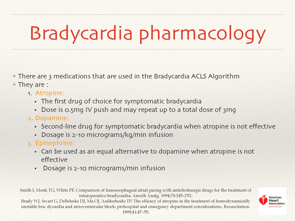
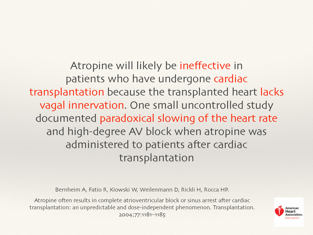
    and Emergency Cardiovascular Care. Circulation. 2010; 112(24 suppl):IV1–IV203 ❖ Smith I, Monk TG, White PF. Comparison of transesophageal atrial pacing with anticholinergic drugs for the treatment of intraoperative bradycardia. Anesth Analg. 1994;78:245–252 ❖ Brady WJ, Swart G, DeBehnke DJ, Ma OJ, Aufderheide TP. The efficacy of atropine in the treatment of hemodynamically unstable bra- dycardia and atrioventricular block: prehospital and emergency department considerations. Resuscitation. 1999;41:47–55 ❖ Swart G, Brady WJJ, DeBehnke DJ, John OM, Aufderheide TP. Acute myocardial infarction complicated by hemodynamically unstable brady- arrhythmia: prehospital and ED treatment with atropine. Am J Emerg Med. 1999;17:647– 652 ❖ Chadda KD, Lichstein E, Gupta PK, Choy R. Bradycardia-hypotension syndrome in acute myocardial infarction. Reappraisal of the overdrive effects of atropine. Am J Med. 1975;59:158–164 ❖ Chadda KD, Lichstein E, Gupta PK, Kourtesis P. Effects of atropine in patients with bradyarrhythmia complicating myocardial infarction: use- fulness of an optimum dose for overdrive. Am J Med. 1977;63:503–510 ❖ Dauchot P, Gravenstein JS. Effects of atropine on the electrocardiogram in different age groups. Clin Pharmacol Ther. 1971;12:274–280 ❖ Bernheim A, Fatio R, Kiowski W, Weilenmann D, Rickli H, Rocca HP Atropine often results in complete atrioventricular block or sinus arrest after cardiac transplantation: an unpredictable and dose-independent phenomenon. Transplantation. 2004;77:1181– 1185 ❖ Morrison LJ, Long J, Vermeulen M, Schwartz B, Sawadsky B, Frank J, Cameron B, Burgess R, Shield J, Bagley P, Mausz V, Brewer JE, Dorian P. A randomized controlled feasibility trial comparing safety and effec- tiveness of prehospital pacing versus conventional treatment: ‘PrePACE.’ Resuscitation. 2008;76:341–349. ❖ http://www.nhs.uk/conditions/heart-block/pages/introduction.aspx ❖ http://highered.mheducation.com/sites/0073520713/student_view0/chapter29/ ecg_rhythm_exercises1/av_heart_blocks/rhythm_strip_quiz_1.html