Upgrade to Pro — share decks privately, control downloads, hide ads and more …

Imaging of Chronic Obstructive Airways Disease

Howard Mann
December 27, 2016

Imaging of Chronic Obstructive Airways Disease

COPD Talk

Howard Mann

December 27, 2016
Tweet

More Decks by Howard Mann

Other Decks in Education

Transcript

  1. Imaging of Chronic Obstructive Pulmonary Disease (COPD) Howard Mann, M.D.

    Department of Radiology and Imaging Sciences University of Utah
  2. Clinical definition1 Lung disease characterized by chronic obstruction of lung

    airflow that interferes with normal breathing and is not fully reversible. 1. Global Initiative for Chronic Obstructive Lung Disease (GOLD)
  3. COPD and associated conditions Chronic Bronchitis The presence of a

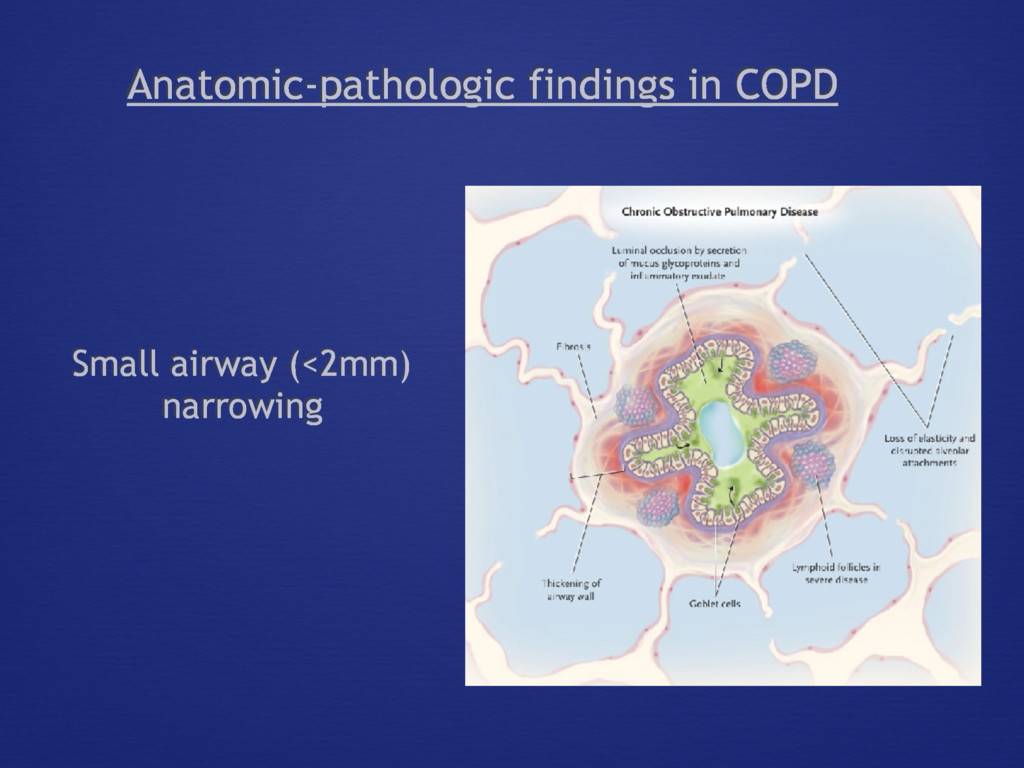
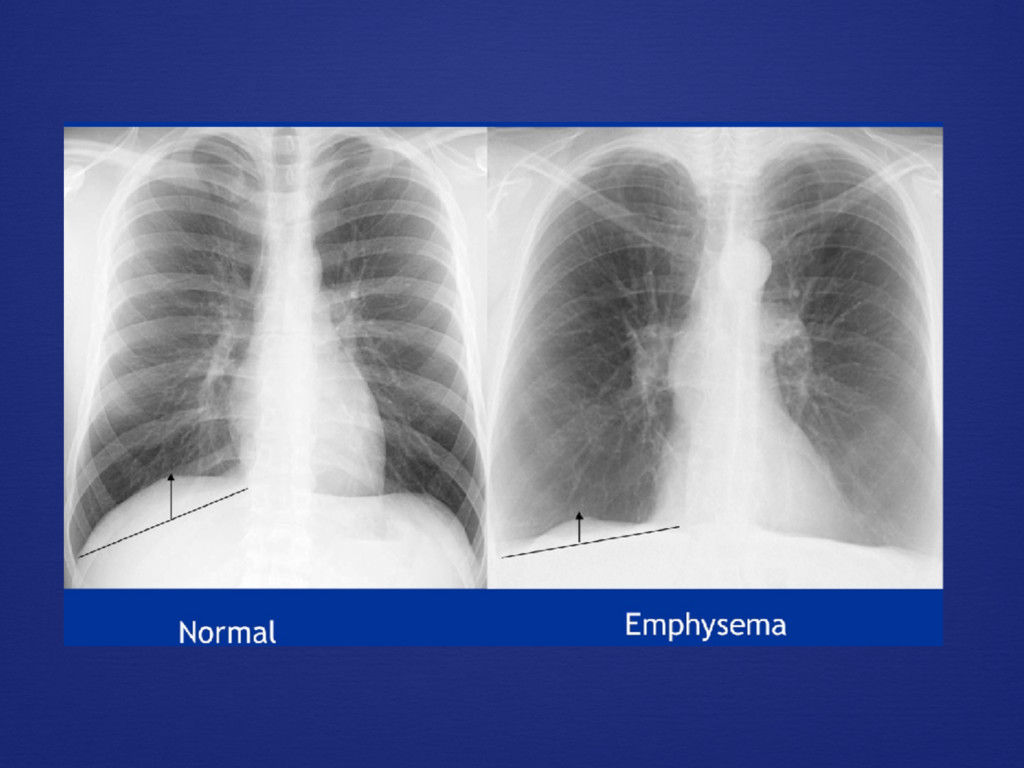
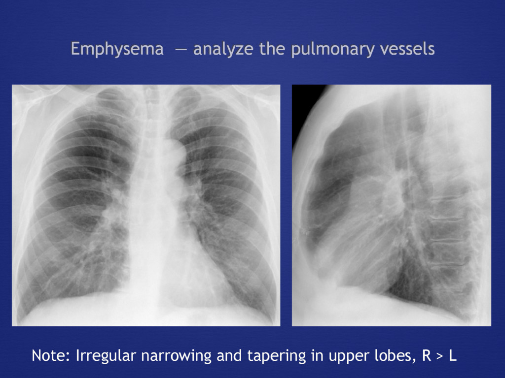
    chronic productive cough for 3 months in each of 2 successive years, provided that other causes of chronic cough have been ruled out Emphysema The destruction of alveolar walls and the permanent enlargement of the airspaces distal to the terminal bronchioles, without obvious fibrosis Asthma Reversible smooth muscle contraction that narrows the airway lumen, limiting expiratory airflow and resulting in symptoms including wheeze, cough, and exertion dyspnea
  4. Imaging in COPD Airway abnormalities • saber-sheath trachea • central

    airway mucus • bronchial outpouchings • bronchial wall thickening • bronchiectasis • [centrilobular ground-glass nodules] • airway malacia and excessive dynamic collapse Airspace abnormalities • large emphysematous bullae • centrilobular emphysema • paraseptal emphysema • panacinar emphysema Hyperinflation (increased FRC) and air-trapping
  5. Hyperinflation in COPD The effects on lung volumes correlate well

    with functional capacity and symptoms Static hyperinflation • diminished elastic recoil of emphysematous lung • significant in severe bullous emphysema • may not be a feature in airway-dominant phenotype Dynamic hyperinflation • inability to exhale all inspired air through narrowed airways
  6. Giant bullous emphysema (vanishing lung disease) • bullae occupy a

    third or more of the lung • may be a severe form of paraseptal emphysema • usually in young smokers • selected patient may be candidates for bullectomy: bullae surrounded by “normal- but-compressed” lung
  7. • Once an area of parenchymal “weakness” in the lung

    reaches a certain size, it will result in a space that will fill preferentially • elastic recoil of surrounding lung will produce retraction of lung away from it and enlarge the space • the efforts of the chest wall to inflate the adjacent lung leads to hyperinflation and symptoms • the effect of bullectomy is akin to effects of darning a sock: to restore its architecture and mechanical linkage with the chest wall, while allowing deflated lung to regain its elastic properties
  8. Lung volume reduction surgery Choosing patients who may benefit Heterogeneous

    distribution of emphysema: Emphysema in the upper lung zones, with emphysema-free lung in the lower lung zones