Upgrade to Pro — share decks privately, control downloads, hide ads and more …

Imaging of Lung Edema

Howard Mann
January 02, 2017

Imaging of Lung Edema

Howard Mann

January 02, 2017
Tweet

More Decks by Howard Mann

Other Decks in Education

Transcript

  1. Definition: An increase in lung water Mechanisms: 1. Increased fluid

    filtration from the pulmonary capillary 2. Impaired clearance of lung water normally passing into the pulmonary interstitium
  2. Mechanisms of fluid filtration from a capillary - the Starling

    equation is the reflection coefficient for movement of proteins across the capillary wall (from 0 if completely permeable to 1 if impermeable)
  3. Clearance and accumulation of filtered fluid In the lung a

    small amount of fluid normally moves from the capillary to the interstitium
  4. Lung edema forms when factors involved in the Starling Equation

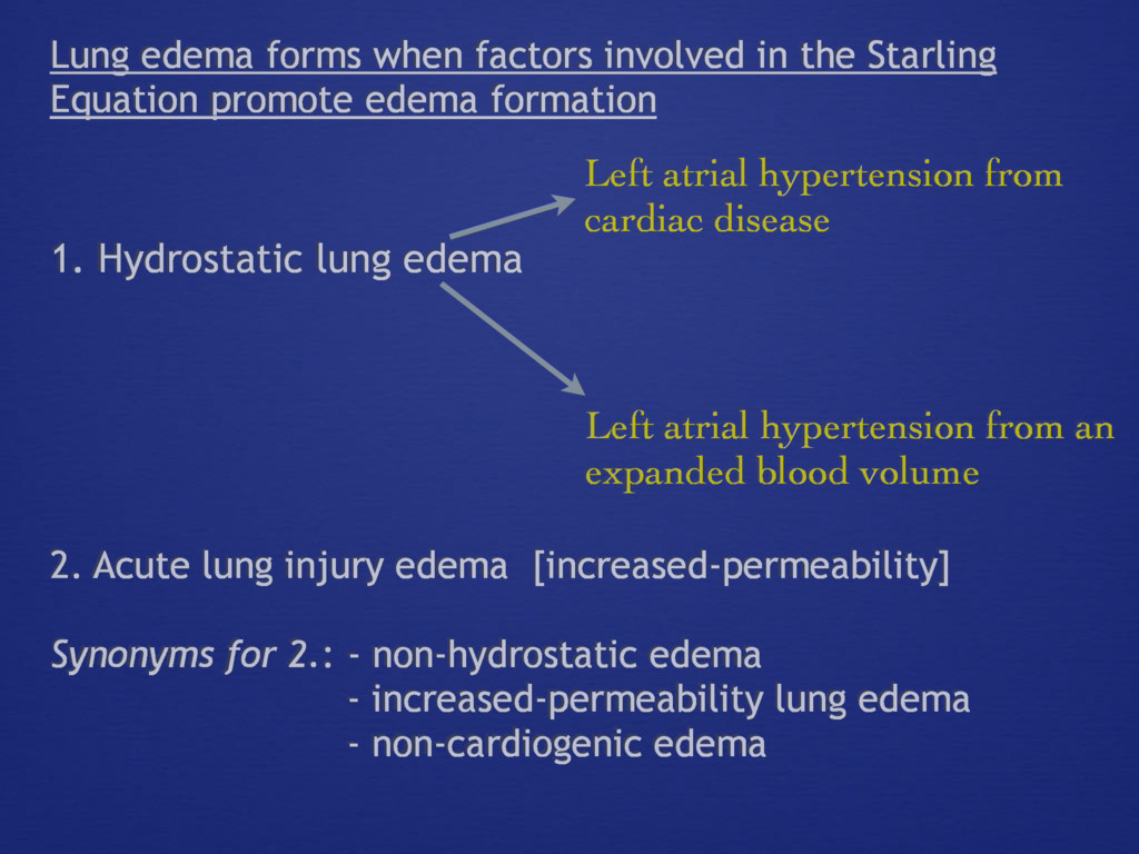
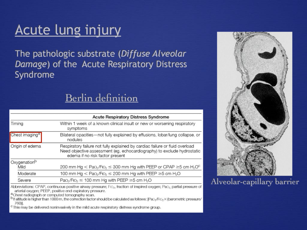
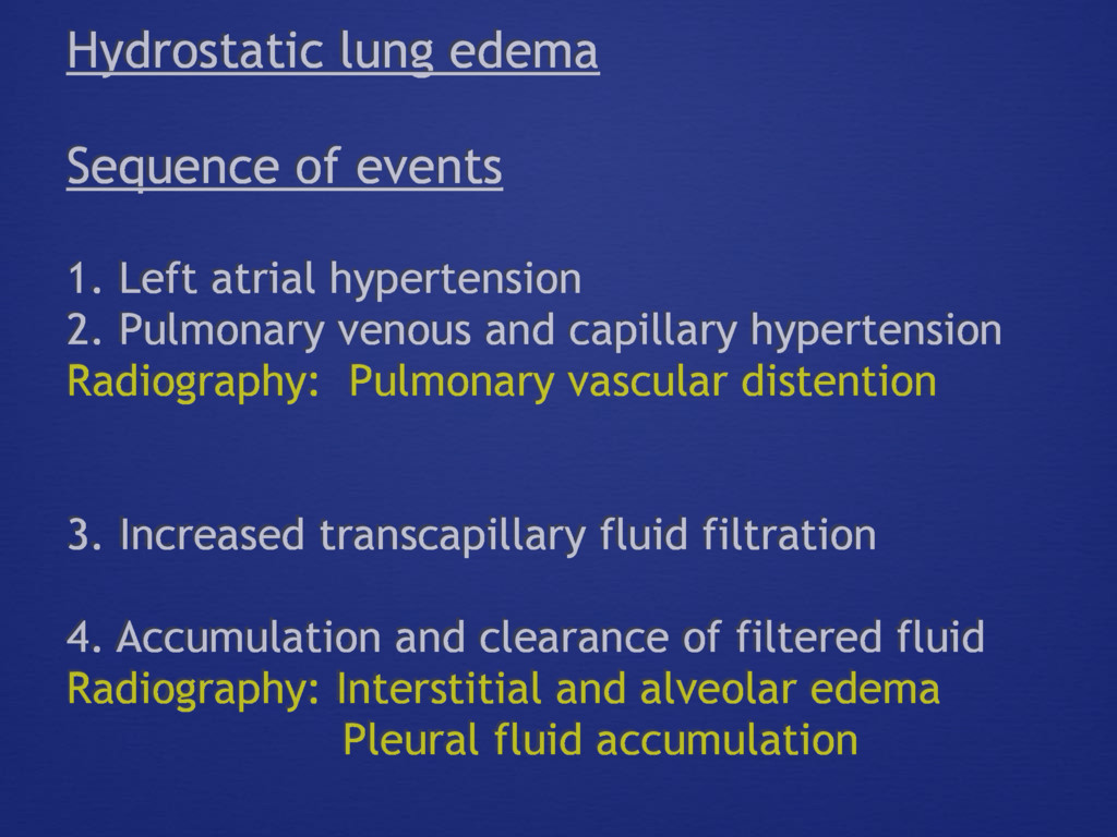
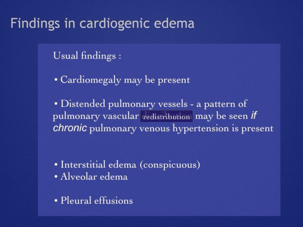
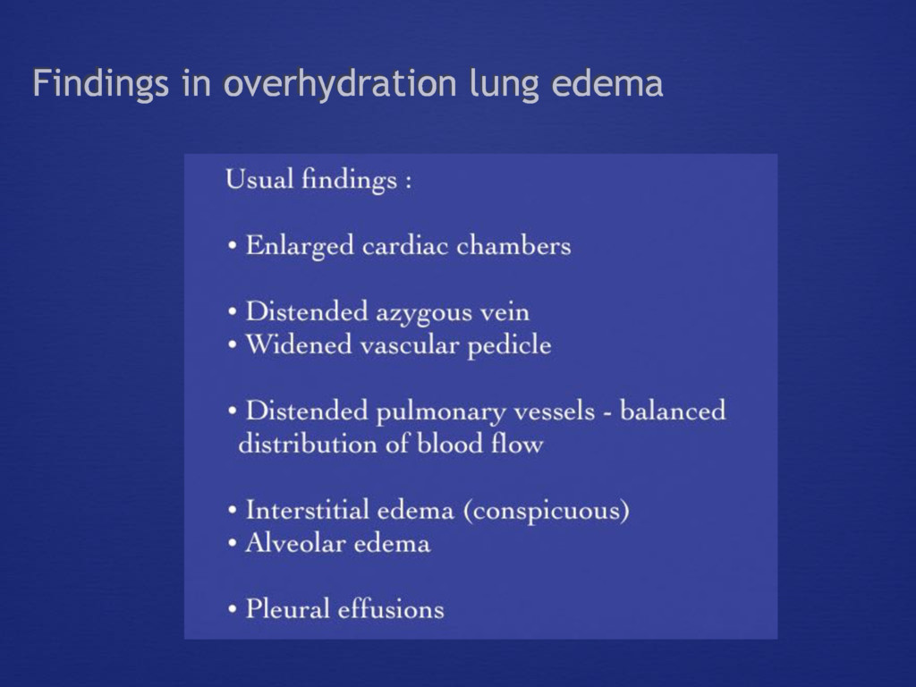
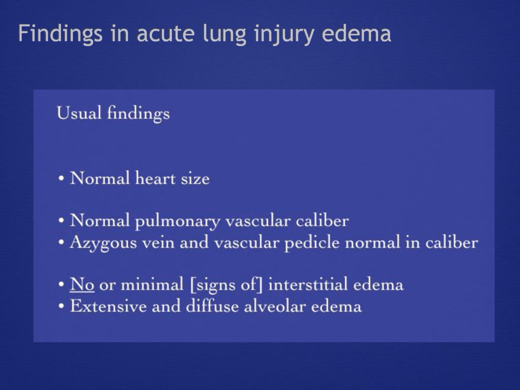
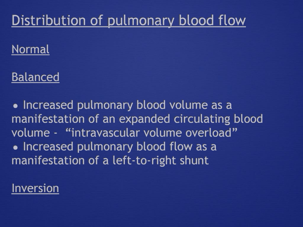
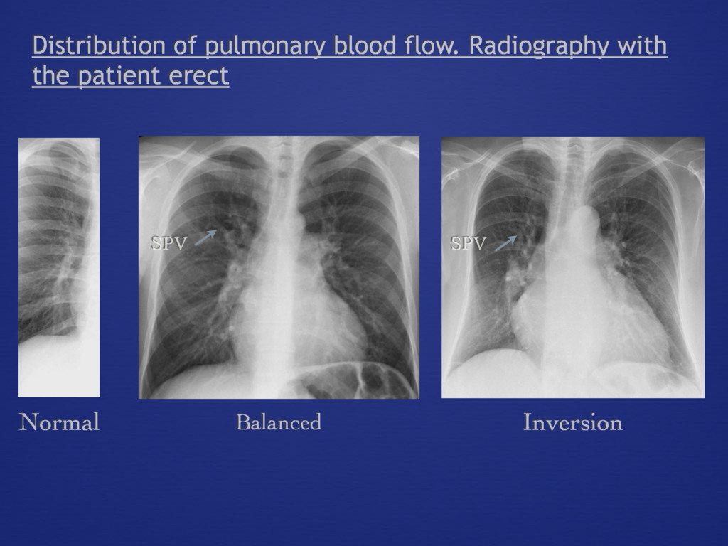
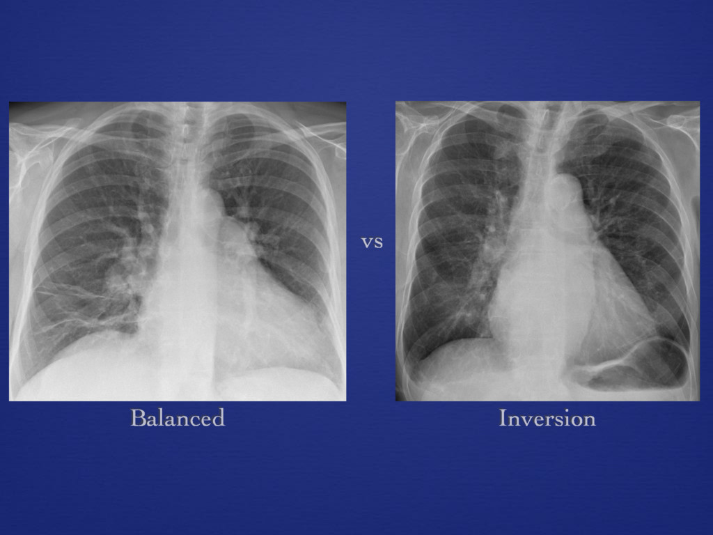
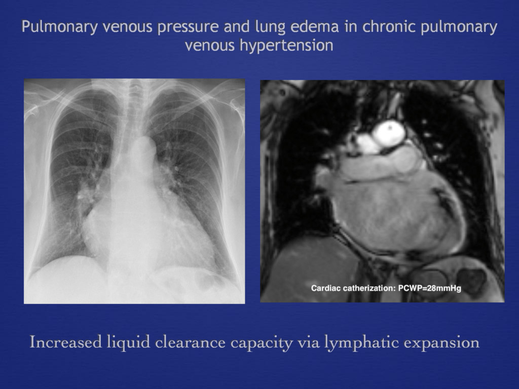
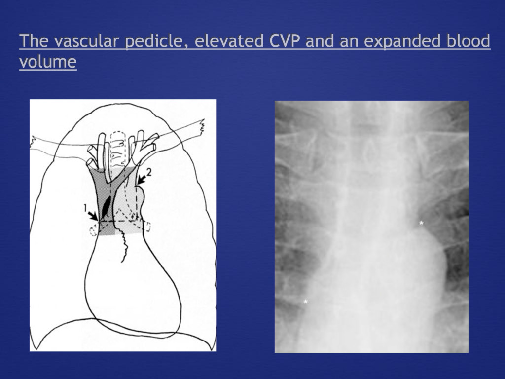
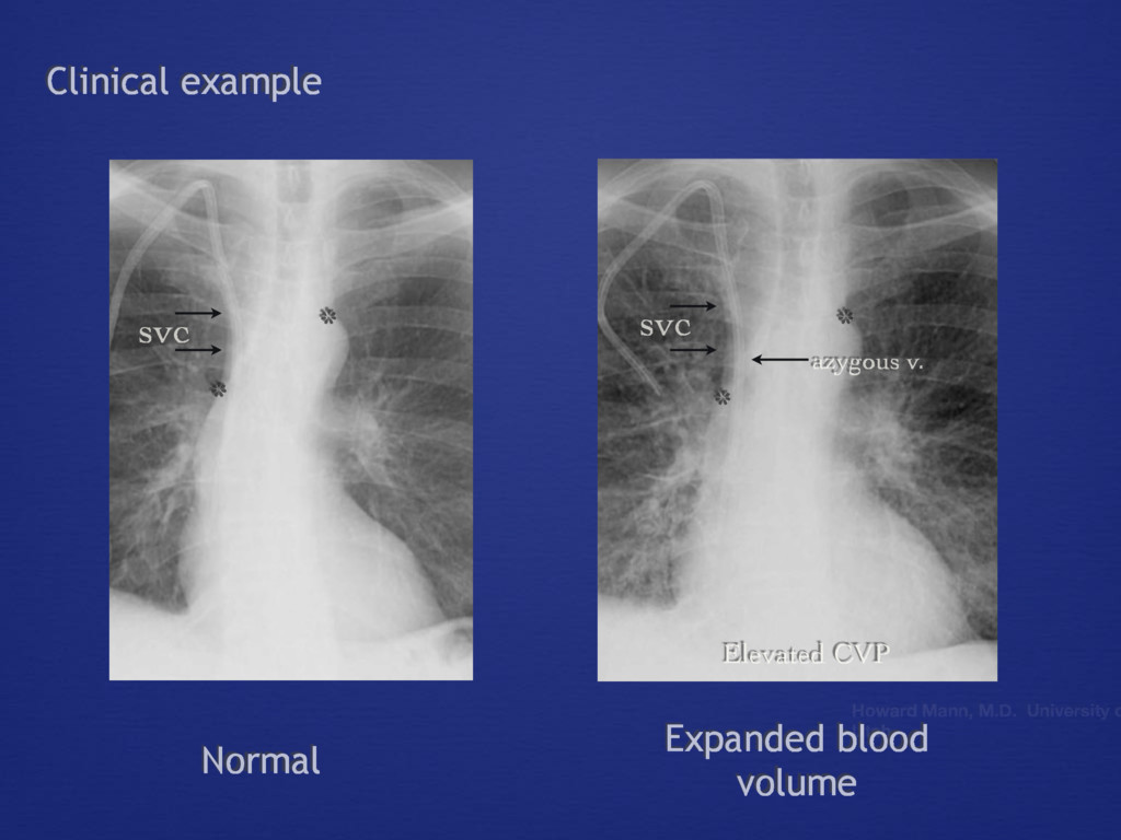
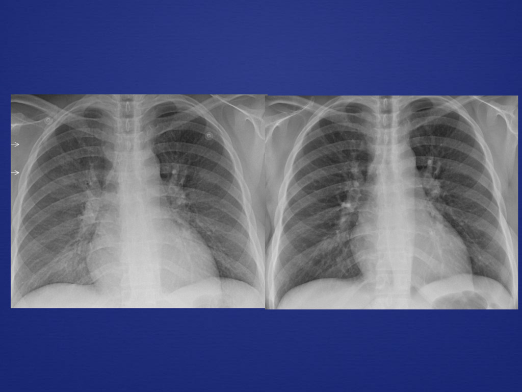
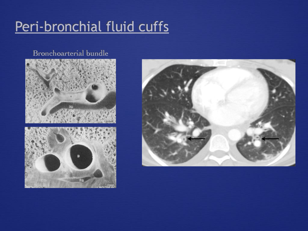
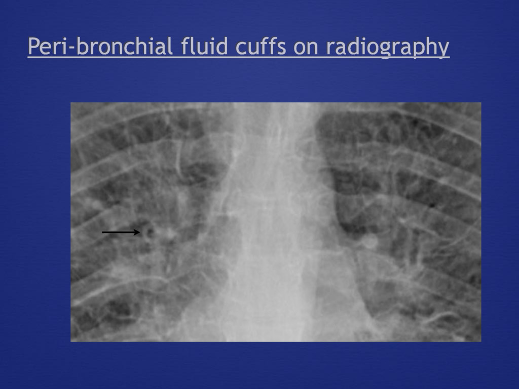
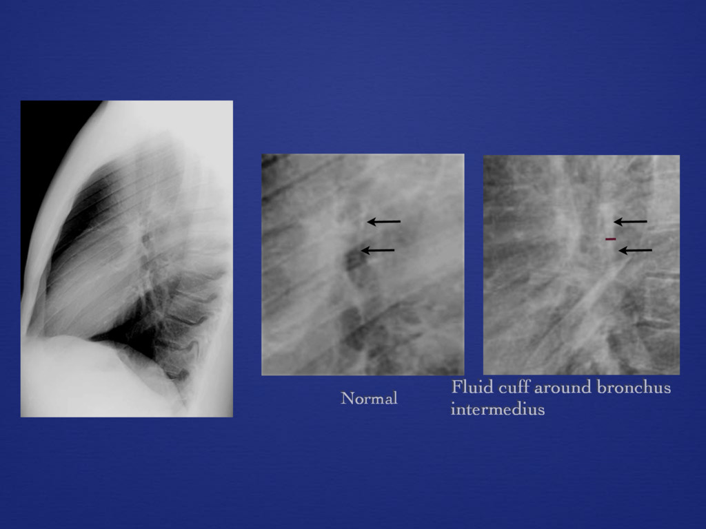
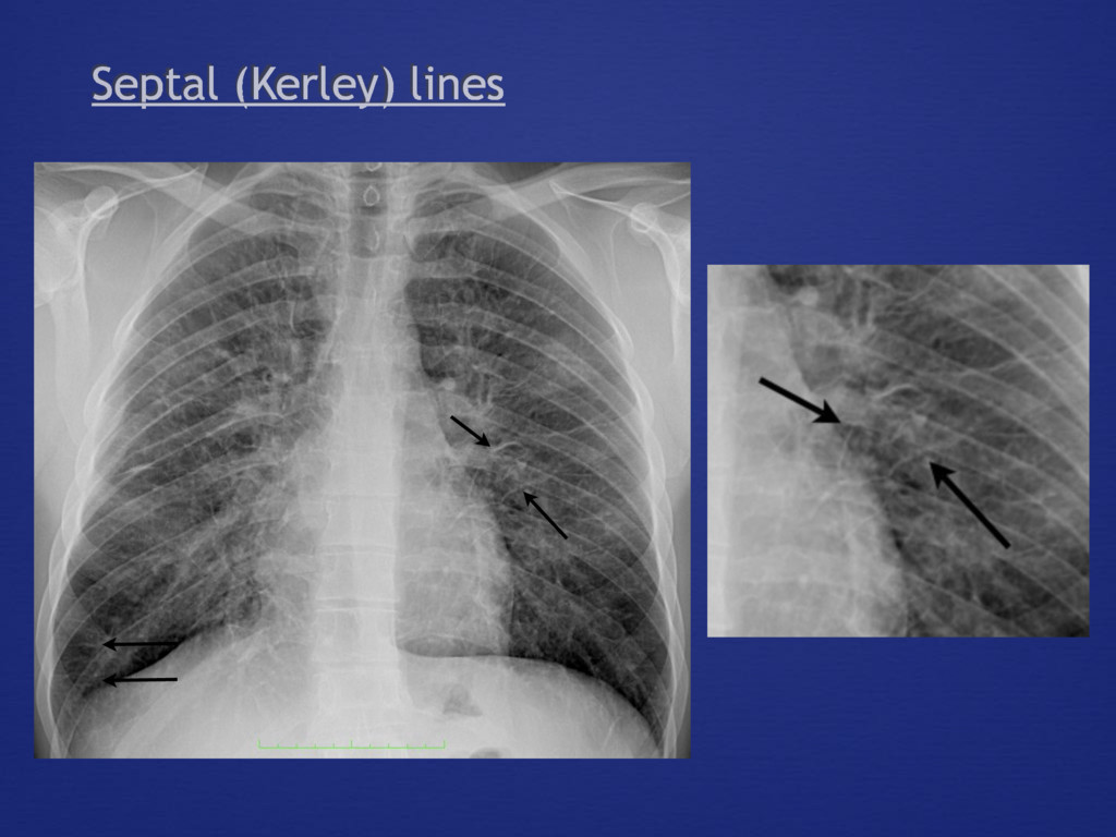
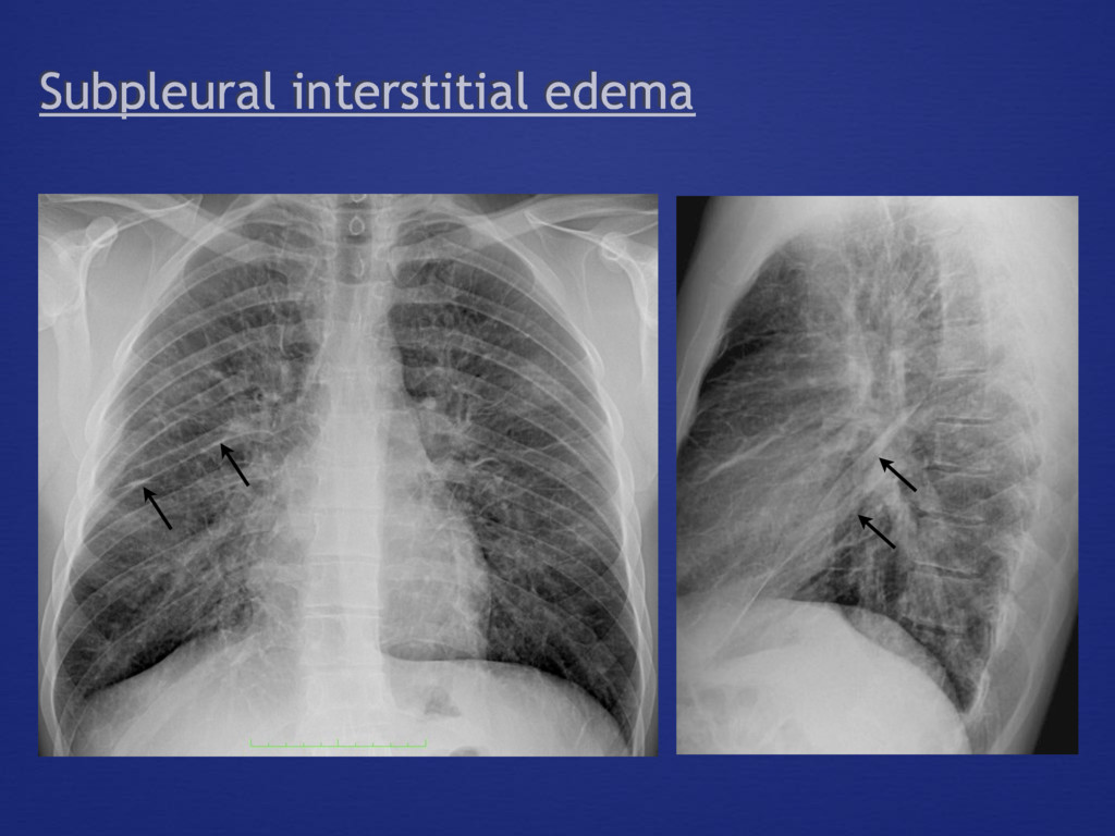
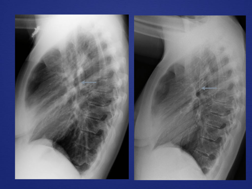
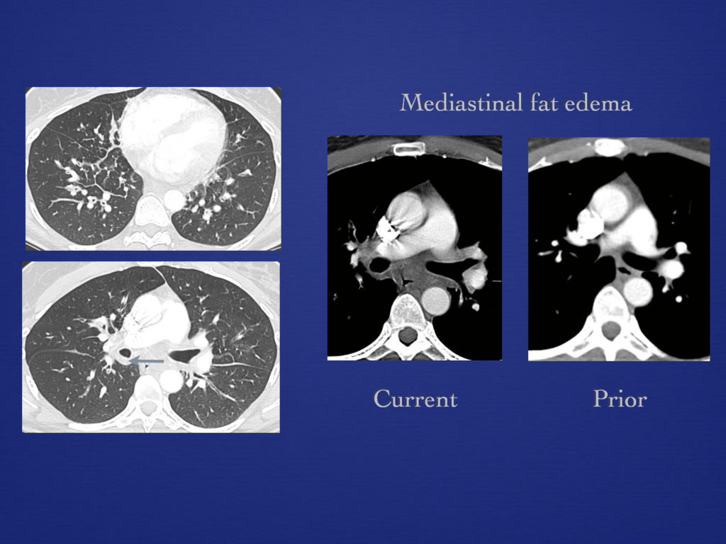
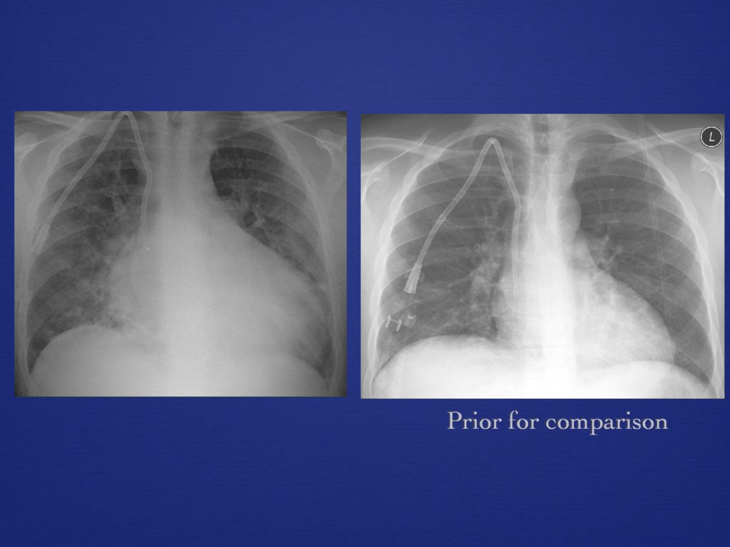
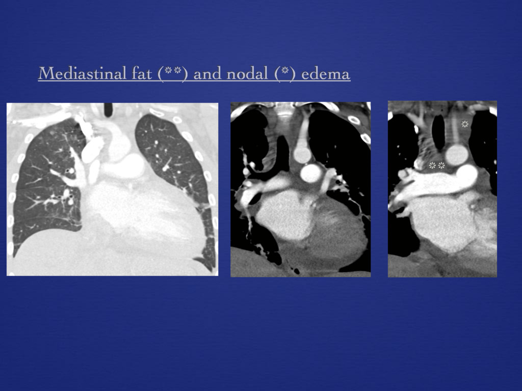
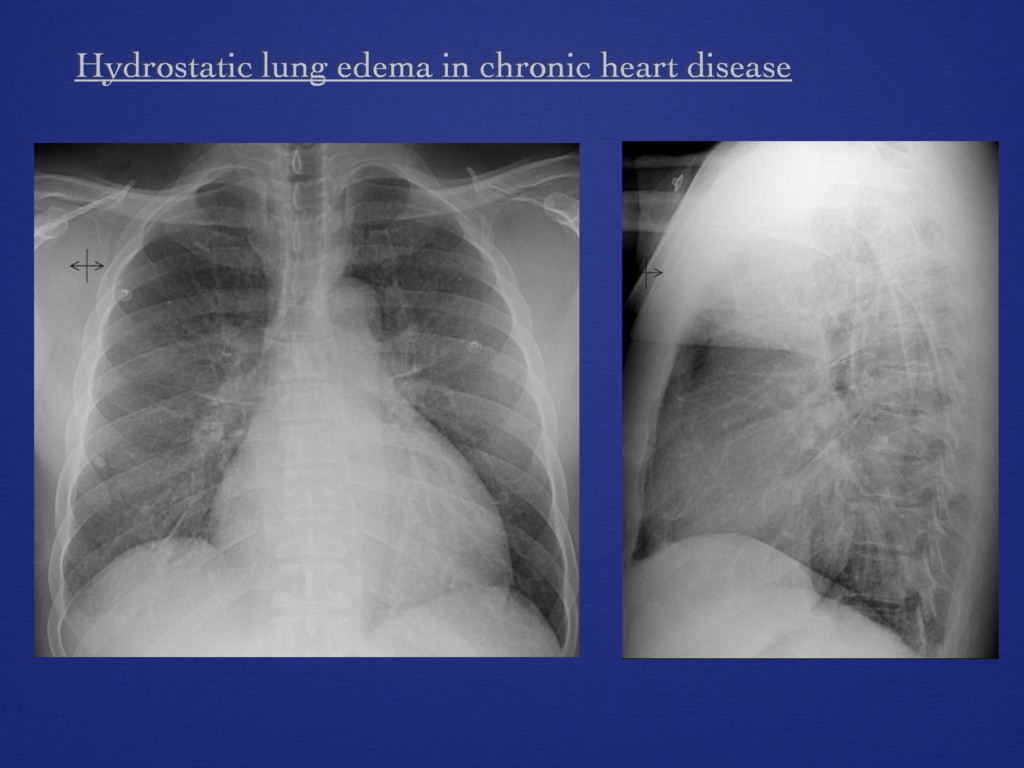
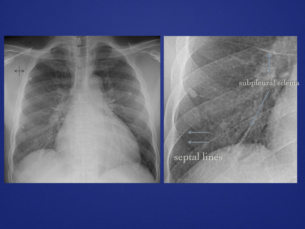
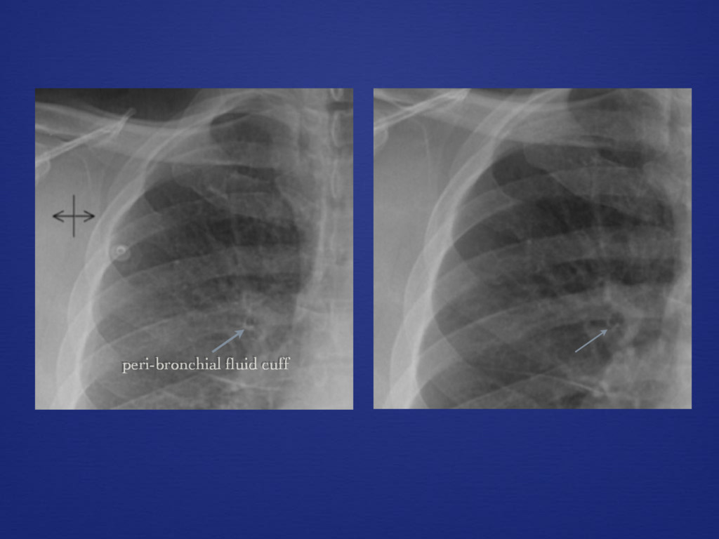
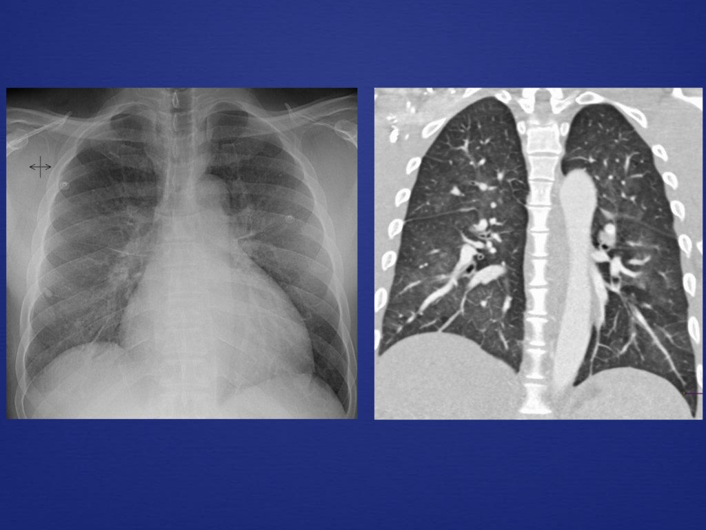
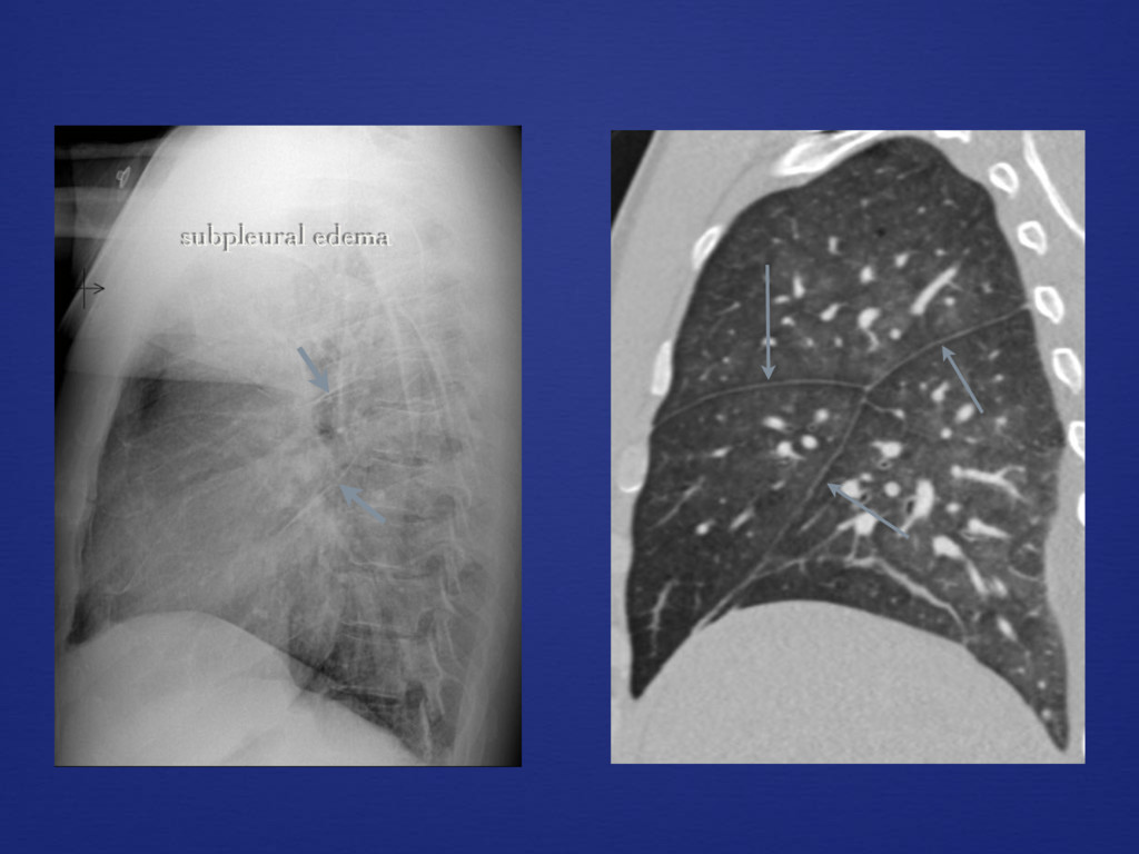
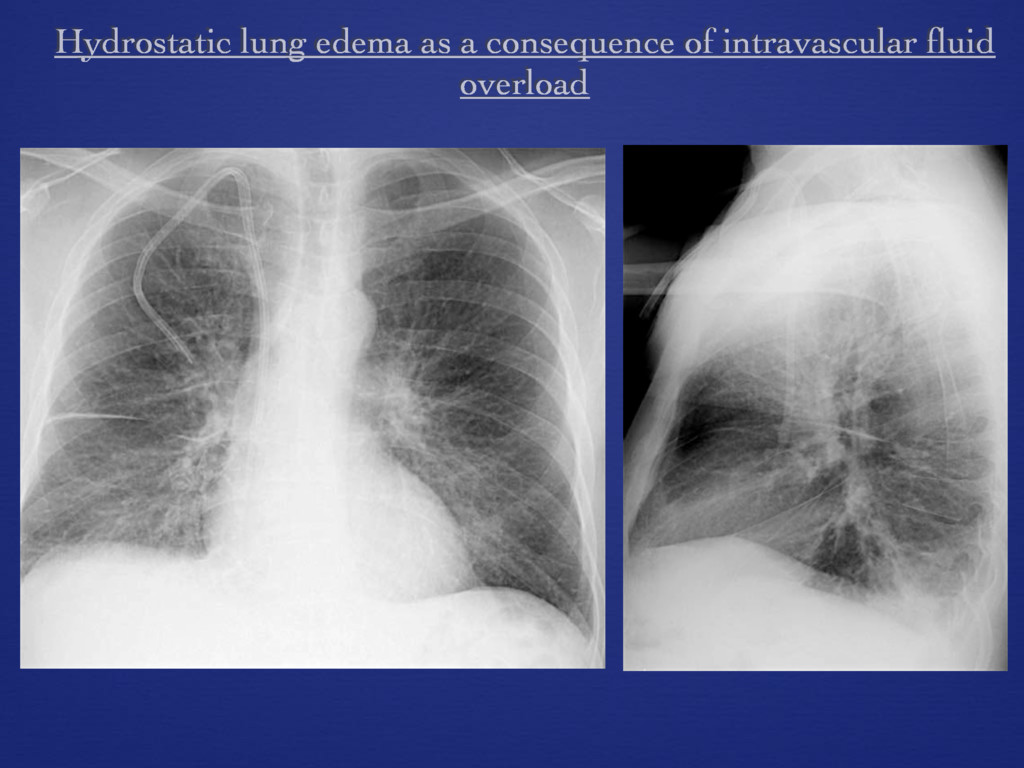
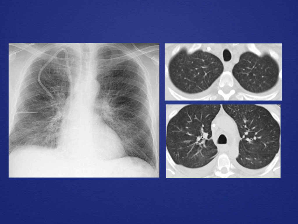
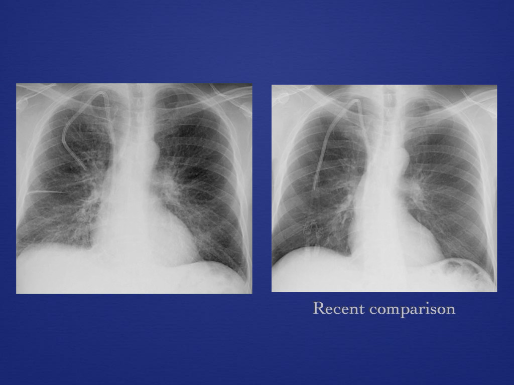
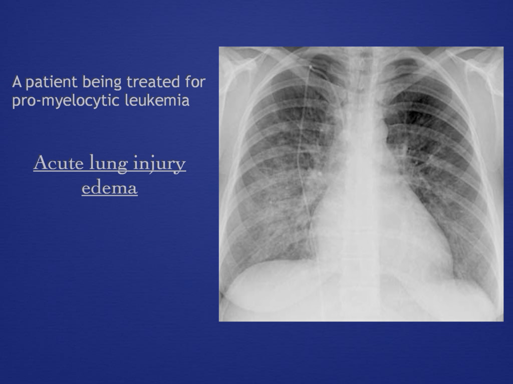
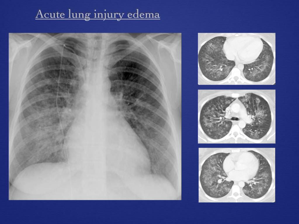
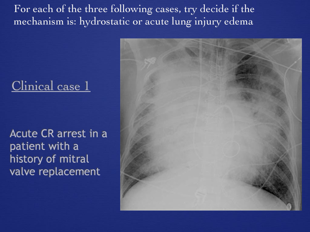
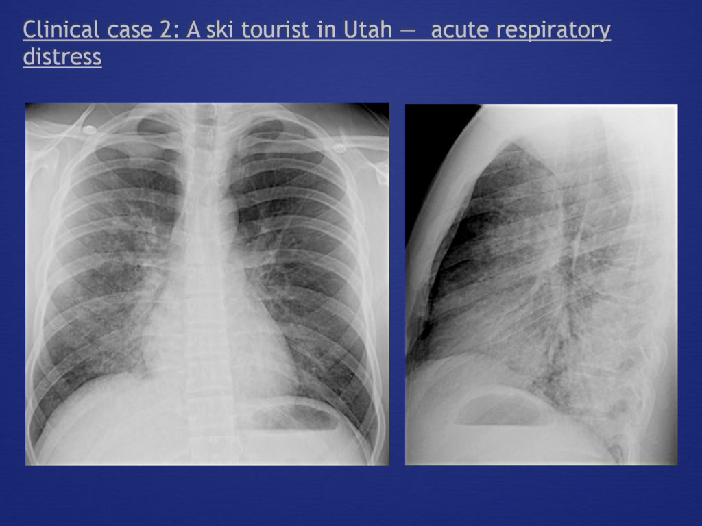
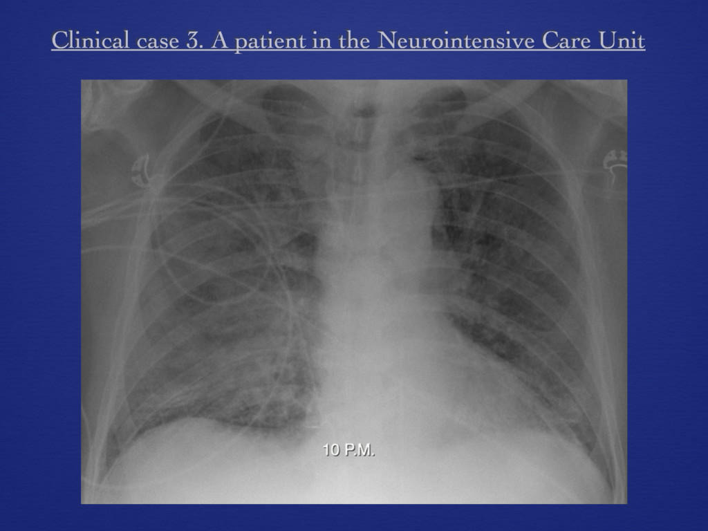
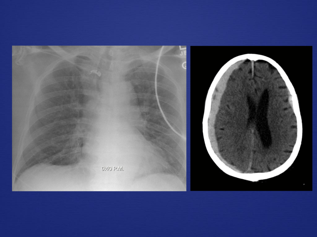
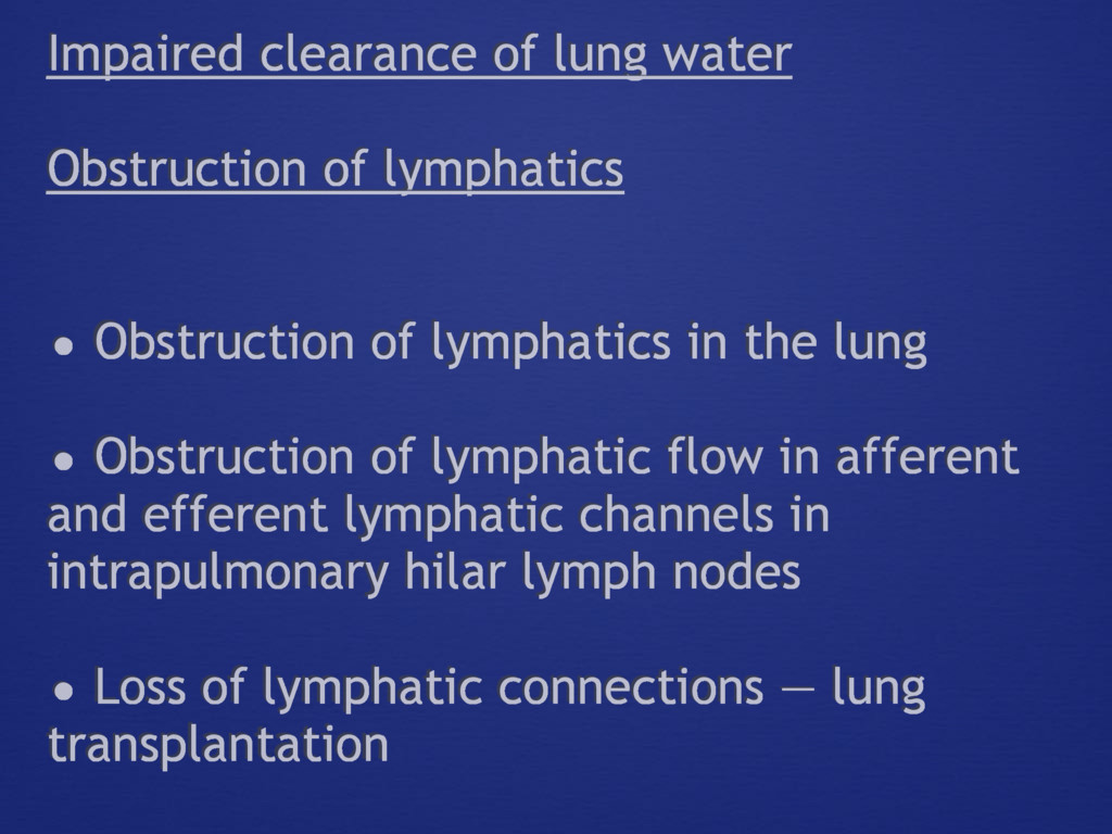
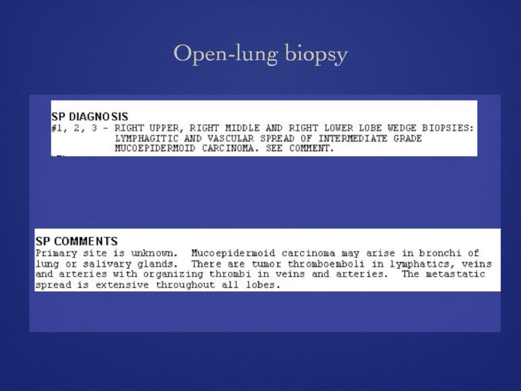
    promote edema formation 1. Hydrostatic lung edema 2. Acute lung injury edema [increased-permeability] Synonyms for 2.: - non-hydrostatic edema - increased-permeability lung edema - non-cardiogenic edema Left atrial hypertension from cardiac disease Left atrial hypertension from an expanded blood volume
  5. Acute lung injury The pathologic substrate (Diffuse Alveolar Damage) of

    the Acute Respiratory Distress Syndrome Alveolar-capillary barrier Berlin definition
  6. Hydrostatic lung edema Sequence of events 1. Left atrial hypertension

    2. Pulmonary venous and capillary hypertension Radiography: Pulmonary vascular distention 3. Increased transcapillary fluid filtration 4. Accumulation and clearance of filtered fluid Radiography: Interstitial and alveolar edema Pleural fluid accumulation
  7. Distribution of pulmonary blood flow Normal Balanced • Increased pulmonary

    blood volume as a manifestation of an expanded circulating blood volume - “intravascular volume overload” • Increased pulmonary blood flow as a manifestation of a left-to-right shunt Inversion
  8. Discerning pulmonary venous hypertension in hydrostatic lung edema Vascular distention

    and blood flow distribution Key Points: • Inversion of pulmonary blood flow is a sequel of chronic pulmonary venous hypertension: chronic and irreversible structural changes in lower lung zone vessels >> increased regional resistance to blood flow. • Inversion of blood flow is not a feature of acute pulmonary venous hypertension
  9. Pulmonary venous pressure and lung edema in chronic pulmonary venous

    hypertension Increased liquid clearance capacity via lymphatic expansion
  10. Howard Mann, M.D. University o Utah Normal Expanded blood volume

    Clinical example * * * * svc svc Elevated CVP azygous v.
  11. Clearance of lung edema fluid 1. Pulmonary lymphatics 2. Hydrostatic

    pressure gradients and water flow in the pulmonary interstitium... 3. Clearance of interstitial water into the mediastinum and pleural space
  12. Pathways for the clearance of interstitial water • along bronchovascular

    sheaths to the mediastinum • along a hydrostatic pressure gradient to the subpleural interstitial compartment... • into the pleural space Key Point: the radiographic signs of interstitial edema depict these pathways
  13. The three “classic” signs of interstitial edema: • peri-bronchial fluid

    cuffs • septal lines • sub[visceral] pleural edema (interlobar fissures)
  14. Acute CR arrest in a patient with a history of

    mitral valve replacement Clinical case 1 For each of the three following cases, try decide if the mechanism is: hydrostatic or acute lung injury edema
  15. Impaired clearance of lung water Obstruction of lymphatics • Obstruction

    of lymphatics in the lung • Obstruction of lymphatic flow in afferent and efferent lymphatic channels in intrapulmonary hilar lymph nodes • Loss of lymphatic connections — lung transplantation
  16. Dx: Pulmonary veno-occlusive disease Pulmonary artery occlusion pressure: normal Key

    points: i. Thrombosis involves small pulmonary veins and venules, and is heterogeneous in distribution ii. A “wedged” PA catheter sees a static column of blood between the tip and left atrium