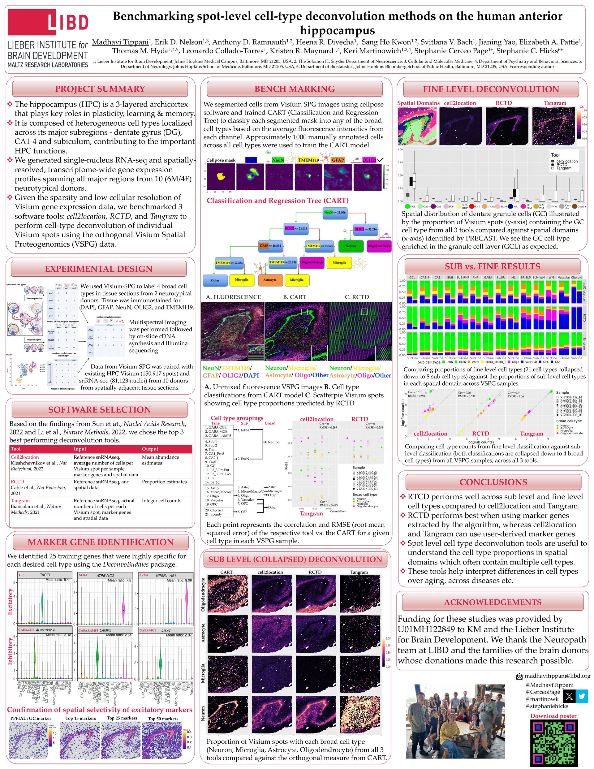
Madhavi Tippani1, Erik D. Nelson1,3, Anthony D. Ramnauth1,2, Heena R. Divecha1, Sang Ho Kwon1,2, Svitlana V. Bach1, Jianing Yao, Elizabeth A. Pattie1, Thomas M. Hyde1,4,5, Leonardo Collado-Torres1, Kristen R. Maynard1,4, Keri Martinowich1,2,4, Stephanie Cerceo Page1+, Stephanie C. Hicks6+ 1. Lieber Institute for Brain Development, Johns Hopkins Medical Campus, Baltimore, MD 21205, USA, 2. The Solomon H. Snyder Department of Neuroscience, 3. Cellular and Molecular Medicine, 4. Department of Psychiatry and Behavioral Sciences, 5. Department of Neurology, Johns Hopkins School of Medicine, Baltimore, MD 21205, USA, 6. Department of Biostatistics, Johns Hopkins Bloomberg School of Public Health, Baltimore, MD 21205, USA. +corresponding author PROJECT SUMMARY v The hippocampus (HPC) is a 3-layered archicortex that plays key roles in plasticity, learning & memory. v It is composed of heterogeneous cell types localized across its major subregions - dentate gyrus (DG), CA1-4 and subiculum, contributing to the important HPC functions. v We generated single-nucleus RNA-seq and spatially- resolved, transcriptome-wide gene expression profiles spanning all major regions from 10 (6M/4F) neurotypical donors. v Given the sparsity and low cellular resolution of Visium gene expression data, we benchmarked 3 software tools: cell2location, RCTD, and Tangram to perform cell-type deconvolution of individual Visium spots using the orthogonal Visium Spatial Proteogenomics (VSPG) data. We identified 25 training genes that were highly specific for each desired cell type using the DeconvoBuddies package. Confirmation of spatial selectivity of excitatory markers PPFIA2 : GC marker Top 15 markers Top 25 markers Top 50 markers Tool Input Output Cell2location Kleshchevnikov et al., Nat Biotechnol, 2022 Reference snRNAseq, average number of cells per Visium spot per sample, marker genes and spatial data Mean abundance estimates RCTD Cable et al., Nat Biotechno, 2021 Reference snRNAseq, and spatial data Proportion estimates Tangram Biancalani et al., Nature Methods, 2021 Reference snRNAseq, actual number of cells per each Visium spot, marker genes and spatial data Integer cell counts Based on the findings from Sun et at., Nuclei Acids Research, 2022 and Li et al., Nature Methods, 2022, we chose the top 3 best performing deconvolution tools. CART cell2location RCTD Tangram Spatial Domains cell2location RCTD Tangram EXPERIMENTAL DESIGN MARKER GENE IDENTIFICATION BENCH MARKING SUB LEVEL (COLLAPSED) DECONVOLUTION SUB vs. FINE RESULTS FINE LEVEL DECONVOLUTION Spatial distribution of dentate granule cells (GC) illustrated by the proportion of Visium spots (y-axis) containing the GC cell type from all 3 tools compared against spatial domains (x-axis) identified by PRECAST. We see the GC cell type enriched in the granule cell layer (GCL) as expected. Proportion of Visium spots with each broad cell type (Neuron, Microglia, Astrocyte, Oligodendrocyte) from all 3 tools compared against the orthogonal measure from CART. ACKNOWLEDGEMENTS We segmented cells from Visium SPG images using cellpose software and trained CART (Classification and Regression Tree) to classify each segmented mask into any of the broad cell types based on the average fluorescence intensities from each channel. Approximately 1000 manually annotated cells across all cell types were used to train the CART model. Cellpose mask DAPI NeuN TMEM119 GFAP OLIG2 SOFTWARE SELECTION Funding for these studies was provided by U01MH122849 to KM and the Lieber Institute for Brain Development. We thank the Neuropath team at LIBD and the families of the brain donors whose donations made this research possible. GABA.MGE GC SUB.1 SUB.2 GABA.LAMP5 GABA.CGE We used Visium-SPG to label 4 broad cell types in tissue sections from 2 neurotypical donors. Tissue was immunostained for DAPI, GFAP, NeuN, OLIG2, and TMEM119. Data from Visium-SPG was paired with existing HPC Visium (150,917 spots) and snRNA-seq (81,123 nuclei) from 10 donors from spatially-adjacent tissue sections. Multispectral imaging was performed followed by on-slide cDNA synthesis and Illumina sequencing A. FLUORESCENCE B. CART C. RCTD Neuron/Microglia/ Astrocyte/ Oligo/Other A. Unmixed fluorescence VSPG images B. Cell type classifications from CART model C. Scatterpie Visium spots showing cell type proportions predicted by RCTD Classification and Regression Tree (CART) Each point represents the correlation and RMSE (root mean squared error) of the respective tool vs. the CART for a given cell type in each VSPG sample. Comparing proportions of fine level cell types (21 cell types collapsed down to 8 sub cell types) against the proportions of sub level cell types in each spatial domain across VSPG samples. Comparing cell type counts from fine level classification against sub level classification (both classifications are collapsed down to 4 broad cell types) from all VSPG samples, across all 3 tools. cell2location RCTD Tangram Inhibitory Excitatory CONCLUSIONS v RTCD performs well across sub level and fine level cell types compared to cell2location and Tangram. v RCTD performs best when using marker genes extracted by the algorithm, whereas cell2location and Tangram can use user-derived marker genes. v Spot level cell type deconvolution tools are useful to understand the cell type proportions in spatial domains which often contain multiple cell types. v These tools help interpret differences in cell types over aging, across diseases etc. Neuron/Microglia/ Astrocyte/Oligo/Other 1. InhN 2. ExcN 3. Astro 4. Micro/Macro/T 5. Oligo 6. Vascular 7. OPC 8. CSF 1. GABA.CGE 2. GABA.MGE 3. GABA.LAMP5 4. Sub.1 5. Sub.2 6. Thal 7. CA1_ProS 8. CA2-4 9. Cajal 10. GC 11. L2_3.Prs.Ent 12. L2_3.PrD.PaS 13. L5 14. L6_6b 15. Astro 16. Micro/Macro/T 17. Oligo 18. Vascular 19. OPC 20. Choroid 21. Ependy Neuron Astro Microglia Oligo Other Fine Sub Broad Cell type groupings Neuron Microglia Astrocyte Oligodendrocyte Cor = 0.51 RMSE = 0.996 Cor = 0.96 RMSE = 0.337 Cor = 0.74 RMSE = 1.06 cell2location RCTD Tangram Cor = 0 RMSE = 0.293 Cor = 0 RMSE = 0.284 Cor = 0 RMSE = 0.413 Download poster
[email protected] @MadhaviTippani @CerceoPage @martinowk @stephaniehicks NeuN/TMEM119/ GFAP/ OLIG2/DAPI GCL CA2-4 CA1 SUB RHP GABA SL.SR ML WM Choroid SUB. RHP Vasc- ular SLM. WM SR. SLM GC