Are the brains of women and men with adult ADHD different?
Our research with structural and functional MRI suggests:
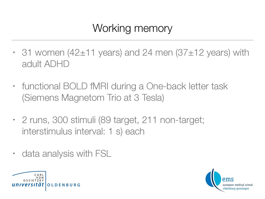
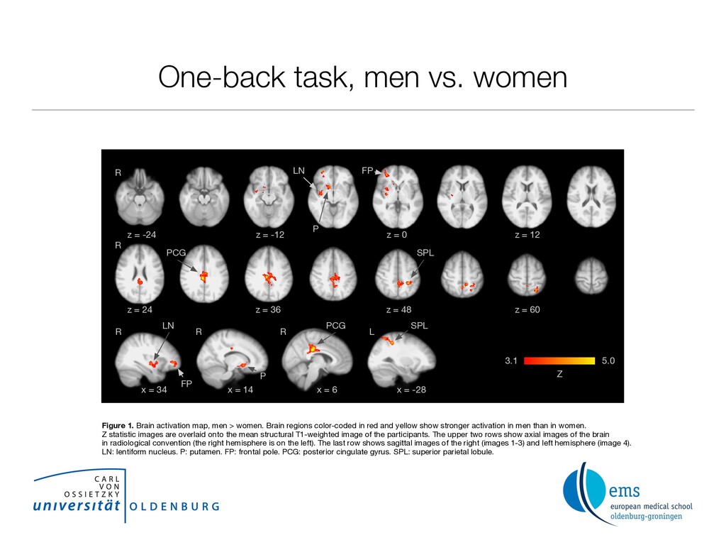
1. Men recruit larger neural resources than women to achieve similar behavioral performance during the one- back letter task.
2. Men show less deactivation of a key area of the default mode network, the posterior cingulate gyrus.
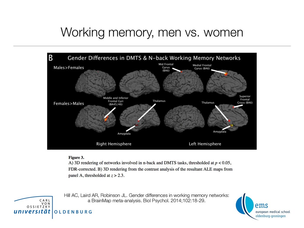
In summary, our results provide evidence that cognitive functions are represented differently in men and women with ADHD.