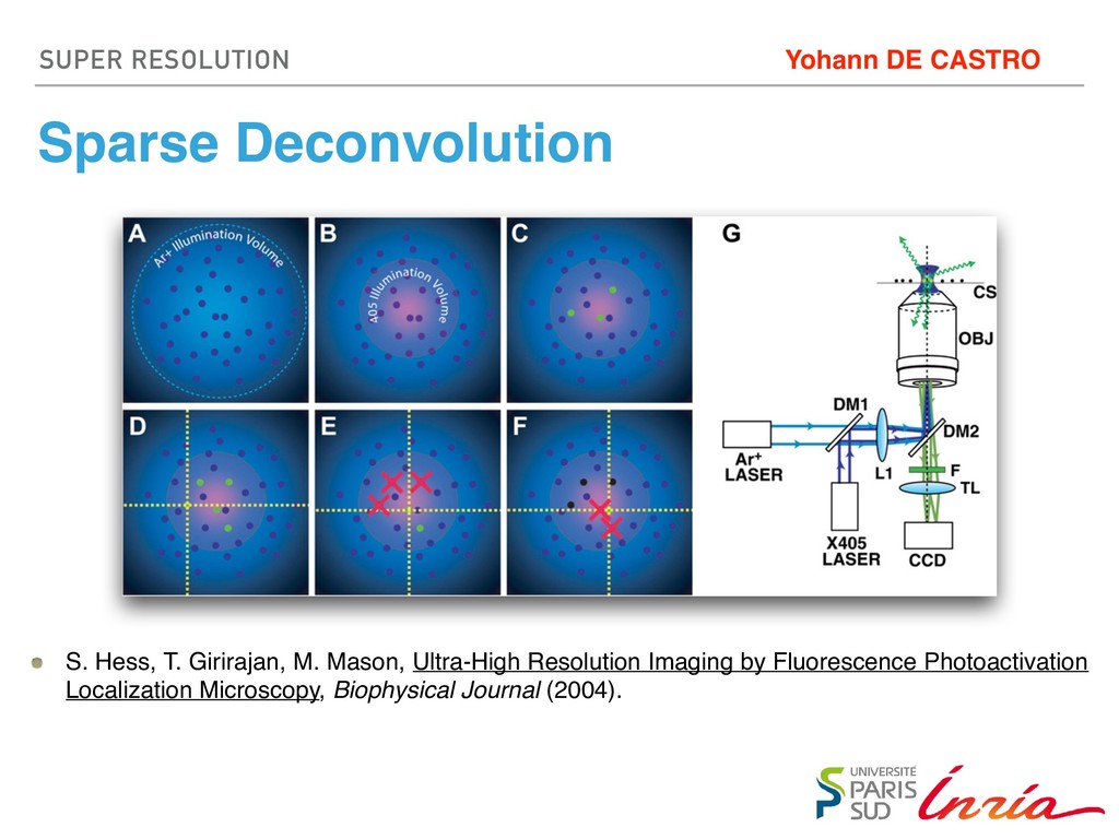
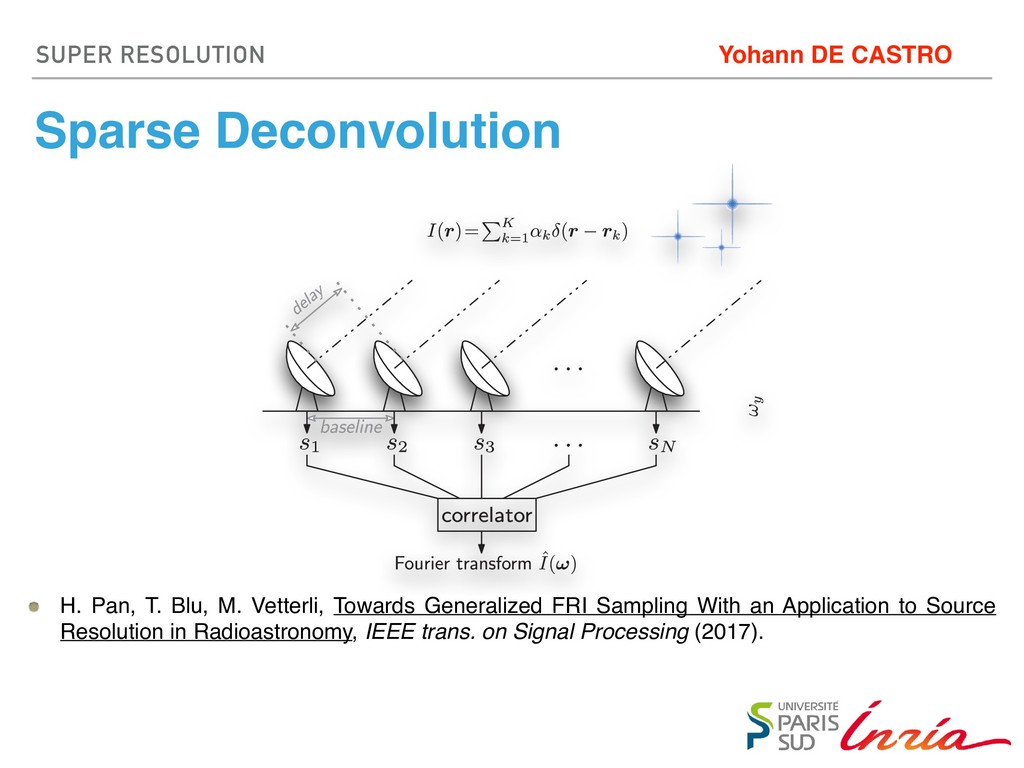
and photobleaching, even if the density of fluorophore-labeled molecules is much higher than one fluorophore per mm2. We present a novel method by which fluorescence micros- copy may be performed to obtain an image with greatly enhanced ability to resolve large numbers of fluorescent 1. The spontaneous interconversion activated (fluorescent) state mus light-controlled activation rate. 2. For irreversible photoactivatio quantum yield must be finite a FIGURE 1 localization area contain (here, PA- neously wit for readout spatial illum a second on diode laser, Within the vation beam blue circles circles) and time, the ac (red Xs) an (black circl then activated, localized, and bleached until a sufficient number of molecules have been analyzed to construct an image. (G) Th the 405-nm activation laser (X405), which is reflected by a dichroic (DM1) to make it collinear with the Ar1 readout laser. A inverted fluorescence microscope is used to focus the lasers, which are reflected upward by a second dichroic mirror (DM2 objective lens (OBJ). The sample, supported by a coverslip (CS), emits fluorescence which is collected by the objective, transm and focused by the tube lens (TL) to form an image on a camera (CCD). Biophysica S. Hess, T. Girirajan, M. Mason, Ultra-High Resolution Imaging by Fluorescence Photoactivation Localization Microscopy, Biophysical Journal (2004). SUPER RESOLUTION
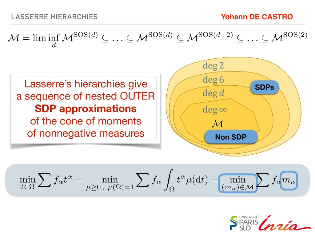
![[Cone] Moments of Nonnegative Measures [Algorithms] Lasserre’s Hierarchies, Frank-Wolfe Yohann](https://files.speakerdeck.com/presentations/c656a853593749db839afcbd1de0ee43/slide_14.jpg)
![[Cone] Moments of Nonnegative Measures [Algorithms] Lasserre’s Hierarchies, Frank-Wolfe Yohann](https://files.speakerdeck.com/presentations/c656a853593749db839afcbd1de0ee43/slide_15.jpg)