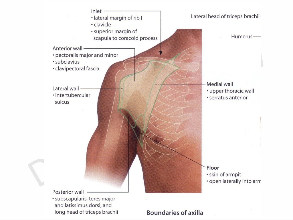
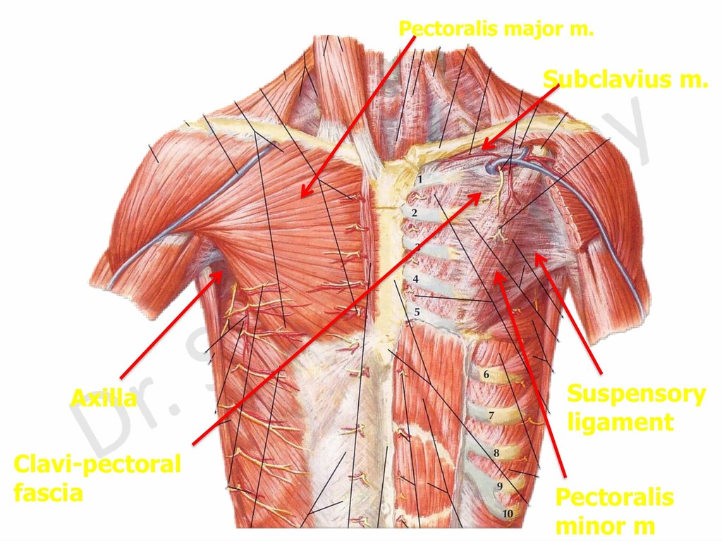
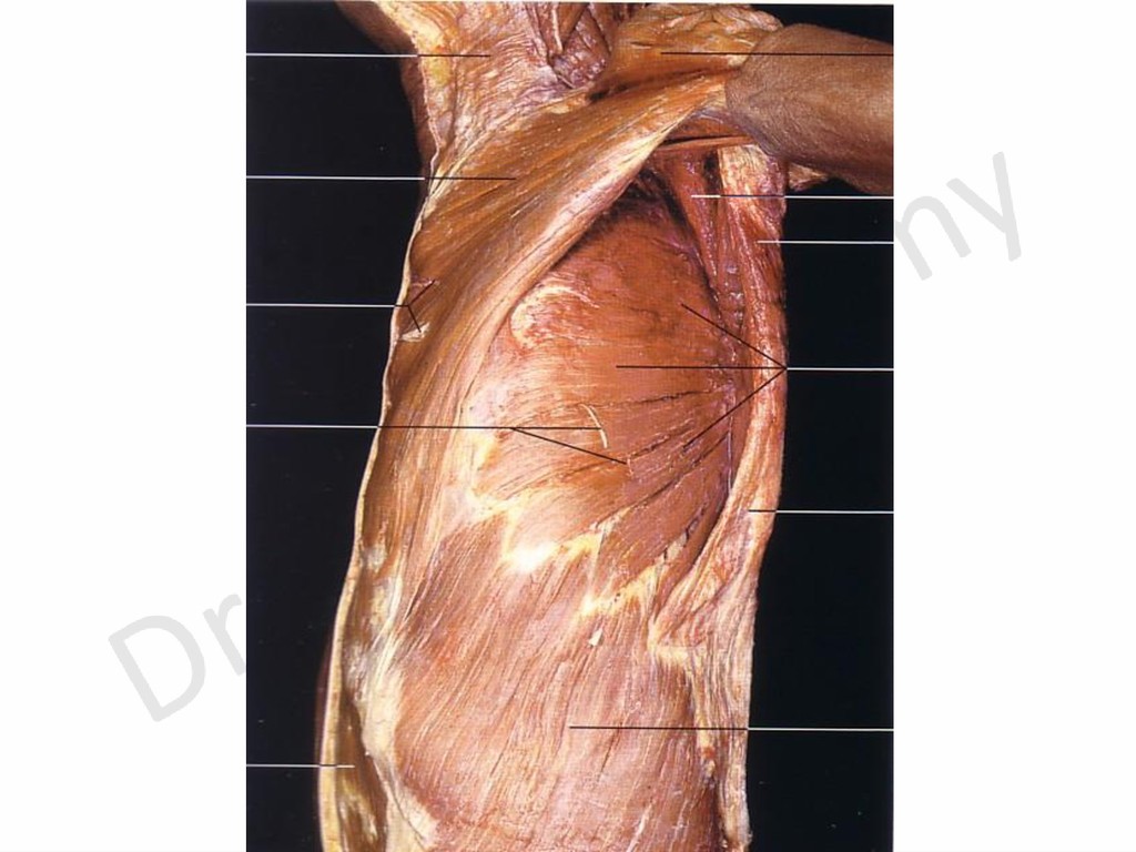
2- Superficial fascia. 3- Deep fascia which is perforated by tail of breast and gives attachment to suspensory ligament of axilla. - Direction of base: downwards
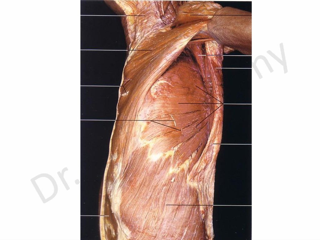
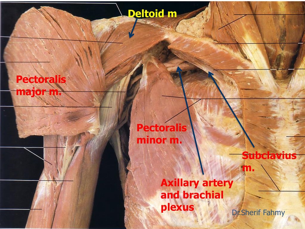
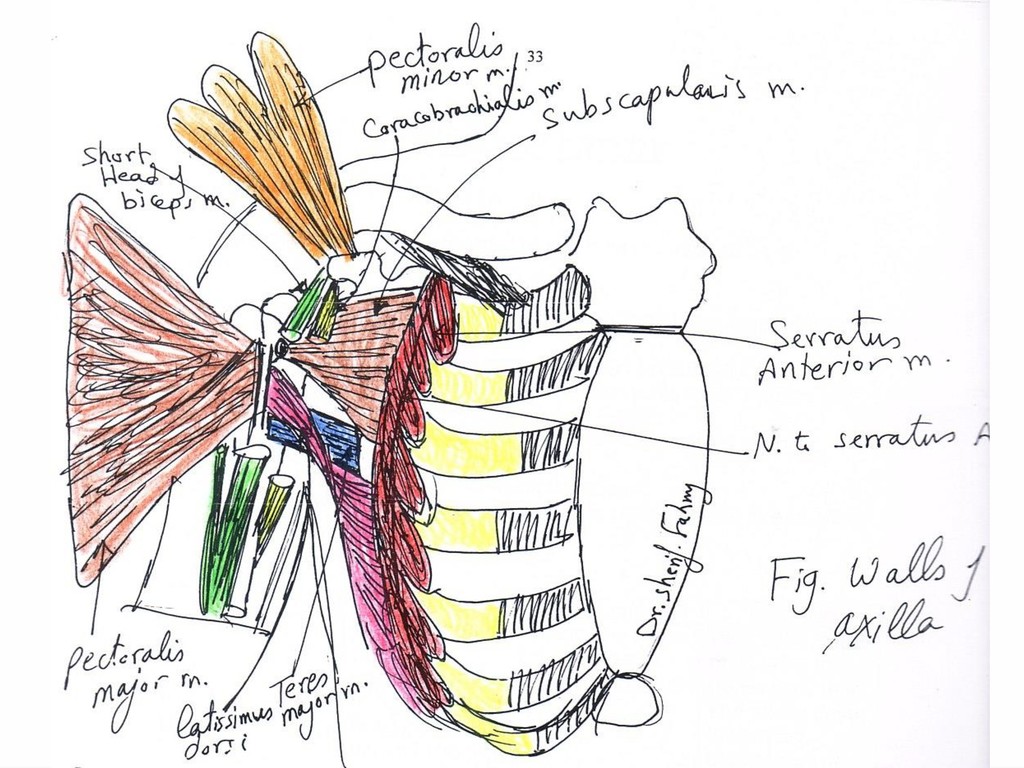
major m. - Deep: Subclavius m. Pectoralis minor m and clavi- pectoral fascia. - Its lower border is called anterior axillary fold and formed of pectoralis major m. 2- Posterior: formed of 3 muscles: - Subscapularis m., Teres major and latissimus dorsi m. - Its lower border is called posterior axillary fold and formed of folded latissimus dorsi around teres major muscle. N.B. Anterior axillary fold is higher than posterior axillary fold.
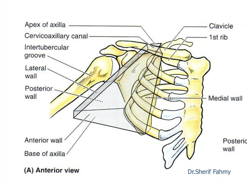
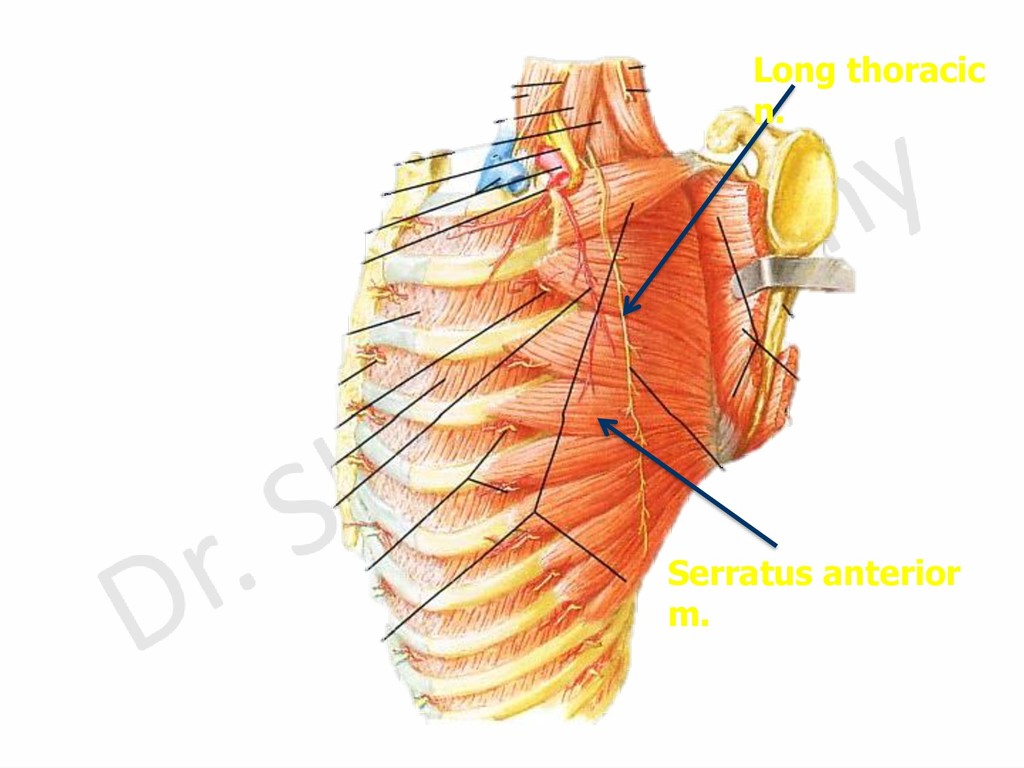
groove. It is the narrowest and most serious. 4- Medial: Formed of upper 4 ribs with upper 4 digitation of serratus anterior m. It is the largest wall.