Upgrade to Pro — share decks privately, control downloads, hide ads and more …

Cubital fossa & muscles of forearm

Cubital fossa & muscles of forearm

This presentation describes Cubital fossa & muscles of forearm of the upper limb

Dr.Sherif Fahmy

July 31, 2019
Tweet

More Decks by Dr.Sherif Fahmy

Other Decks in Science

Transcript

  1. Cubital fossa Site: Infront elbow joint and upper 1/3 of

    forearm. Shape: inverted triangular depression.
  2. Pronator teres Brachio-radialis Imaginary line between 2 epicondyles Apex is

    the overlap of brachio- radialis m. over pronator teres m. Lateral Medial
  3. Medial Lateral Basilic vein &medial cutaneous n of forearm Cephalic

    vein and lateral cutaneous n. of forearm Median cubital vein Bicipital aponeurosis Epitrochlear lymph nodes
  4. Tendon of biceps brachii m. Radial nerve Superficial radial nerve

    Deep radial nerve (posterior interosseous nerve) Brachial art. Median nerve Ulnar art. Radial art. Brachio-radialis (Reflected) Medial Lateral
  5. Muscles of the Front of the Forearm (8 muscles-Superficial &

    Deep) ➢2 pronator (teres & quadratus). ➢2 flexor digitorum (superficialis & profundus). ➢2 flexor carpi (radialis & ulnaris). ➢1 flexor pollicis longus. ➢1 Palmaris longus.
  6. Arrangement of Muscles of the front of Forearm Superficial Group

    A- Superficial layer: 1) Pronator teres. 2) Flexor carpi radialis. 3) Palmaris longus. 4) Flexor carpi ulnaris. B- Dee layer: 1) Flexor digitorum superficialis. Deep Group A- Superficial layer: 1) Flexor pollicis longus. 2) Flexor digitorum profundus. B- Deep layer: 1) Pronator quadratus
  7. Medial supracondylar ridge (pronator teres) Front of medial epicondyle (Common

    flexor origin) Medial margin of coronoid process (Flexor digitorum superficialis & pronator teres) Common flexor origin Elbow joint Superior radio-ulnar joint
  8. Anterior oblique line of radius Coronoid process Olecranon process Superior

    radio-ulnar joint Anterior surface of shaft of radius Anterior surface of ulna Interosseous membrane
  9. Tubercle of scaphoid Crest of trapezium Groove of trapezium Pisiform

    bone Hook of hamate metacarpal bones Flexor retinaculum Phalanges
  10. Wrist joint Carpo-metacarpal joint of thumb Metacarpo-phalangeal joint of thumb

    Interphalangeal joint of thumb Proximal interphalangeal joints Distal interphalangeal joint
  11. Common rules of superficial flexors ➢Muscles are named after their

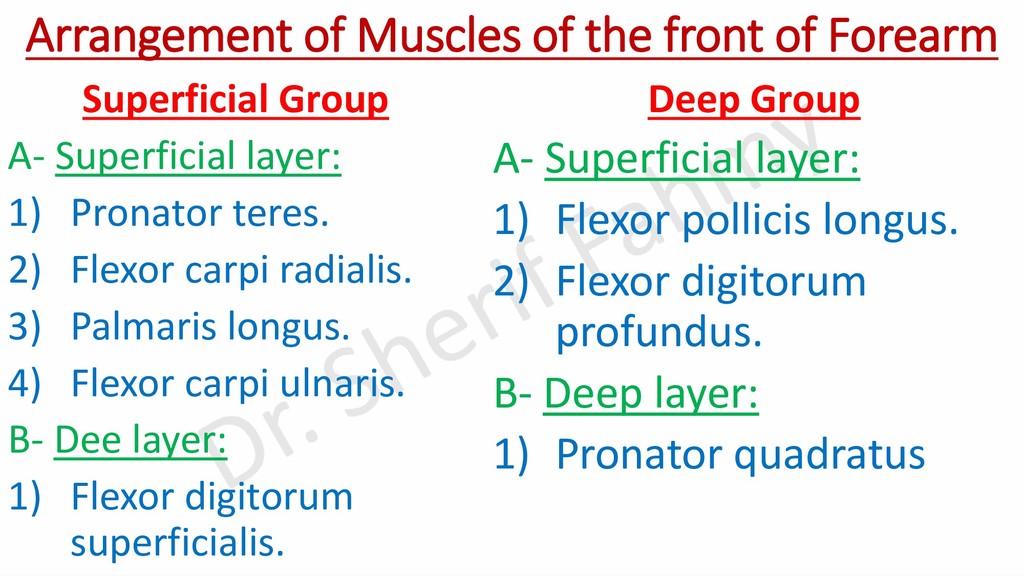
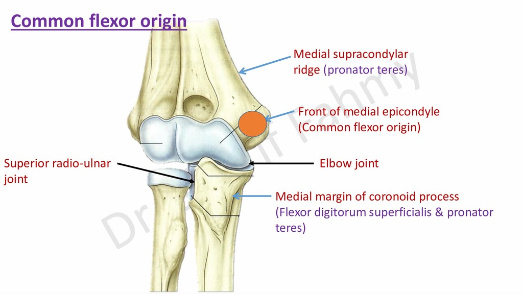
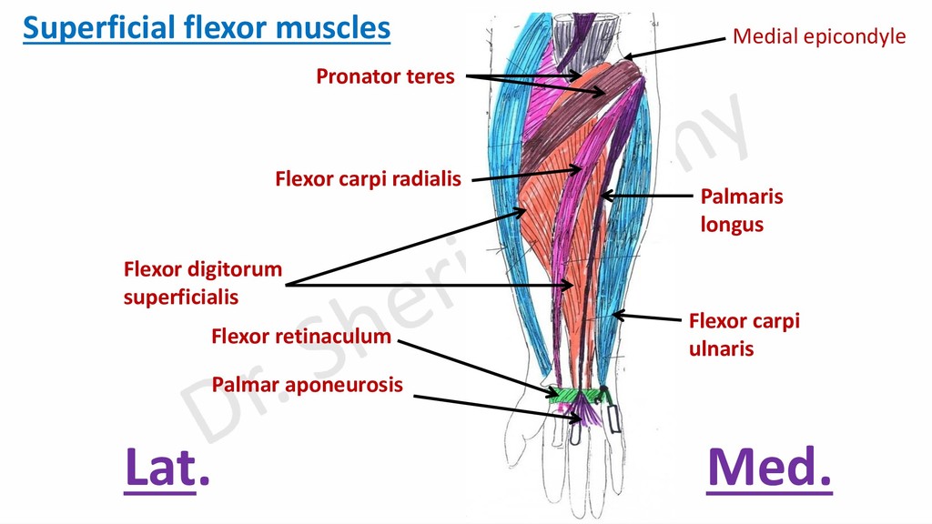
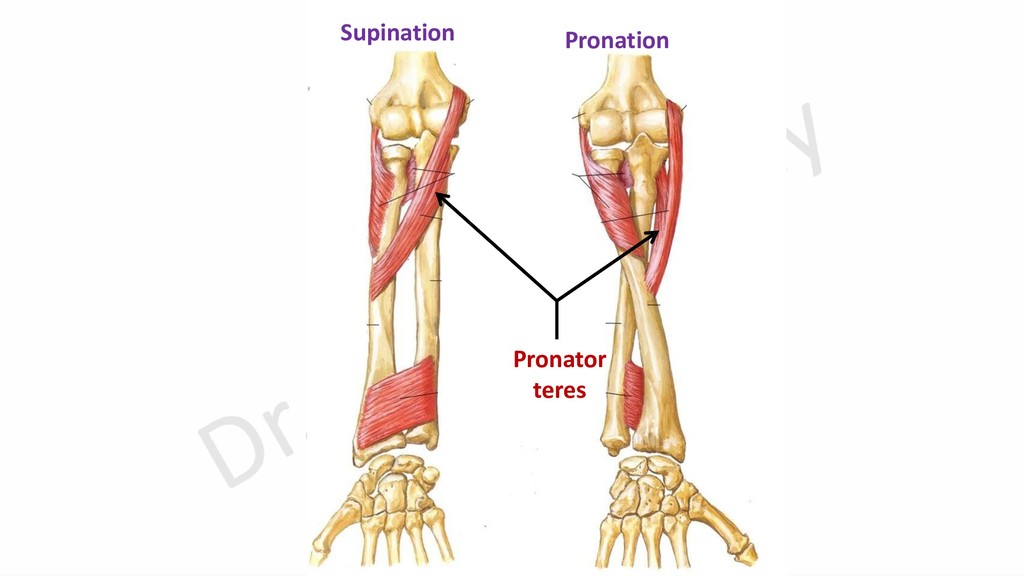
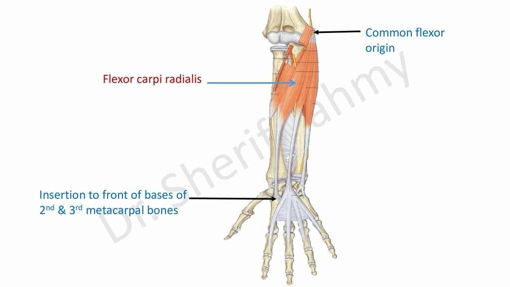
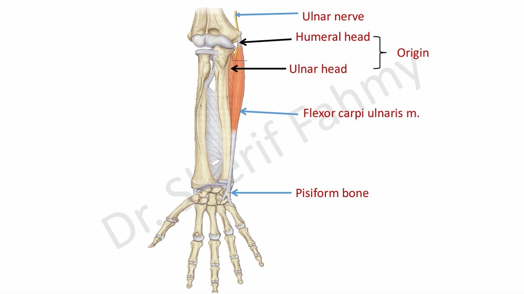
    action. ➢All muscles arises partially or totally from common flexor origin (front of medial epicondyle). ➢All muscles have 2 heads except flexor carpi radialis and palmaris longus. ➢Each 2 headed muscle has a nerve that passes and supplies the muscle. Nerve supply: ➢Median nerve except flexor carpi ulnaris is supplied by ulnar nerve. Common action: ➢All are week flexors of elbow. ➢All are flexors of wrist except pronator teres.
  12. Pronator teres Flexor carpi radialis Palmaris longus Flexor carpi ulnaris

    Flexor digitorum superficialis Flexor retinaculum Palmar aponeurosis Superficial flexor muscles Med. Lat. Medial epicondyle
  13. Relations of 8 flexor tendons to flexor retinaculum: ➢Tendons of

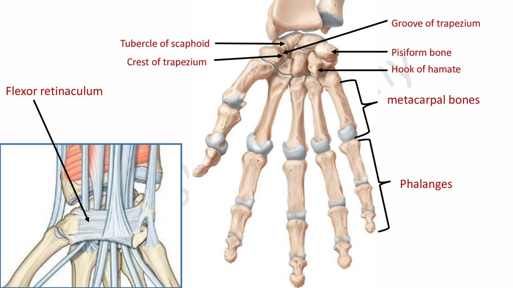
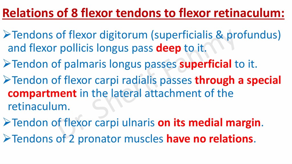
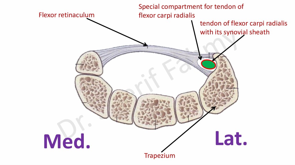
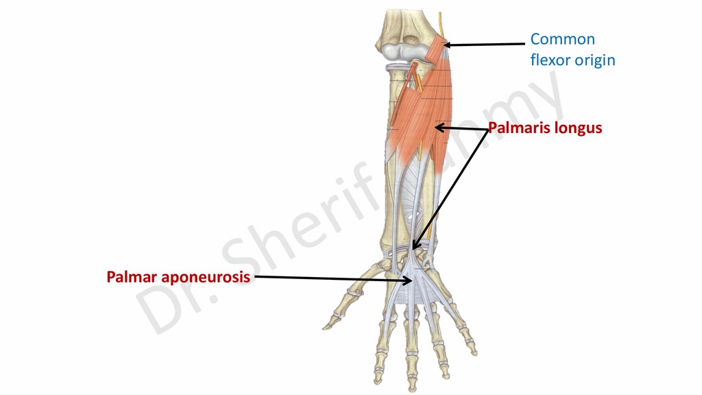
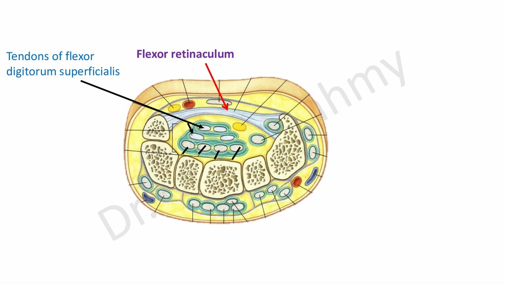
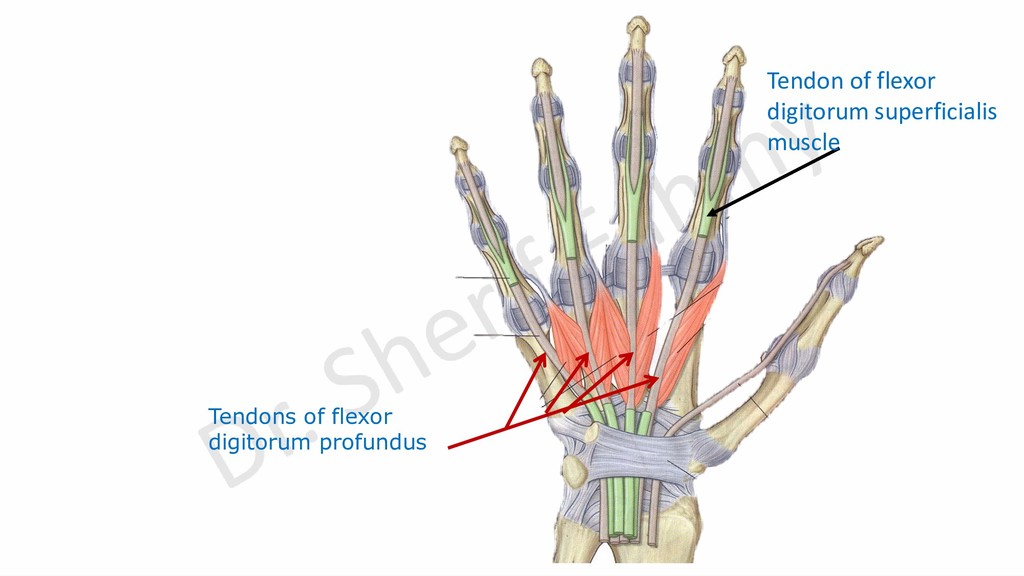
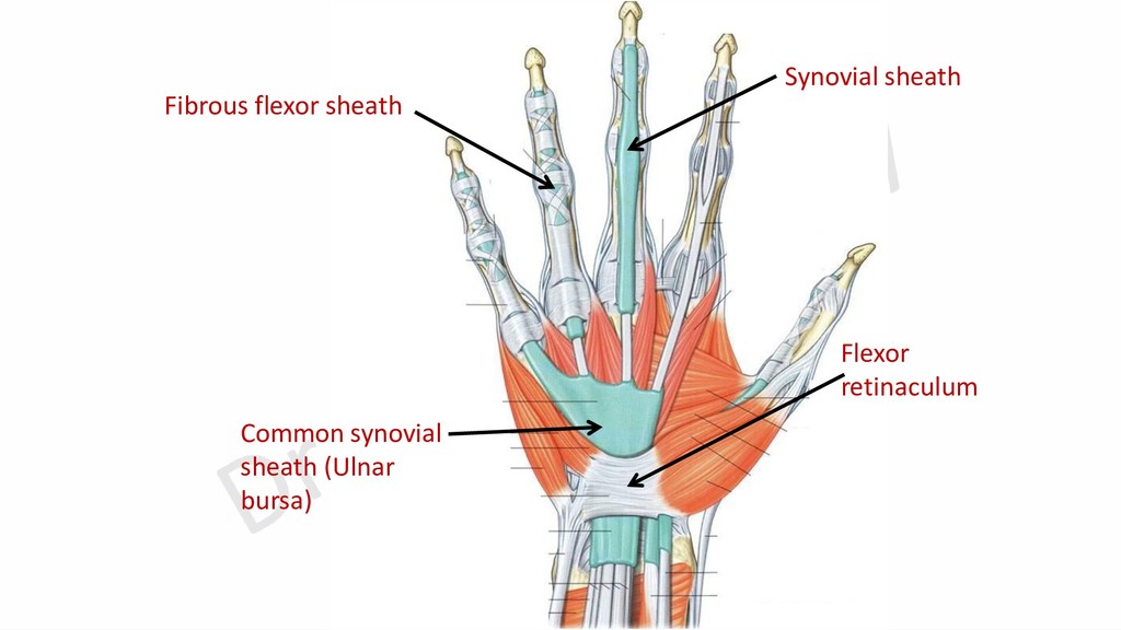
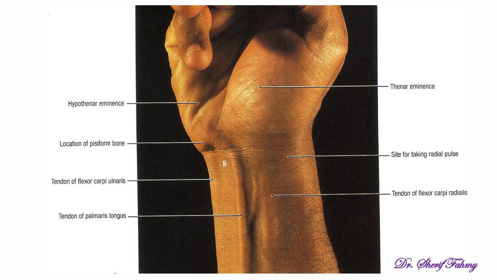
    flexor digitorum (superficialis & profundus) and flexor pollicis longus pass deep to it. ➢Tendon of palmaris longus passes superficial to it. ➢Tendon of flexor carpi radialis passes through a special compartment in the lateral attachment of the retinaculum. ➢Tendon of flexor carpi ulnaris on its medial margin. ➢Tendons of 2 pronator muscles have no relations.
  14. Superficial head Deep head Insertion of pronator teres Median nerve

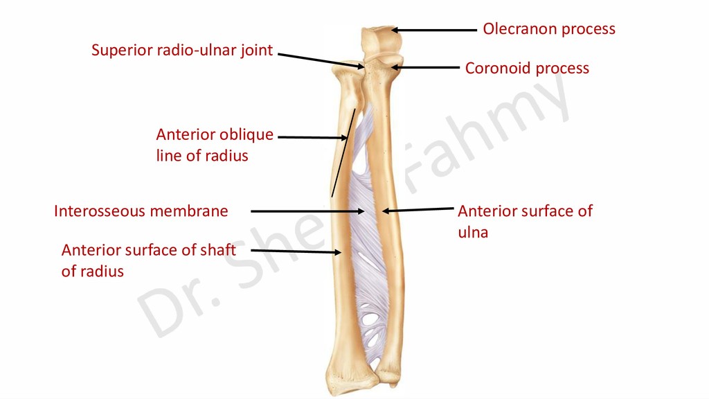
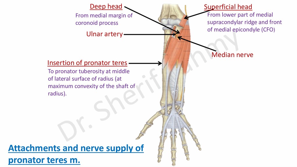
    Attachments and nerve supply of pronator teres m. From lower part of medial supracondylar ridge and front of medial epicondyle (CFO) From medial margin of coronoid process To pronator tuberosity at middle of lateral surface of radius (at maximum convexity of the shaft of radius). Ulnar artery
  15. Trapezium Flexor retinaculum Special compartment for tendon of flexor carpi

    radialis Med. Lat. tendon of flexor carpi radialis with its synovial sheath
  16. Nerve supply: -Median nerve. Action: -Weak flexion of wrist and

    elbow. -It makes the palmar aponeurosis more tense to protect underlying vessels and nerves.
  17. Radial head of flexor digitorum superficialis Humero-ulnar head of flexor

    digitorum superficialis Origin of flexor digitorum superficialis Anterior oblique line
  18. Humero-ulnar head Radial head Tendinous arch Flexor retinaculum Insertion of

    flexor digitorum superficialis Flexor digitorum superficialis Attachments of flexor digitorum superficialis
  19. Nerve supply: Median nerve. Action: -Flexion of medial 4 fingers

    at proximal inter- phalangeal joints and metacarpo-phalangeal joints. -Flexion of wrist. -Weak flexion of elbow.