Upgrade to Pro — share decks privately, control downloads, hide ads and more …

Radial & Ulnar Arteries

Radial & Ulnar Arteries

This presentation describes Radial & Ulnar Arteries of upper limb

Dr.Sherif Fahmy

August 02, 2019
Tweet

More Decks by Dr.Sherif Fahmy

Other Decks in Science

Transcript

  1. Brachialis Pronator teres Flexor carpi radialis Palmaris longus Flexor carpi

    ulnaris Flexor digitorum superficialis Flexor retinaculum Med. Lat. Supinator m Brachioradialis m Tendon of biceps brachii
  2. Pronator teres Flexor carpi radialis (cut) Palmaris longus (cut) Flexor

    carpi ulnaris (Cut) Flexor digitorum superficialis (cut) Flexor digitorum profundus Flexor pollicis longus Pronator quadratus Flexor retinaculum Brachioradialis (cut) Deep muscles of forearm Medial Lateral Interosseous membrane
  3. Brachial artery Tendon of biceps It leaves forearm by passing

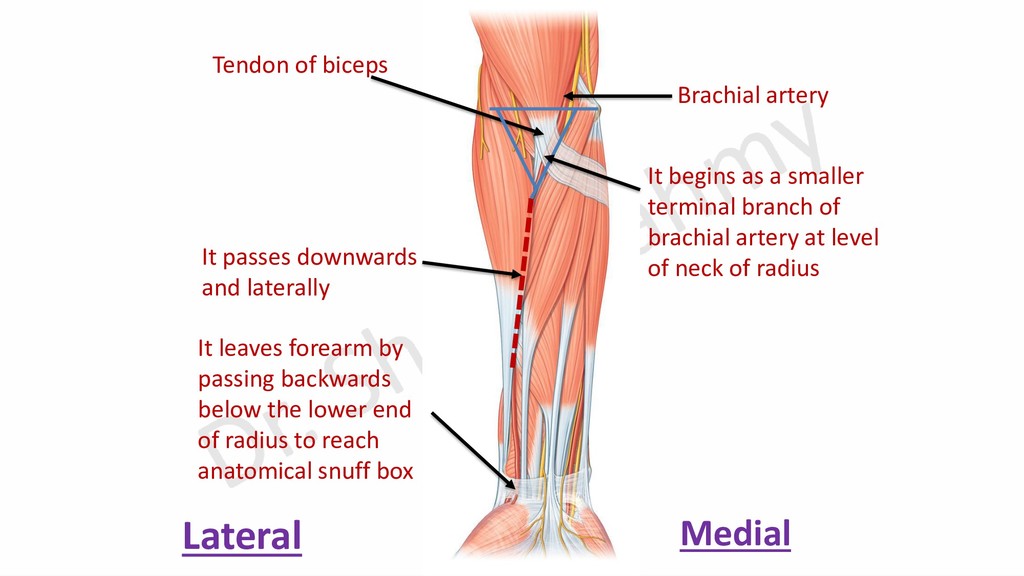
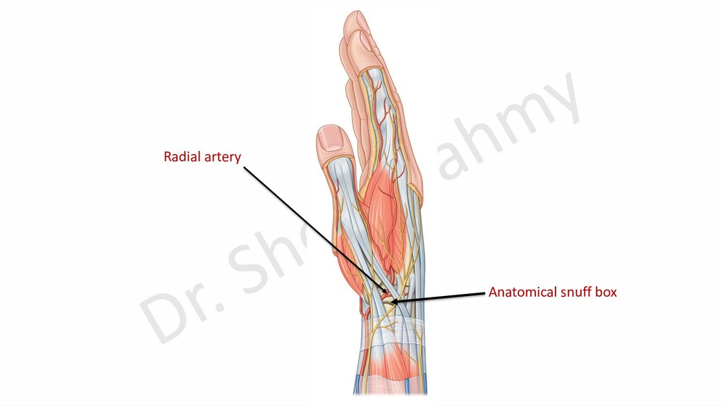
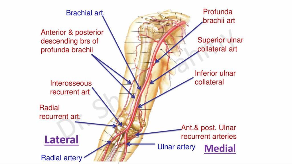
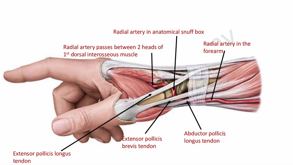
    backwards below the lower end of radius to reach anatomical snuff box Lateral Medial It begins as a smaller terminal branch of brachial artery at level of neck of radius It passes downwards and laterally
  4. Point 1: 1 cm below line of elbow (flexion crease),

    medial to tendon of biceps brachii Point 2: Lower end of radius, lateral to tendon of flexor carpi radialis
  5. Radial artery Brachioradialis m. (upper 2/3 of forearm) Lateral Medial

    Skin & Fascia (cubital fossa) Skin & fascia (lower 1/3 of forearm)
  6. 1-Tendon of biceps m. 2-Supinator m. 3-Pronator teres m. 4-Radial

    head of flexor digitorum superficialis m. 5-Flexor pollicis longus m. 6-Pronator quadratus m. 7-Lower end of radius Lateral Medial
  7. Brachial artery Ulnar artery & nerve Tendon of biceps Pisiform

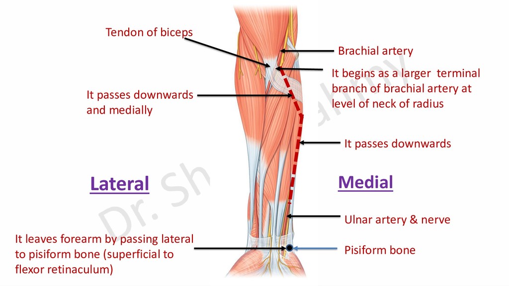
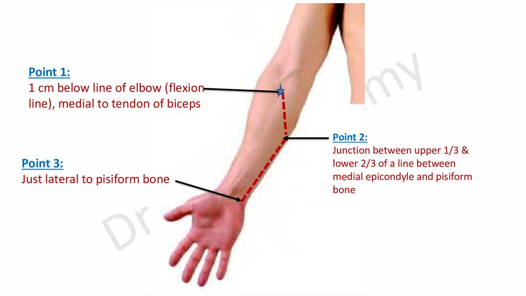
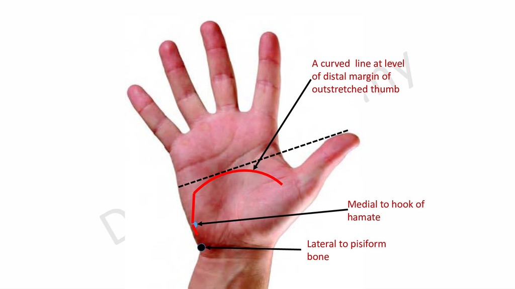
    bone It begins as a larger terminal branch of brachial artery at level of neck of radius It leaves forearm by passing lateral to pisiform bone (superficial to flexor retinaculum) Lateral Medial It passes downwards and medially It passes downwards
  8. Point 1: 1 cm below line of elbow (flexion line),

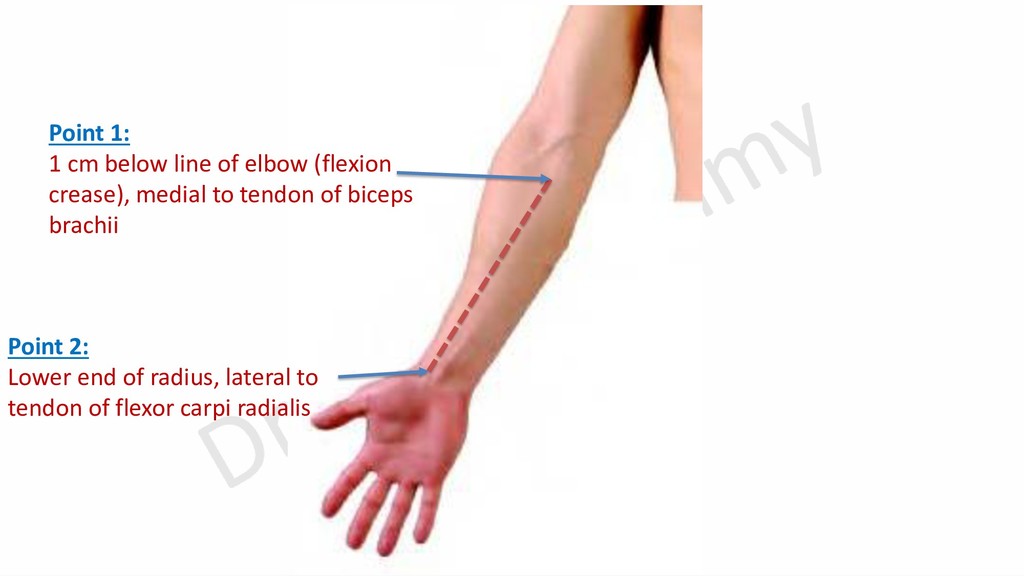
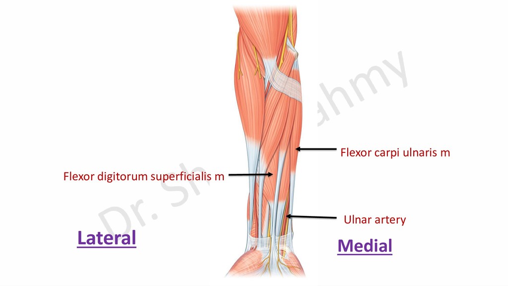
    medial to tendon of biceps Point 3: Just lateral to pisiform bone Point 2: Junction between upper 1/3 & lower 2/3 of a line between medial epicondyle and pisiform bone
  9. Pronator teres m Skin & fascia Flexor carpi radialis m

    Palmaris longus m. Flexor digitorum superficialis m. Flexor carpi ulnaris m. Lateral Medial Upper 1/3 Middle 1/3 Lower 1/3
  10. Radial Artery Ulnar Artery In the Forearm 1- Radial recurrent

    1- Anterior & posterior ulnar recurrent 2- Anterior carpal 2- Anterior & posterior carpal 3- Muscular 3- Muscular 4- Superficial palmar 4- Common interosseous Dr. Sherif Fahmy
  11. Branches of radial & ulnar arteries in forearm Radial recurrent

    br. Anterior carpal br. Superficial palmar br. Anterior & posterior ulnar recurrent brs. Anterior carpal br. Posterior carpal br. Common interosseous art. Radial Artery Ulnar artery Anterior interosseous art. Posterior interosseous art Interosseous membrane
  12. Course & branches of anterior interosseous artery Common interosseous art.

    Posterior interosseous artery Median artery Descending br. Anterior interosseous artery Anterior carpal arch Nutrient to radius Nutrient to ulna Ulnar artery Interosseous membrane Median nerve Lateral Medial
  13. Interosseous recurrent br. Posterior interosseous art. Course & branches of

    posterior interosseous artery Lateral epicondyle Posterior descending br. of profunda brachii Supinator m. Abductor pollicis longus Posterior interosseous nerve End of posterior interosseous artery Lateral
  14. Profunda brachii art Superior ulnar collateral art Inferior ulnar collateral

    Ulnar artery Radial artery Anterior & posterior descending brs of profunda brachii Ant.& post. Ulnar recurrent arteries Radial recurrent art. Interosseous recurrent art Brachial art. Lateral Medial
  15. Arterial Arches in the Hand 1- Anterior carpal arch: gives

    articular blood supply to wrist joint. 2- Superficial palmar arch: gives 3 common palmar digital arteries. 3- Posterior carpal arch: gives 2nd, 3rd and 4th dorsal metacarpal arteries. 4- Deep palmar arch: gives 3 palmar metacarpal arteries.
  16. Ulnar artery Flexor retinaculum Deep br. of ulnar artery Thenar

    eminence Superficial palmar arch Superficial palmar branch from radial artery Pisiform bone Superficial branch of ulnar artery Hook of hamate Ulnar nerve
  17. Lateral to pisiform bone Medial to hook of hamate A

    curved line at level of distal margin of outstretched thumb
  18. Relations of Superficial Palmar Arch Superficial: Skin, superficial fascia and

    palmar aponeurosis. Deep: Terminal branches of median nerve, flexor digitorum tendons, common synovial sheath and lumbricals.
  19. Superficial palmar arch Terminal branches of median nerve Tendons of

    flexor digitorum muscles & lumbricals Common synovial sheath
  20. Branches of Superficial Palmar Arch 1- Palmar digital branch of

    little finger. 2- Three common palmar digital branches.
  21. Palmar metacarpal branch Communication between Branches of Deep Palmar Arch

    and branches of superficial palmar arch Deep palmar arch Common palmar digital branches
  22. Radial artery in the forearm Radial artery in anatomical snuff

    box Radial artery passes between 2 heads of 1st dorsal interosseous muscle Abductor pollicis longus tendon Extensor pollicis brevis tendon Extensor pollicis longus tendon
  23. Transverse head of adductor pollicis m Oblique head of adductor

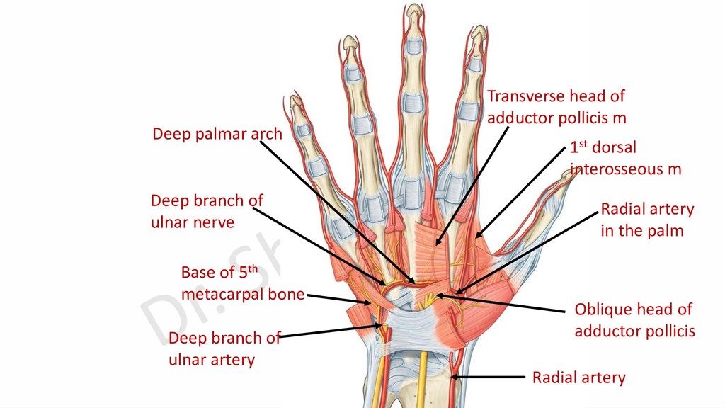
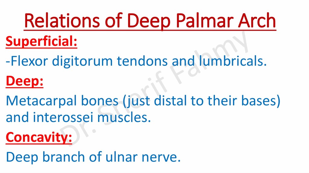
    pollicis Deep palmar arch Base of 5th metacarpal bone Deep branch of ulnar artery Radial artery in the palm Radial artery 1st dorsal interosseous m Deep branch of ulnar nerve
  24. Curved line 1/2 inch proximal to superficial palmar arch A

    curved line at level of distal margin of outstretched thumb
  25. Relations of Deep Palmar Arch Superficial: -Flexor digitorum tendons and

    lumbricals. Deep: Metacarpal bones (just distal to their bases) and interossei muscles. Concavity: Deep branch of ulnar nerve.
  26. Branches of Radial Artery in the Hand Dorsum of hand:

    1- Dorsal (posterior) carpal branch. 2- Dorsal digital branch. 3- First dorsal metacarpal branch. In the 1st intermetacarpal space: 1- Radialis indices. 2- Princips pollicis. In the palm (Deep palmar arch): 1- Three palmar metacarpal branches. 2- Proximal perforating arteries. 3- Recurrent branch to anterior carpal arch.
  27. 2- Dorsal digital branch 3-1st dorsal metacarpal br. 1-Posterior carpal

    branch Radial artery Branches of Radial Artery on the Dorsum Dorsal (posterior) branch of ulnar artery Dorsal (posterior) carpal arch 2nd, 3rd and 4th dorsal metacarpal arteries
  28. Perforating arteries: A- Proximal perforating arteries: Between dorsal metacarpal arteries

    and deep palmar arch. B- Distal perforating arteries: Between dorsal metacarpal arteries and palmar metacarpal arteries.
  29. 2- Priceps pollicis artery 1- Radialis indices Branches of Radial

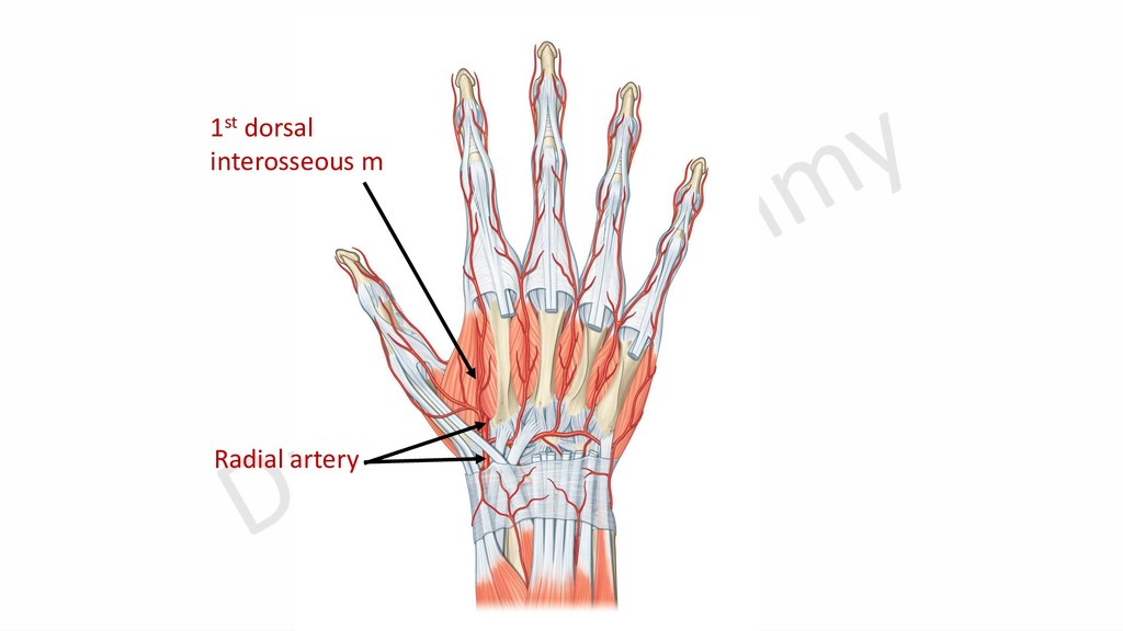
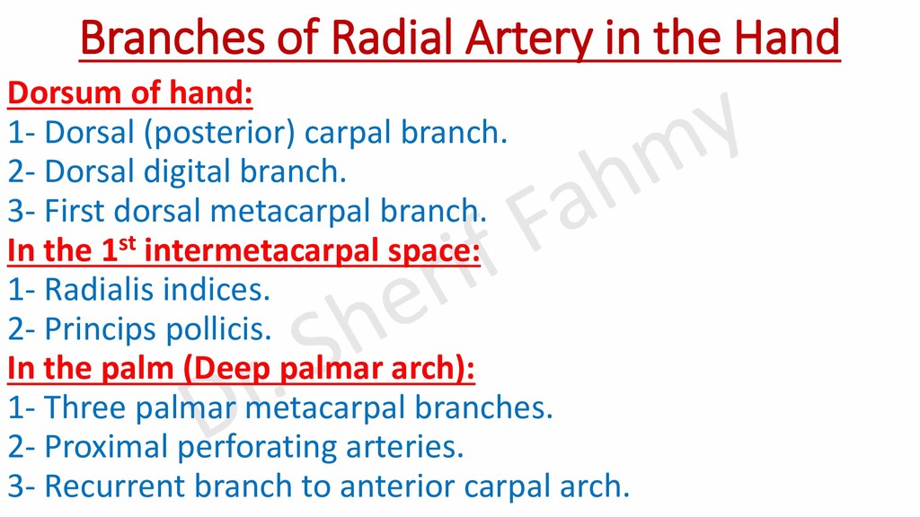
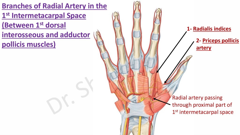
    Artery in the 1st Intermetacarpal Space (Between 1st dorsal interosseous and adductor pollicis muscles) Radial artery passing through proximal part of 1st intermetacarpal space
  30. Arterial Arches in the Hand Superficial to flexor digitorum tendons.

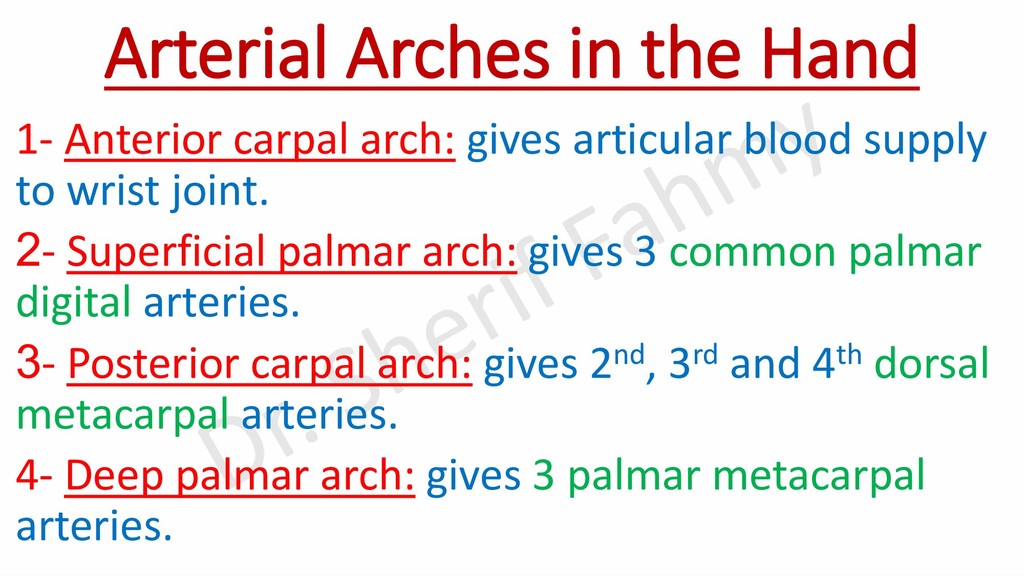
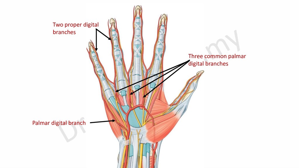
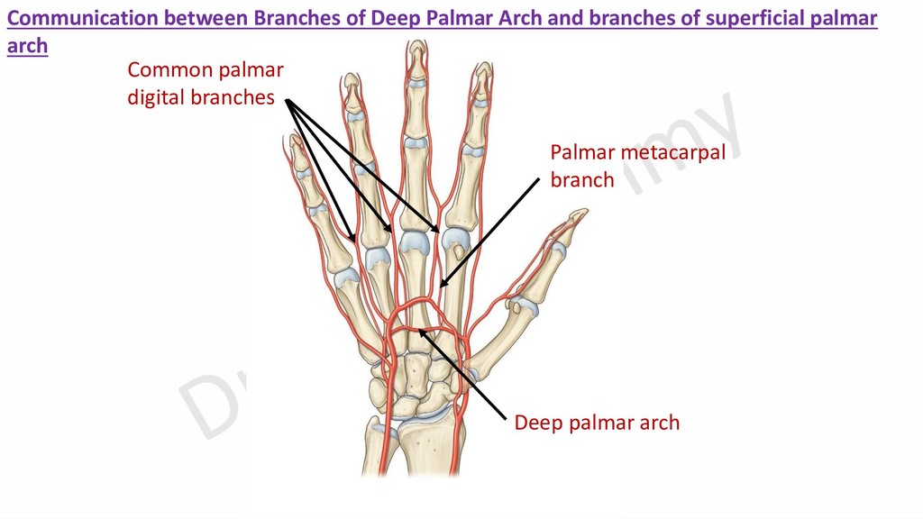
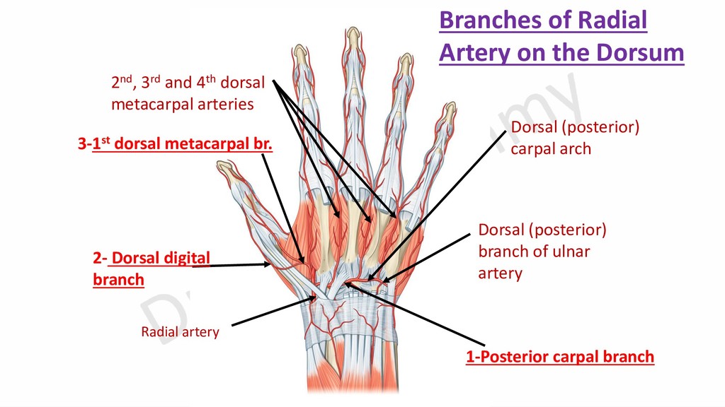
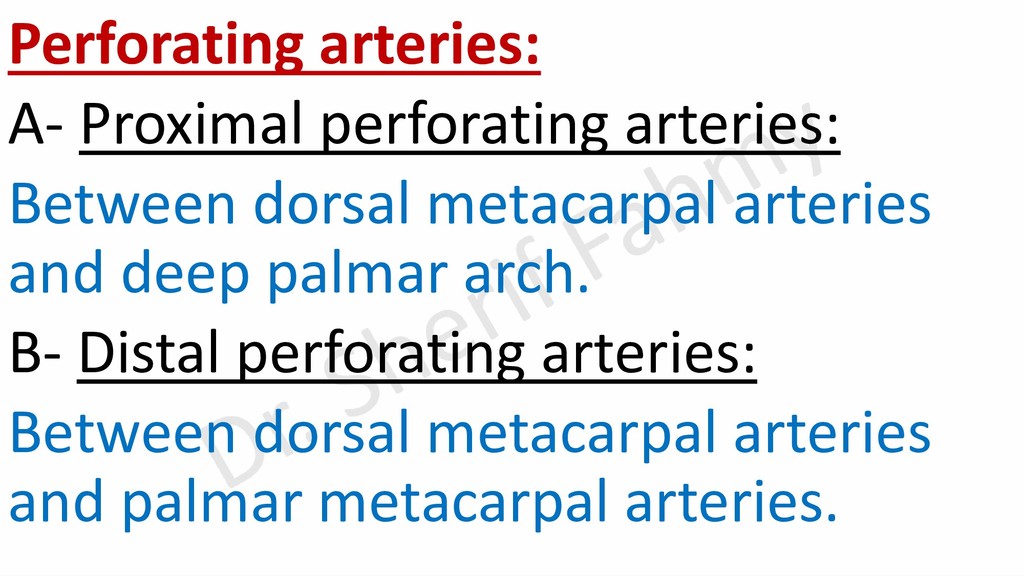
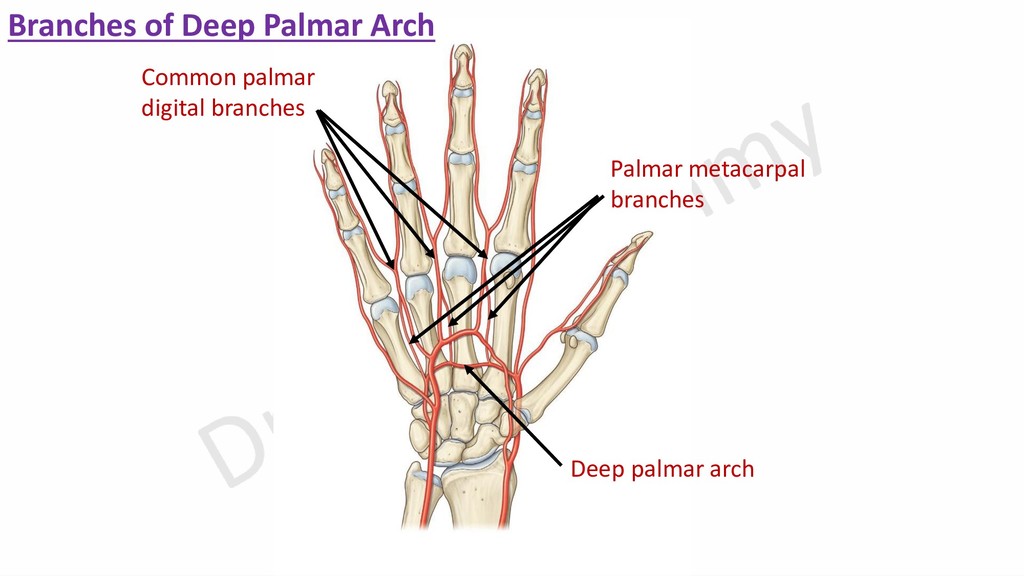
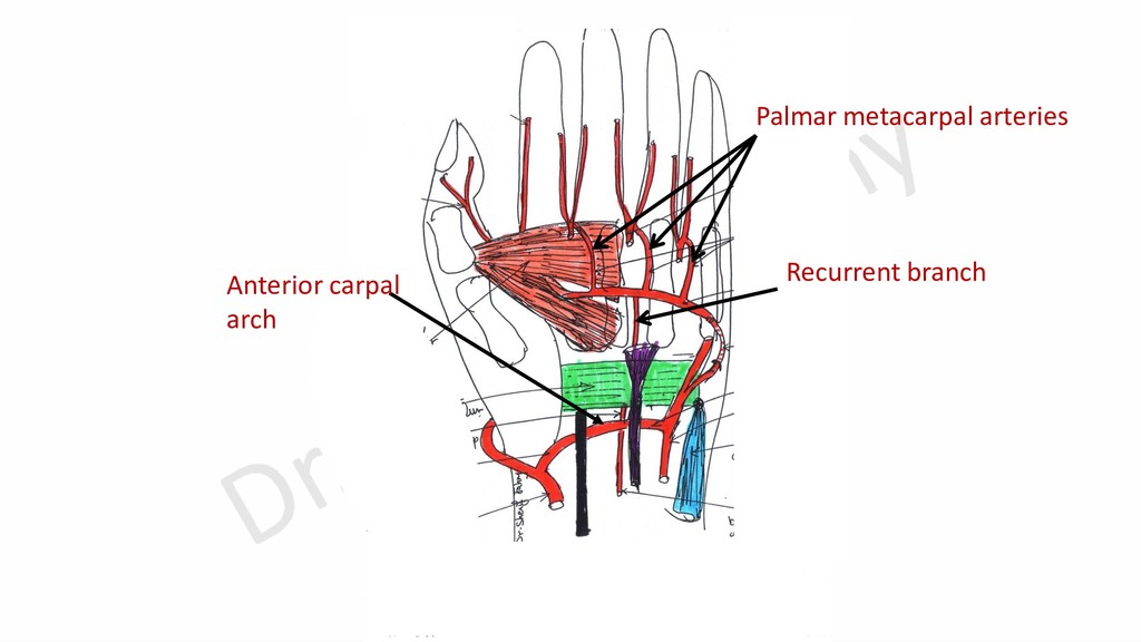
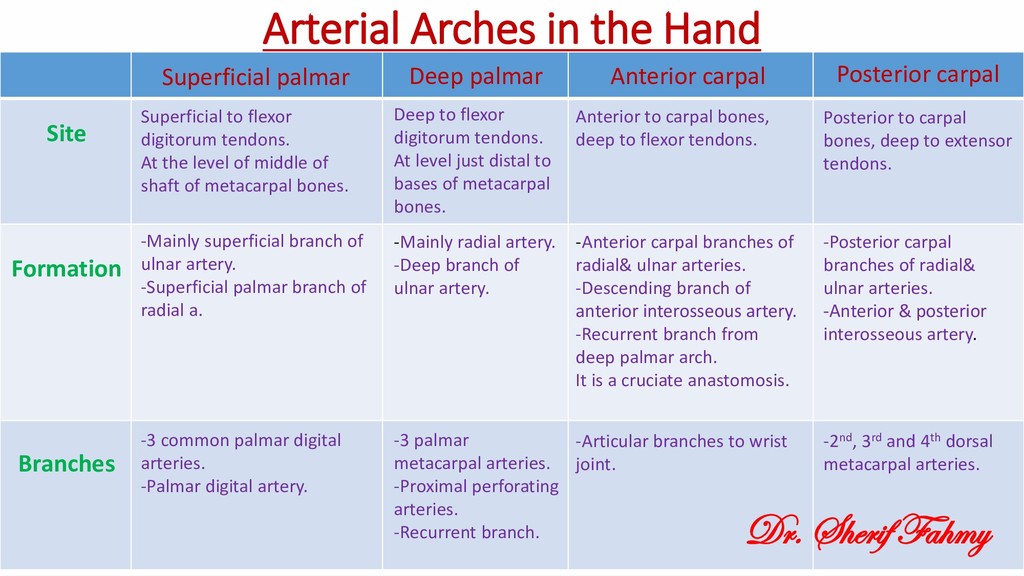
    At the level of middle of shaft of metacarpal bones. Deep to flexor digitorum tendons. At level just distal to bases of metacarpal bones. Anterior to carpal bones, deep to flexor tendons. Posterior to carpal bones, deep to extensor tendons. -Mainly superficial branch of ulnar artery. -Superficial palmar branch of radial a. -Mainly radial artery. -Deep branch of ulnar artery. -Anterior carpal branches of radial& ulnar arteries. -Descending branch of anterior interosseous artery. -Recurrent branch from deep palmar arch. It is a cruciate anastomosis. -Posterior carpal branches of radial& ulnar arteries. -Anterior & posterior interosseous artery. -3 common palmar digital arteries. -Palmar digital artery. -3 palmar metacarpal arteries. -Proximal perforating arteries. -Recurrent branch. -Articular branches to wrist joint. -2nd, 3rd and 4th dorsal metacarpal arteries. Site Formation Branches Superficial palmar Deep palmar Anterior carpal Posterior carpal Dr. Sherif Fahmy