Upgrade to Pro — share decks privately, control downloads, hide ads and more …

A Beginner’s Guide to Critical Understandings in Pulmonary Function Testing

A Beginner’s Guide to Critical Understandings in Pulmonary Function Testing

Introduction
A key component of respiratory therapy is pulmonary function testing (PFT), which is essential for both diagnosing and tracking lung problems. Knowing the ins and outs of PFT is crucial whether you’re a seasoned respiratory therapist looking for last-minute CEUs or are brand-new to the profession. This beginner’s guide will go into the fundamental concepts of PFT and provide you with information, advice, and tools to help you succeed in this important area of respiratory treatment.

Section 1: What is Pulmonary Function Testing?
Subtitle: Unraveling the Basics

A battery of tests called pulmonary function testing, or PFT, determines how effectively your lungs are working. These exams assess the capacity, airflow, and gas exchange of the lungs, among other aspects of lung health. PFTs are crucial in the diagnosis and treatment of respiratory conditions such pulmonary fibrosis, COPD, and asthma.

Section 2: Types of Pulmonary Function Tests
Subtitle: Exploring the Varieties

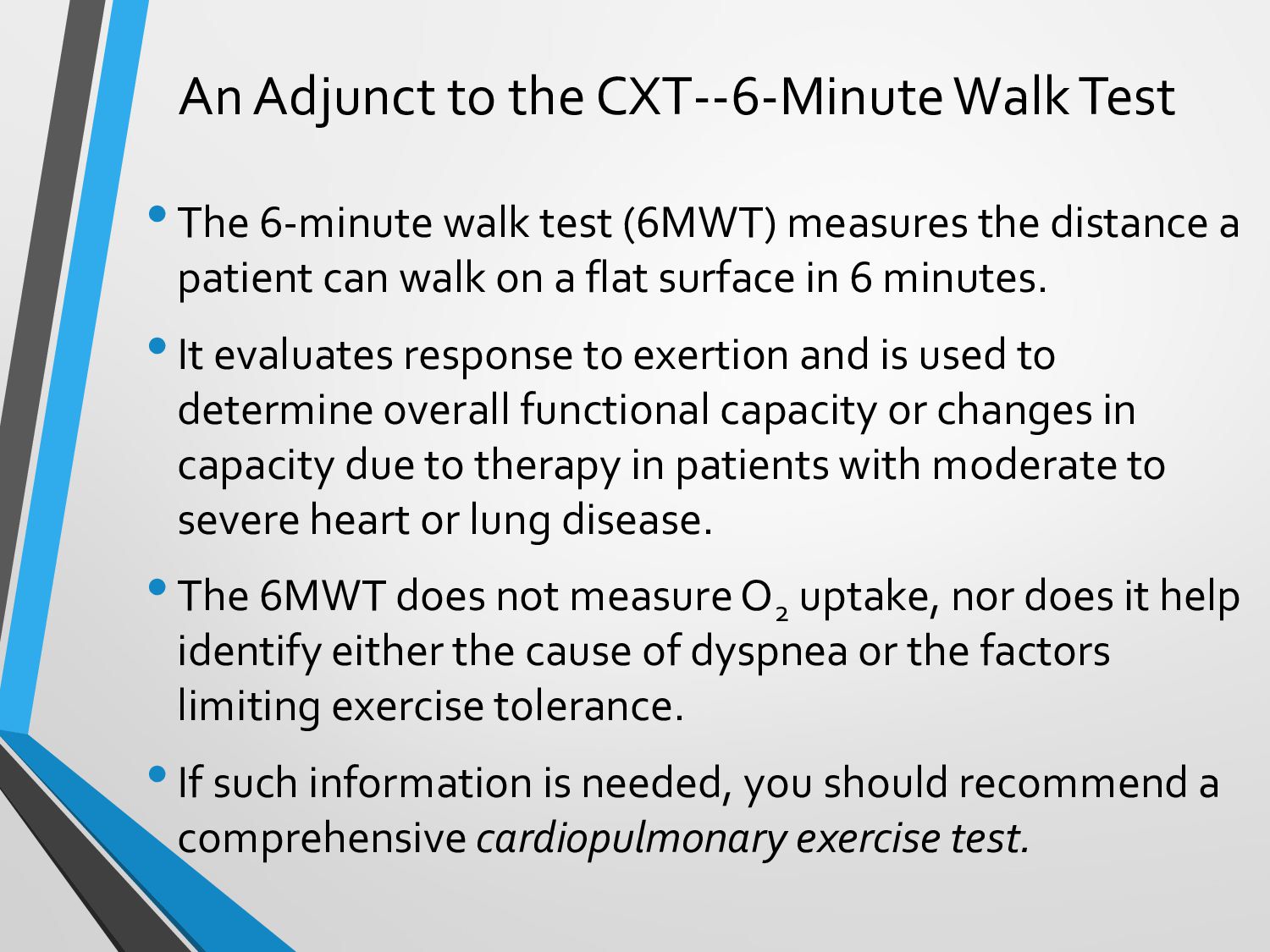
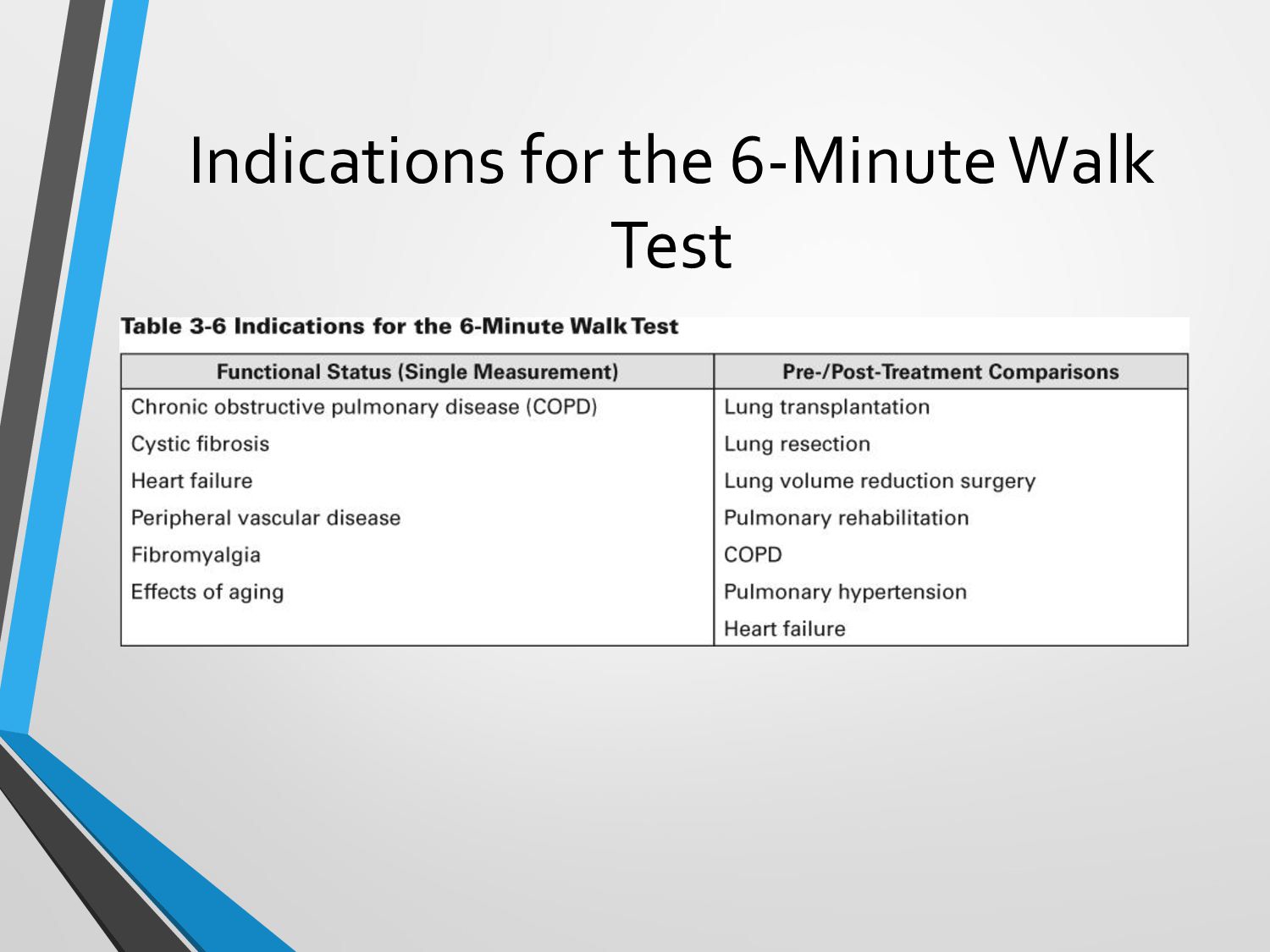
PFTs come in a variety of forms, each with a distinct function. Spirometry, lung volume testing, and gas exchange tests are a few examples of typical PFTs. For instance, lung volume tests identify lung capacity whereas spirometry assesses lung function and airflow. Effective patient care requires a thorough understanding of the various PFT types.

Section 3: Importance of PFTs in Respiratory Care
Subtitle: The Clinical Significance

PFTs provide insightful information about a patient’s respiratory health. They support the diagnosis of lung disorders, evaluate the severity of diseases, and track the success of treatments. PFT findings are used by respiratory therapists to create individualized care programs and monitor patients’ advancement over time.

Section 4: Interpreting PFT Results
Subtitle: Decoding the Numbers

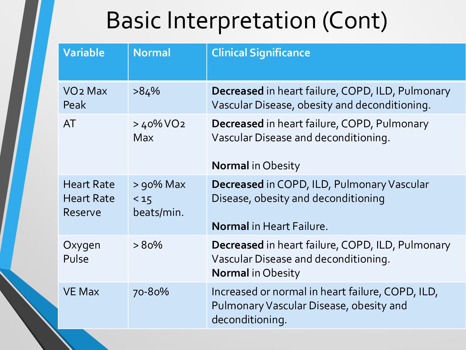
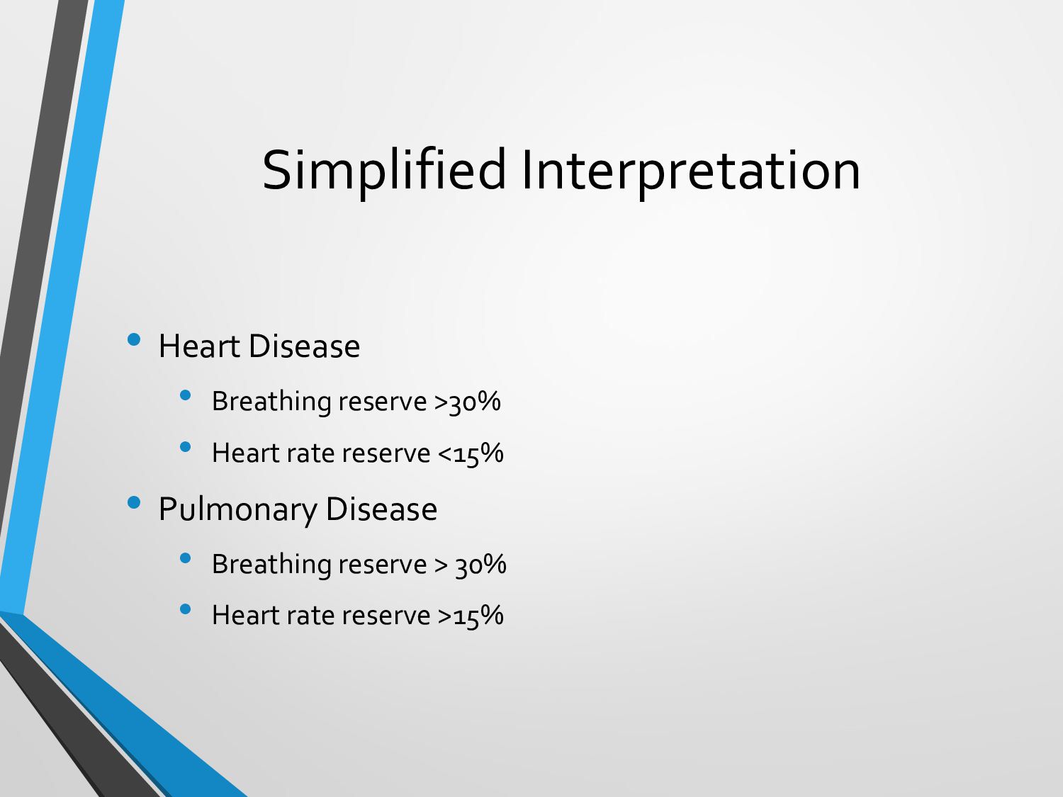
Although it might be challenging, understanding PFT results is an essential ability for respiratory therapists. The forced vital capacity (FVC), forced expiratory volume in one second (FEV1), and the FEV1/FVC ratio are important measurements to look for. These figures offer important details about lung health and the presence of any blockages.

Section 5: Common Pitfalls and Challenges
Subtitle: Navigating Roadblocks

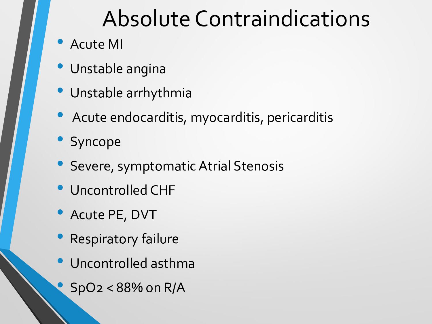
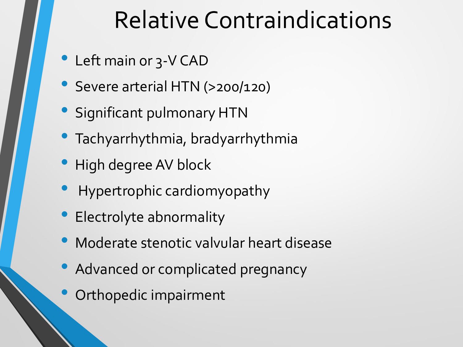
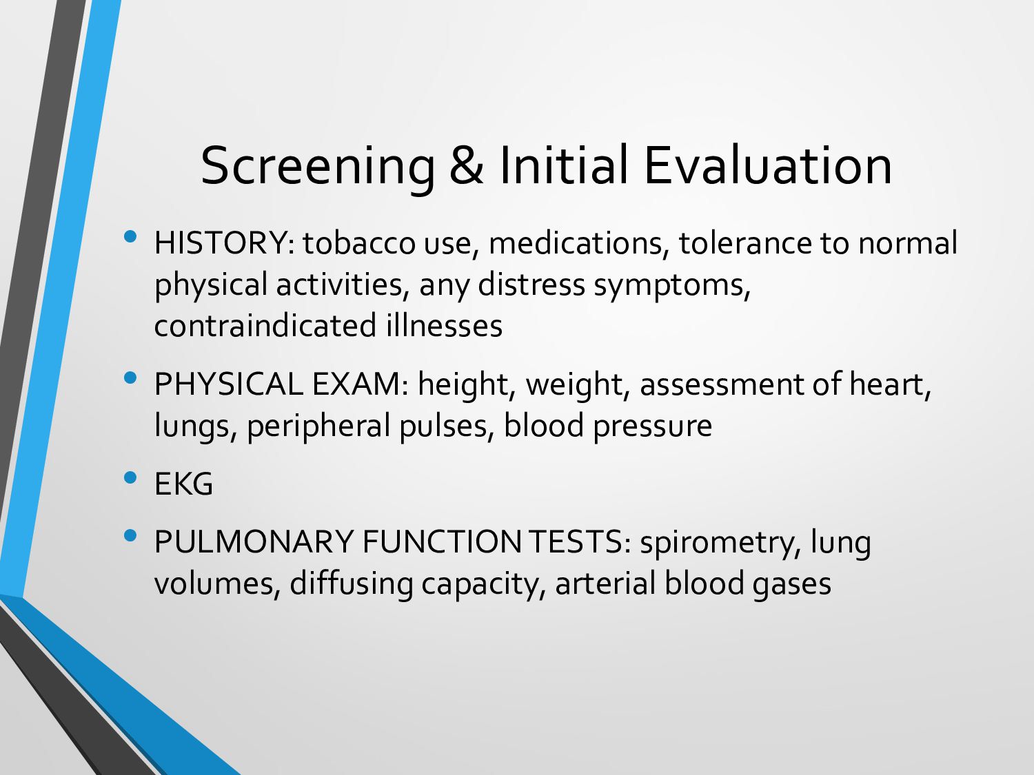
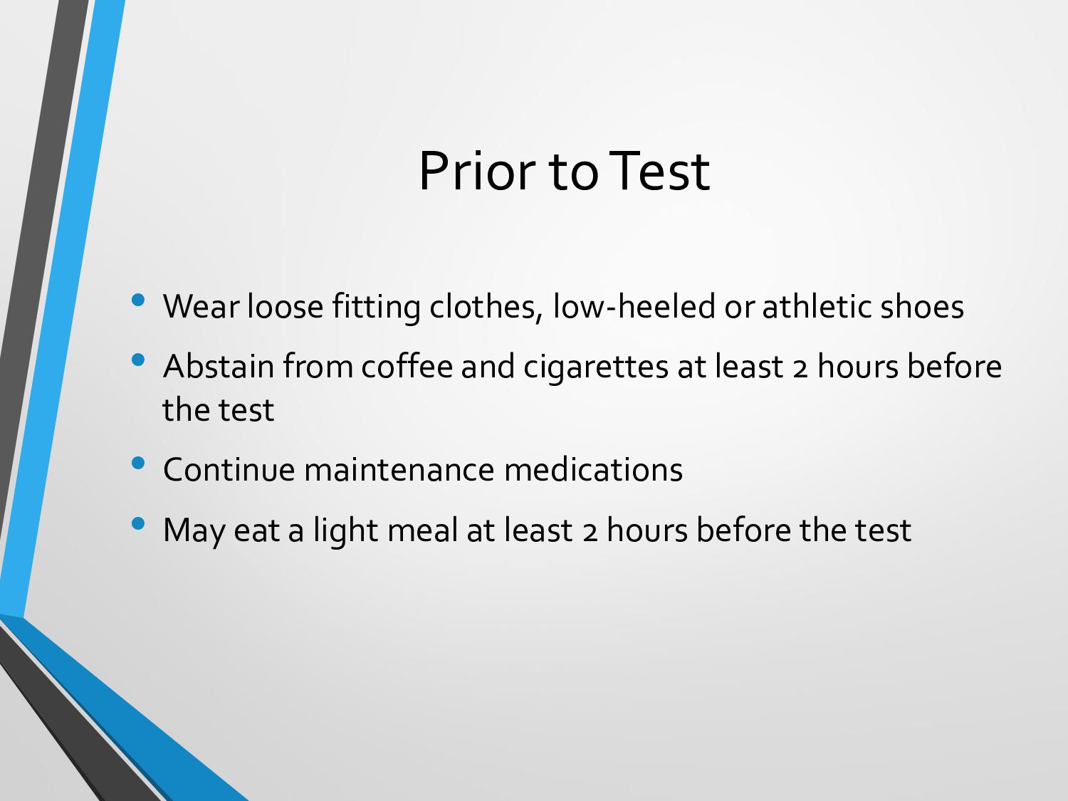
PFTs have their difficulties, much as any diagnostic test. The outcomes can be affected by patient cooperation, sound technique, and external variables. To achieve accurate assessments, it’s critical to be aware of potential dangers and work toward reducing inaccuracies.

Section 6: Resources for Respiratory Therapists
Subtitle: CEUs and Learning Opportunities

It’s essential for respiratory therapists to keep learning. There are tools accessible if you’re trying to find CEUs (Continuing Education Units) at a discount or last-minute CEUs. Courses for respiratory therapists are available on websites like “CEUs for Less”. Furthermore, the AARC (American Association for Respiratory Care) offers free CEUs for respiratory therapists that have been approved by the AARC, guaranteeing that you stay up to speed with the most recent developments in pulmonary function testing.

Conclusion:
Knowing How the Pulmonary System Works A crucial component of respiratory therapy is testing. These tests are an essential tool in the field because they offer crucial information for identifying and treating lung problems. To improve your knowledge and abilities as a respiratory therapist, keep in mind to evaluate results cautiously, face problems with confidence, and utilize tools like AARC-approved free CEUs.

visit our site : unlimited ceu

atrespiratory lectures

September 11, 2023
Tweet

Other Decks in Education

Transcript

  1. An Overview of Exercise Stress Testing And It's Value In

    Patient Management Al Heuer, PhD, MBA, RRT-ACCS, RPFT, FAARC Rutgers – School of Health Professions Co-Owner A & T Lectures
  2. Learning Objectives Review Key Definitions Describe the Indications and Contraindications

    Examine the Equipment Review the Set-up and Procedure Evaluate and Interpret Test Results Brief Review of 6-Minute Walk Test Examine Key References 2
  3. Definition and Goal of Cardiopulmonary Stress Testing (CPET) • Definition:

    CARDIO-PULMONARY exercise testing evaluates, characterizes and quantifies the physiologic response of the heart lungs and muscles to an increase in physical stress. • Goal: The goal of CARDIO-PULMONARY exercise testing is to evaluate the physiologic response of the heart lungs and muscles to an increase in physical stress
  4. Major Indications • Evaluation of dyspnea • Distinguish cardiac vs

    pulmonary vs peripheral limitation vs others • Detection of exercise-induced bronchoconstriction detection of exertional desaturation • Detection of exertional desaturation • Disability determination • Pulmonary Rehabilitation • Exercise intensity/prescription • Response to participation • Pre-op evaluation and risk stratification • Assess response to therapy
  5. Absolute Contraindications • Acute MI • Unstable angina • Unstable

    arrhythmia • Acute endocarditis, myocarditis, pericarditis • Syncope • Severe, symptomatic Atrial Stenosis • Uncontrolled CHF • Acute PE, DVT • Respiratory failure • Uncontrolled asthma • SpO2 < 88% on R/A
  6. Relative Contraindications • Left main or 3-V CAD • Severe

    arterial HTN (>200/120) • Significant pulmonary HTN • Tachyarrhythmia, bradyarrhythmia • High degree AV block • Hypertrophic cardiomyopathy • Electrolyte abnormality • Moderate stenotic valvular heart disease • Advanced or complicated pregnancy • Orthopedic impairment
  7. Screening & Initial Evaluation • HISTORY: tobacco use, medications, tolerance

    to normal physical activities, any distress symptoms, contraindicated illnesses • PHYSICAL EXAM: height, weight, assessment of heart, lungs, peripheral pulses, blood pressure • EKG • PULMONARY FUNCTION TESTS: spirometry, lung volumes, diffusing capacity, arterial blood gases
  8. Prior to Test • Wear loose fitting clothes, low-heeled or

    athletic shoes • Abstain from coffee and cigarettes at least 2 hours before the test • Continue maintenance medications • May eat a light meal at least 2 hours before the test
  9. Exercise Modalities • Advantages of cycle ergometer • Cheaper •

    Safer - Less danger of fall/injury • Can stop anytime • Direct power calculation • Independent of weight • Holding bars has no effect • Little training needed • Easier BP recording, blood draw • Requires less space - less noise • Advantages of treadmill • Attain higher VO2 • More functional
  10. Equipment & Instrumentation • Treadmill or cycle ergometer can be

    used for exercise • Cycle preferred because: • Ease of measurements (ABGs, BP) • Better patient safety (falling not likely) • Power can be directly measured • Ability to continuously monitor and print 12-lead ECG • Manual or automatic BP device • Pulse oximeter
  11. Equipment & Instrumentation (cont.) • Gas analyzers for O2 and

    CO2 • Mass Spectrometer – gold standard • Breathing valves • Ability to measure volume
  12. Summary of the Procedure • After screening, the patient is

    connected to monitoring equipment, including BP, HR, RR, inspired FIO2 and exhaled CO2, VT and Minute Ventilation. • They are then subjected to and increasing workload, per a specified protocol. • The Bruce protocol: Multiple exercise stages of three minutes each. At each stage, the gradient and speed of the treadmill are elevated to increase work output, called METS. Stage 1 of the Bruce protocol is performed at 1.7 miles per hour and a 10% gradient. Stage 2 is 2.5 mph and 12%, while Stage 3 goes to 3.4 mph and 14%. • Parameters are measured at each step of the protocol and calculations are then done, which are then interpreted. • The test is halted if the patient can not continue per the protocol or if adverse responses are noted.
  13. Key Features of the Test • Symptom-limited exercise test •

    Measures airflow, SpO2 , and expired oxygen and carbon dioxide • Allows calculation of peak oxygen consumption, anaerobic threshold
  14. Cardiopulmonary Exercise Testing— Work Unit=MET •Standard work unit used for

    exercise testing is the metabolic equivalent of task (MET): 1 MET = 3.5 mL O2 consumption/kg of body weight, about equal to normal resting O2 consumption/minute. •MET levels are varied during exercise by altering treadmill speeds and inclinations. Most protocols increase exercise intensity by 1–2 METs at each step-up in workload.
  15. Indications to Terminate • Patient’s request: fatigue, dyspnea, pain •

    Ischemic ECG changes • 2 mm ST depression • Chest pain suggestive of ischemia • Significant ectopy • 2 nd or 3rd degree heart block • BP sys >240-250, • BP dias >110-120 • Fall in BPsys >20 mmHg • SpO2 < 85%
  16. Mechanism to Exercise Limitation • Pulmonary • Ventilatory impairment •

    Respiratory muscle dysfunction • Impaired gas exchange • Cardiovascular • Reduced stroke volume • Abnormal HR response • Circulatory abnormality • Blood abnormality • Peripheral • Inactivity - Atrophy • Neuromuscular dysfunction • Malnutrition • Other- Perceptual, Motivational, Environmental
  17. CPET Measurements • Work • VO2 • VCO2 • HR

    • ECG • BP • RR • SpO2 • Anaerobic Threshold (AT) • ABG Lactate • Dyspnea
  18. Pulmonary Parameters • MINUTE VENTILATION • NORMAL = 5 –

    6 liters/ min --AT EXERCISE = 100 liters/min • Increase is due to stimulation of the respiratory centers by brain motor cortex, joint propioceptors and chemoreceptors • ANAEROBIC THRESHOLD (AT) the minute ventilation increases more than the workload • TIDAL VOLUME : NORMAL = 500 ml • DURING EXERCISE = 2.3 – 3 liters • Increases early in the exercise • BREATHING RATE • NORMAL = 12 – 16 / min AT EXERCISE = 40 – 50 / min • DEAD SPACE / TIDAL VOLUME ratio • NORMAL = 0.20 – 0.40 -- AT EXERCISE = 0.04 – 0.20 • Decrease is due to increased tidal volume with constant dead space
  19. Pulmonary Parameters (Cont.) • PULMONARY CAPILLARY BLOOD TRANSIT TIME NORMAL

    = 0.75 second • AT EXERCISE = 0.38 second • Decrease is due to increased cardiac output • ALVEOLAR-ARTERIAL OXYGEN DIFFERENCE • NORMAL = 10 mm Hg --AT EXERCISE = 20 – 30 mm Hg • Changes very little until a heavy workload is achieved • OXYGEN TRANSPORT • Increase in temperature, PCO2 and relative acidosis in the muscles -> increase in release of Oxygen by blood for use by the tissues for metabolism.
  20. Cardiovascular Parameters • CARDIAC OUTPUT NORMAL = 4 – 6

    liters / min • AT EXERCISE = 20 liters / min • increase is linear with increase in workload during exercise until the point of exhaustion. • STROKE VOLUME NORMAL = 50 – 80 ml • AT EXERCISE = double • Increase is linear with increase in workload • HEART RATE NORMAL = 60 – 100 /min AT EXERCISE = 2.5 – 4 times the resting HR • HR max is achieved just prior to total exhaustion, physiologic endpoint of an individual • HR max = 220 – age or HR max = 210 – (0.65 x age) • OXYGEN PULSE= VO2/ HR NORMAL = 2.5 – 4 ml O2 / heartbeat • AT EXERCISE = 10 – 15 ml • With increasing muscle work during exercise, each heart contraction must deliver a greater quantity of oxygen out to the body • BLOOD PRESSURE DURING EXERCISE: • Systolic BP increases (to 200 mm Hg) • Diastolic BP is relatively stable (up to 90 mmHg) • Increase in Pulse Pressure (difference between Systolic and Diastolic pressures) • ARTERIAL – VENOUS OXYGEN CONTENT DIFFERENCE • mL of O2 / 100 ml of blood • NORMAL = 5 vol % • AT EXERCISE = 2.5 – 3 times higher the increase is due to the greater amounts of Oxygen that are extracted by the working muscle tissue
  21. Metabolic Parameters • OXYGEN CONSUMPTION • NORMAL = 250 ml

    / min 3.5 – 4 ml / min / kg • Increases directly with the level of muscular work until exhaustion occurs and until individual reaches VO2max • NORMAL RANGE = 1,700 – 5,800 ml / min • CARBON DIOXIDE PRODUCTION • NORMAL = 200 ml / min 2.8 ml / min / kg • AT EXERCISE initial phase, increases at same rate as VO2 • Once Anaerobic Threshold (AT) is reached, increases at a faster rate than VO2 due to increased acid production • ANAEROBIC THERSHOLD (AT) • NORMAL: occurs at about 60% of VO2 max • Followed by breathlessness, burning sensation begins in working muscles • RESPIRATORY QUOTIENT (RQ) • RESTING LEVEL = 0.8 • AT = 1.0 or more may exercise for a short time on 1.5 • RER = CO2 produced / O2 consumed = VCO2 / VO2
  22. The relationship of physiologic dead space (VD) to VT is

    commonly called the VD/VT ratio. In healthy individuals this ratio is approximately 0.35 (or 35%) at rest and falls to between 0.05 and 0.25 (or 5% and 25%) during exercise.
  23. The VT increases up to approximately 60% of the patient’s

    VC and levels off, while f increases up to 50 to 60 breaths/min. In patients with restrictive processes the relationship between f and VT is shifted upward and to the left (dashed line). This is a result of their rapid shallow breathing pattern.
  24. Systolic blood pressure increases 80 to 150 mmHg during exercise

    in healthy individuals. The diastolic blood pressure generally remains near resting levels.
  25. CO2 production (VCO2 ) increases linearly with work up to

    approximately the point anaerobic metabolism begins, after which there is increased CO2 production from buffering lactic acid.
  26. Basic Interpretation Variable Normal Clinical Significance SaO2 & PaO2 >

    95% 80-100 torr < 4% decrease Decreased in ILD and Pulmonary Vascular Disease Normal in heart failure, obesity and deconditioning. P (A-a) O2 < 35 mm Hg Increased in COPD, ILD and Pulmonary Vascular Disease. Normal in Heart failure & Deconditioned. VD/VT < 0.28 Increased in Heart Failure, COPD, ILD and Pulmonary Vascular Disease Normal in Obesity & Deconditioned. VE/VCO2 at AT < 34 Increased in Heart Failure, COPD, ILD and Pulmonary Vascular Disease Normal in Obesity & Deconditioned.
  27. Basic Interpretation (Cont) Variable Normal Clinical Significance VO2 Max Peak

    >84% Decreased in heart failure, COPD, ILD, Pulmonary Vascular Disease, obesity and deconditioning. AT > 40% VO2 Max Decreased in heart failure, COPD, Pulmonary Vascular Disease and deconditioning. Normal in Obesity Heart Rate Heart Rate Reserve > 90% Max < 15 beats/min. Decreased in COPD, ILD, Pulmonary Vascular Disease, obesity and deconditioning Normal in Heart Failure. Oxygen Pulse > 80% Decreased in heart failure, COPD, ILD, Pulmonary Vascular Disease and deconditioning. Normal in Obesity VE Max 70-80% Increased or normal in heart failure, COPD, ILD, Pulmonary Vascular Disease, obesity and deconditioning.
  28. Simplified Interpretation • Heart Disease • Breathing reserve >30% •

    Heart rate reserve <15% • Pulmonary Disease • Breathing reserve > 30% • Heart rate reserve >15%
  29. An Adjunct to the CXT--6-Minute Walk Test •The 6-minute walk

    test (6MWT) measures the distance a patient can walk on a flat surface in 6 minutes. •It evaluates response to exertion and is used to determine overall functional capacity or changes in capacity due to therapy in patients with moderate to severe heart or lung disease. •The 6MWT does not measure O2 uptake, nor does it help identify either the cause of dyspnea or the factors limiting exercise tolerance. •If such information is needed, you should recommend a comprehensive cardiopulmonary exercise test.
  30. • Contraindications: • Absolute: A recent myocardial infarction (MI) or

    experienced unstable angina. • Relative: A resting heart rate > 120 beats/min; BP > 180/100 mm Hg • The American Thoracic Society (ATS) standardized protocol (Excerpt) • The object of this test is to have the patient walk as far as possible for 6 minutes. The patient will probably get out of breath or become exhausted. The patient is permitted to slow down, stop or r as necessary. able. • Equipment: a stopwatch, a movable chair, a recording worksheet, a sphygmomanometer, and a visual Borg Scale to assess the patient’s dyspnea and level of exertion. If used, a pulse oximeter m be lightweight and not have to be held by the patient while walking. • Stop the test if the patient develops chest pain, severe dyspnea, etc. • Outcomes Measures: Distance, Borg Dyspnea Level, SPO2 • Interpretation: • Most 6MWTs will be done before and after intervention, and the primary question is whether the patient has experienced a clinically significant improvement. • Until further research is available, The ATS recommend that’s pre/post changes in 6MWD be expressed as an absolute value (e.g., the patient walked 50 m farther). 6-Minute Walk Test - Procedure
  31. Take Home Messages • Cardio Pulmonary Exercises Stress Testing Can

    be Very Valuable • Distinguish among causes of exertional dyspnea • Disability determination • Screening for Pulmonary Rehabilitation • Pre-op evaluation and risk stratification • Assess response to therapy • However, it can be expensive and inconvenient. • It should be ordered with discretion and adjuncts such as the 6 Min Walk text considered.
  32. • Kacmarek, Stoller, Heuer, Egan’s Fundamentals of Respiratory Care, ed

    12 2021. • Heuer & Scanlan, Clinical Assessment in Respiratory Care, ed. 9, 2021. • Culver B, Grahan B, Coates A, et al, Recommendations for a Standardized Pulmonary Function Report. An Official American Thoracic Society Technical Statement, Vol 196, (11) 2018. • Mezzani A Cardiopulmonary Exercise Testing: Basics of Methodology and Measurements, Ann Am Thorac Soc, 2018 Jul;14(Supplement-1):S3-S11 • .. Selected Sources & References