Share
Embed
Light-CT atlas of images
Charles Brossollet
January 30, 2013
Transcript
29 slides
-
© LLTech 2012 – www.lltechimaging.com - No copy without authorization
1 LLTech Light-CT Scanner Atlas of images 2012 -
© LLTech 2012 – www.lltechimaging.com - No copy without authorization
2 • BREAST Page 6 • BRAIN Page 24 • SKIN Page 34 • LUNG Page 58 • GI Page 64 • URO GENITAL Page 69 • NEEDLE CORE Page 78 • THYROID Page 83 TABLE OF CONTENTS • FAT CELLS Page 86 • CORNEA AND RETINA Page 88 • DEVELOPMENT BIOLOGY Page 99 • PLANT IMAGING Page 112 • CONTACTS Page 118 Table of contents -
© LLTech 2012 – www.lltechimaging.com - No copy without authorization
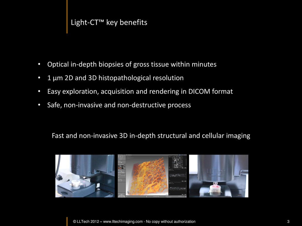
3 • Optical in-depth biopsies of gross tissue within minutes • 1 µm 2D and 3D histopathological resolution • Easy exploration, acquisition and rendering in DICOM format • Safe, non-invasive and non-destructive process Light-CT™ key benefits Fast and non-invasive 3D in-depth structural and cellular imaging -
© LLTech 2012 – www.lltechimaging.com - No copy without authorization
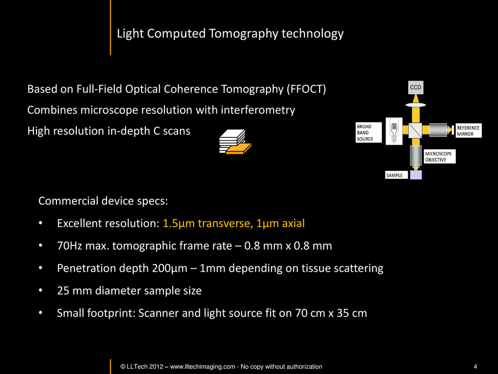
4 Based on Full-Field Optical Coherence Tomography (FFOCT) Combines microscope resolution with interferometry High resolution in-depth C scans Commercial device specs: • Excellent resolution: 1.5µm transverse, 1µm axial • 70Hz max. tomographic frame rate – 0.8 mm x 0.8 mm • Penetration depth 200µm – 1mm depending on tissue scattering • 25 mm diameter sample size • Small footprint: Scanner and light source fit on 70 cm x 35 cm Light Computed Tomography technology -
© LLTech 2012 – www.lltechimaging.com - No copy without authorization
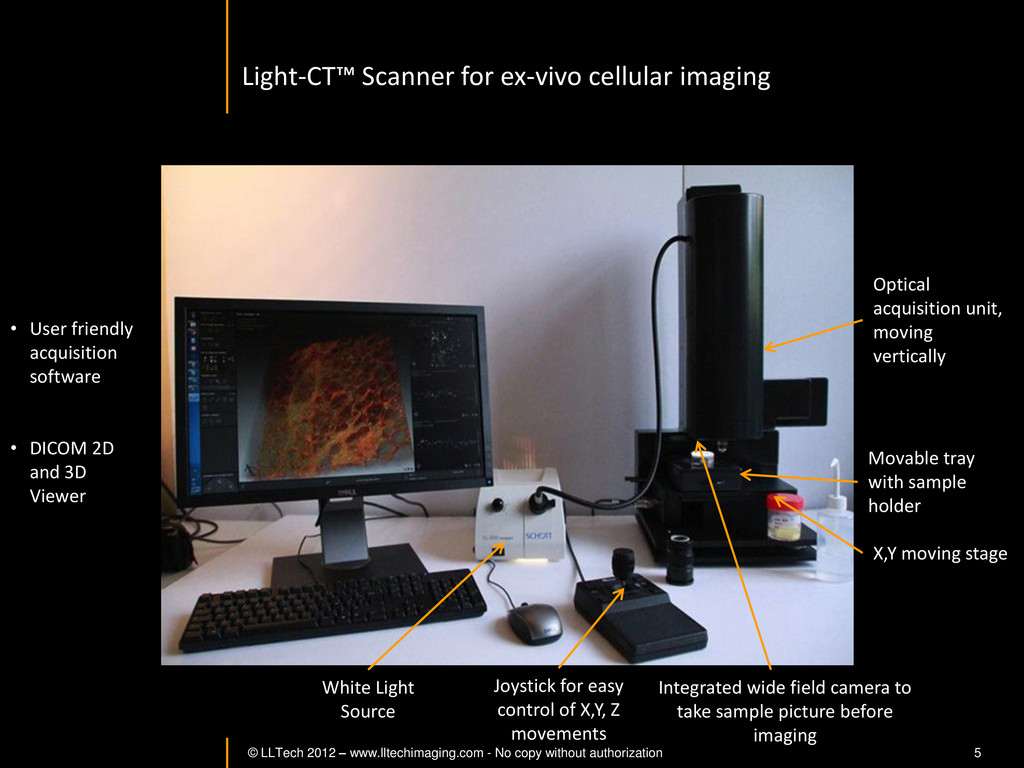
5 Optical acquisition unit, moving vertically • User friendly acquisition software • DICOM 2D and 3D Viewer Movable tray with sample holder X,Y moving stage Joystick for easy control of X,Y, Z movements White Light Source Integrated wide field camera to take sample picture before imaging Light-CT™ Scanner for ex-vivo cellular imaging -
© LLTech 2012 – www.lltechimaging.com - No copy without authorization
6 BREAST IMAGING BREAST IMAGING Back to table of contents -
© LLTech 2012 – www.lltechimaging.com - No copy without authorization
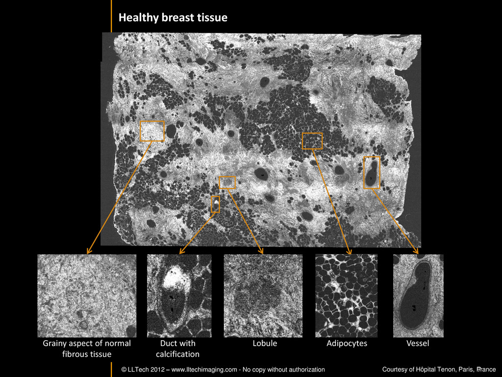
7 Grainy aspect of normal fibrous tissue Duct with calcification Lobule Adipocytes Vessel Courtesy of Hôpital Tenon, Paris, France Healthy breast tissue -
© LLTech 2012 – www.lltechimaging.com - No copy without authorization
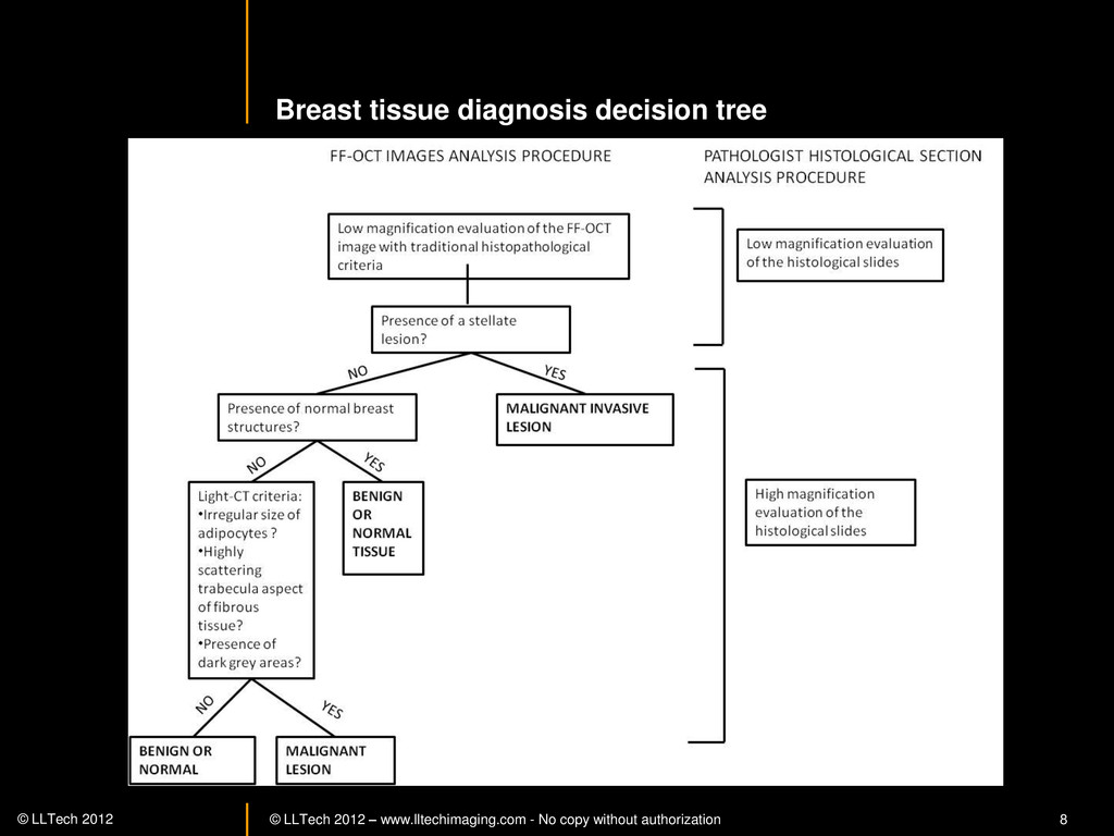
8 © LLTech 2012 Figure 3 : Diagnosis decision tree for FF-OCT images on human breast tissue Breast tissue diagnosis decision tree -
© LLTech 2012 – www.lltechimaging.com - No copy without authorization
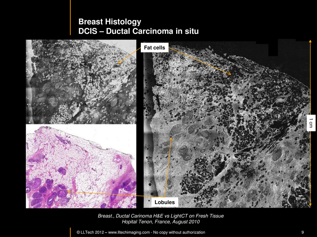
9 Breast Histology DCIS – Ductal Carcinoma in situ Breast., Ductal Carinoma H&E vs LightCT on Fresh Tissue Hopital Tenon, France, August 2010 Lobules Fat cells 1 cm -
© LLTech 2012 – www.lltechimaging.com - No copy without authorization
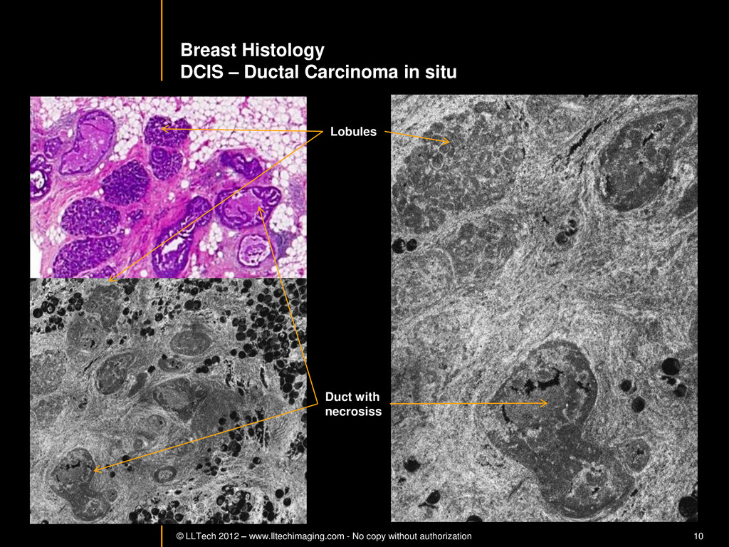
10 Breast Histology DCIS – Ductal Carcinoma in situ Lobules Duct with necrosiss -
© LLTech 2012 – www.lltechimaging.com - No copy without authorization
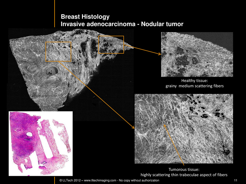
11 Breast Histology Invasive adenocarcinoma - Nodular tumor Tumorous tissue: highly scattering thin trabeculae aspect of fibers Healthy tissue: grainy medium scattering fibers -
© LLTech 2012 – www.lltechimaging.com - No copy without authorization
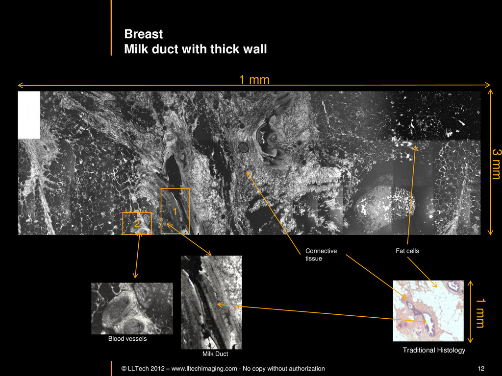
12 Breast Milk duct with thick wall 1 mm 3 mm 1 Milk Duct 2 Blood vessels Traditional Histology 1 mm Connective tissue Fat cells -
© LLTech 2012 – www.lltechimaging.com - No copy without authorization
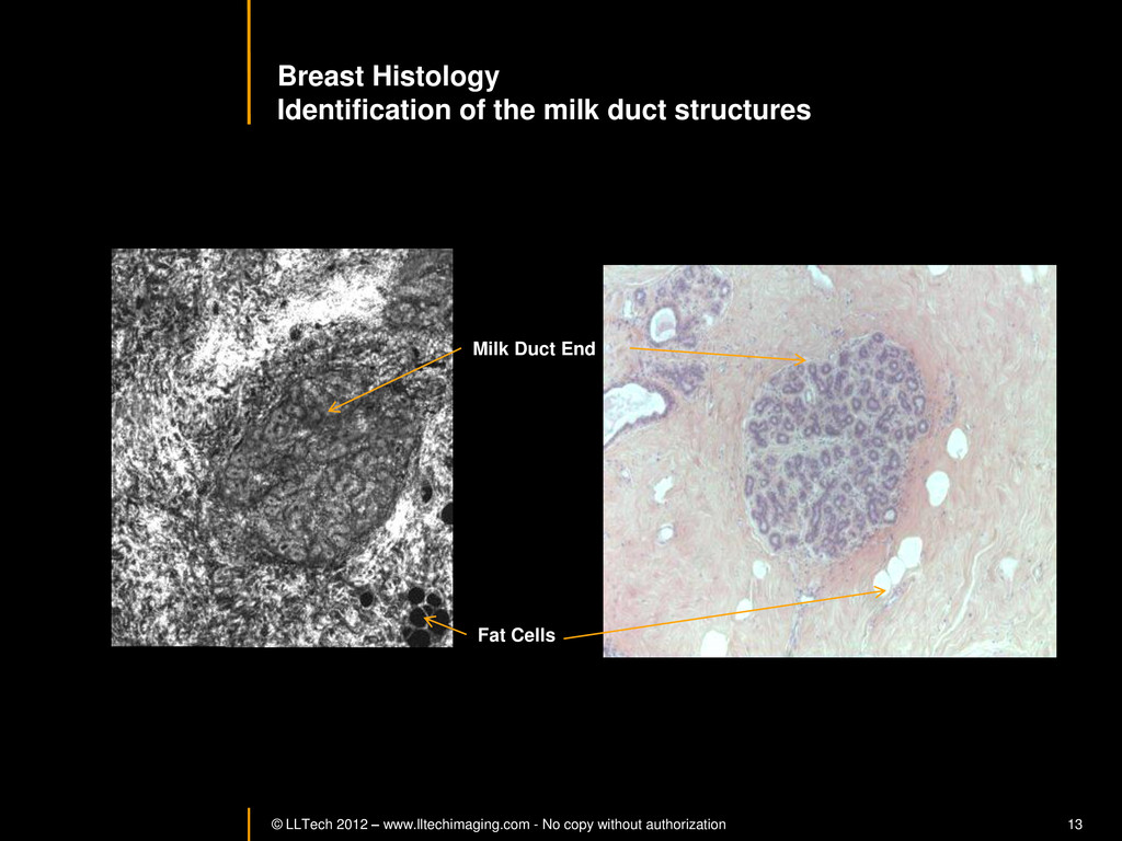
13 Breast Histology Identification of the milk duct structures Milk Duct End Fat Cells -
© LLTech 2012 – www.lltechimaging.com - No copy without authorization
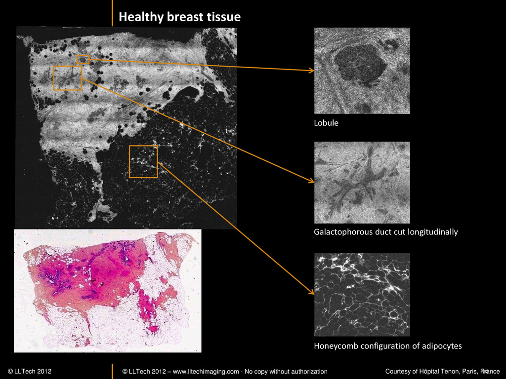
14 Healthy breast tissue Lobule Galactophorous duct cut longitudinally Honeycomb configuration of adipocytes © LLTech 2012 Courtesy of Hôpital Tenon, Paris, France -
© LLTech 2012 – www.lltechimaging.com - No copy without authorization
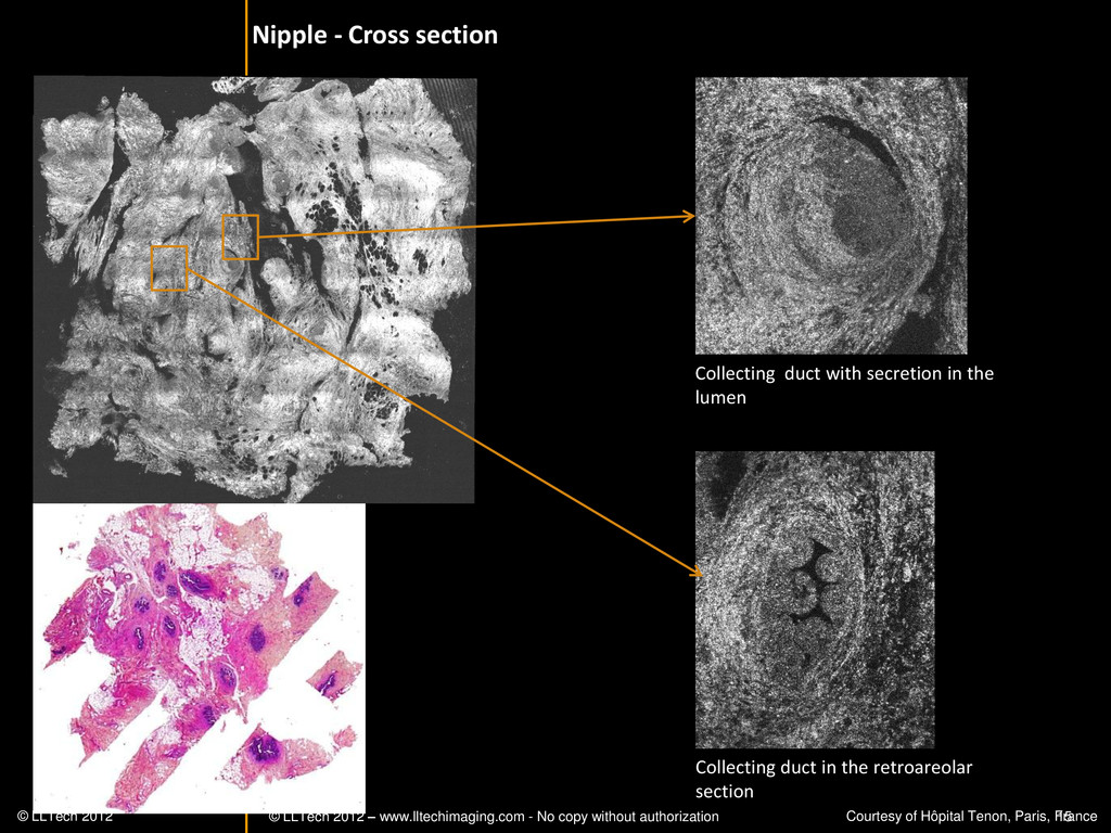
15 Collecting duct with secretion in the lumen Collecting duct in the retroareolar section Nipple - Cross section © LLTech 2012 Courtesy of Hôpital Tenon, Paris, France -
© LLTech 2012 – www.lltechimaging.com - No copy without authorization
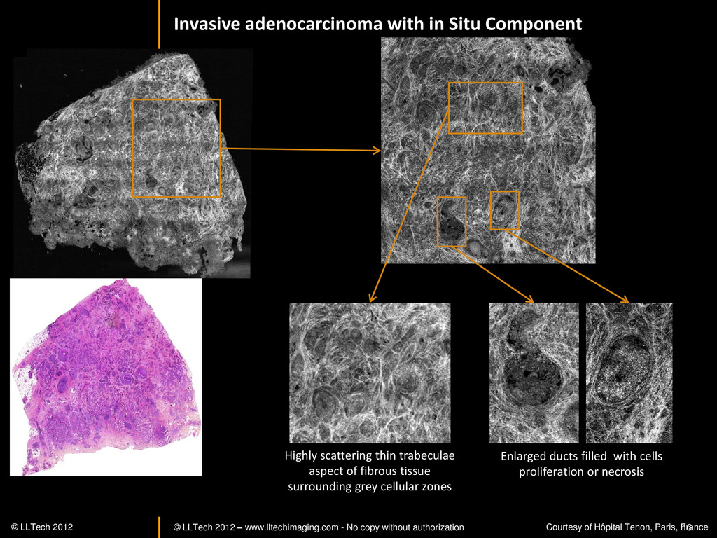
16 Invasive adenocarcinoma with in Situ Component Enlarged ducts filled with cells proliferation or necrosis Highly scattering thin trabeculae aspect of fibrous tissue surrounding grey cellular zones © LLTech 2012 Courtesy of Hôpital Tenon, Paris, France -
© LLTech 2012 – www.lltechimaging.com - No copy without authorization
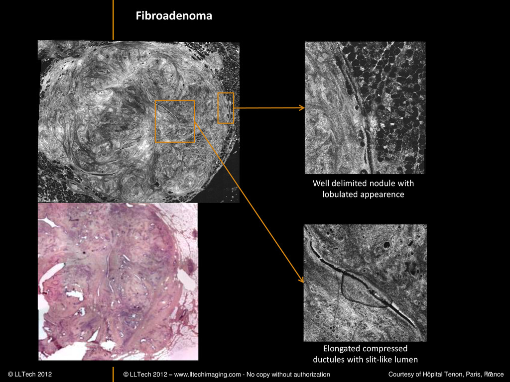
17 Elongated compressed ductules with slit-like lumen Well delimited nodule with lobulated appearence Fibroadenoma © LLTech 2012 Courtesy of Hôpital Tenon, Paris, France -
© LLTech 2012 – www.lltechimaging.com - No copy without authorization
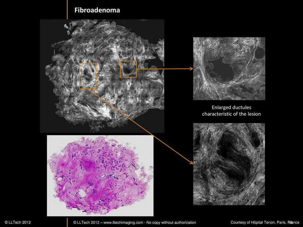
18 Fibroadenoma Enlarged ductules characteristic of the lesion © LLTech 2012 Courtesy of Hôpital Tenon, Paris, France -
© LLTech 2012 – www.lltechimaging.com - No copy without authorization
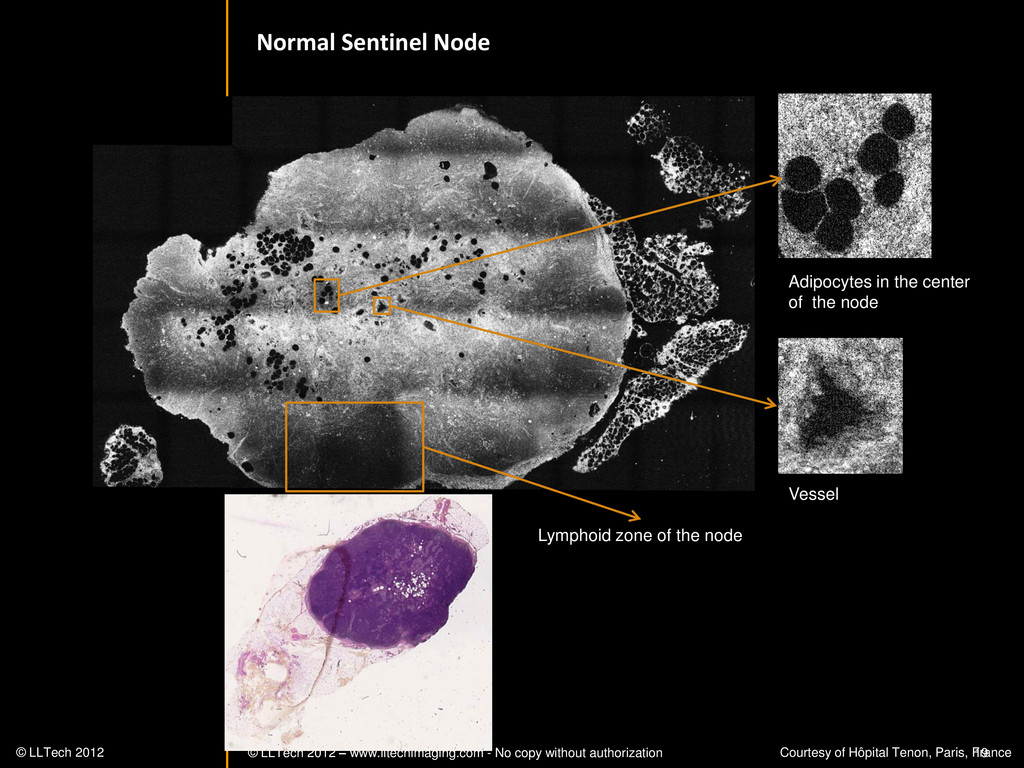
19 Lymphoid zone of the node Vessel Adipocytes in the center of the node Normal Sentinel Node © LLTech 2012 Courtesy of Hôpital Tenon, Paris, France -
© LLTech 2012 – www.lltechimaging.com - No copy without authorization
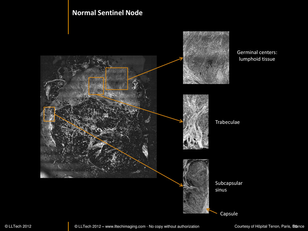
20 Subcapsular sinus Capsule Germinal centers: lumphoid tissue Trabeculae Normal Sentinel Node © LLTech 2012 Courtesy of Hôpital Tenon, Paris, France -
© LLTech 2012 – www.lltechimaging.com - No copy without authorization
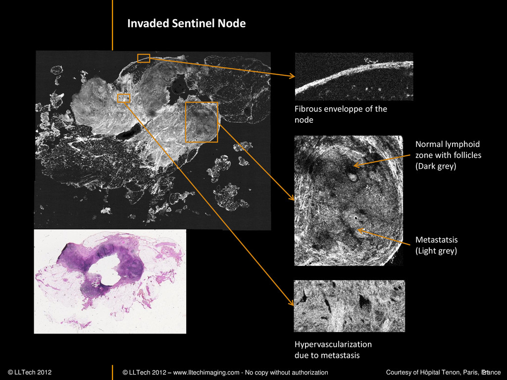
21 Normal lymphoid zone with follicles (Dark grey) Metastatsis (Light grey) Hypervascularization due to metastasis Fibrous enveloppe of the node Invaded Sentinel Node © LLTech 2012 Courtesy of Hôpital Tenon, Paris, France -
© LLTech 2012 – www.lltechimaging.com - No copy without authorization
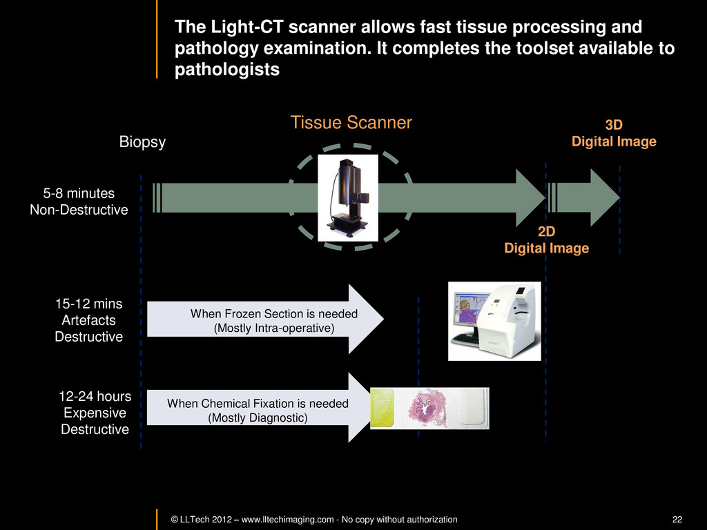
22 The Light-CT scanner allows fast tissue processing and pathology examination. It completes the toolset available to pathologists When Chemical Fixation is needed (Mostly Diagnostic) When Frozen Section is needed (Mostly Intra-operative) Biopsy 3D Digital Image 2D Digital Image Slide 15-12 mins Artefacts Destructive 12-24 hours Expensive Destructive 5-8 minutes Non-Destructive Tissue Scanner -
© LLTech 2012 – www.lltechimaging.com - No copy without authorization
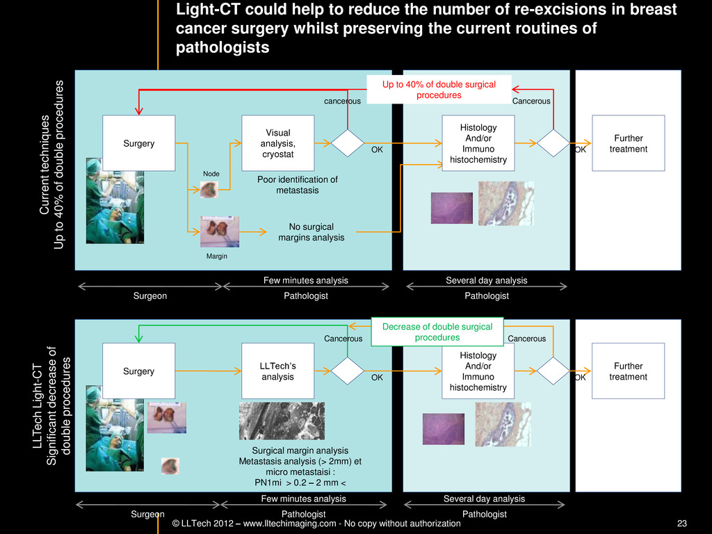
23 Light-CT could help to reduce the number of re-excisions in breast cancer surgery whilst preserving the current routines of pathologists Per surgery LLTech’s analysis Histology And/or Immuno histochemistry Further treatment Post surgery Surgical margin analysis Metastasis analysis (> 2mm) et micro metastaisi : PN1mi > 0.2 – 2 mm < Surgery OK OK Per surgery Post Surgery Current techniques Up to 40% of double procedures LLTech Light-CT Significant decrease of double procedures Visual analysis, cryostat Further treatment Surgery OK Cancerous cancerous OK Poor identification of metastasis Up to 40% of double surgical procedures Several day analysis Few minutes analysis Surgeon Pathologist Pathologist Decrease of double surgical procedures Cancerous Cancerous No surgical margins analysis Histology And/or Immuno histochemistry Node Margin Several day analysis Few minutes analysis Surgeon Pathologist Pathologist -
© LLTech 2012 – www.lltechimaging.com - No copy without authorization
24 BRAIN IMAGING Back to table of contents -
© LLTech 2012 – www.lltechimaging.com - No copy without authorization
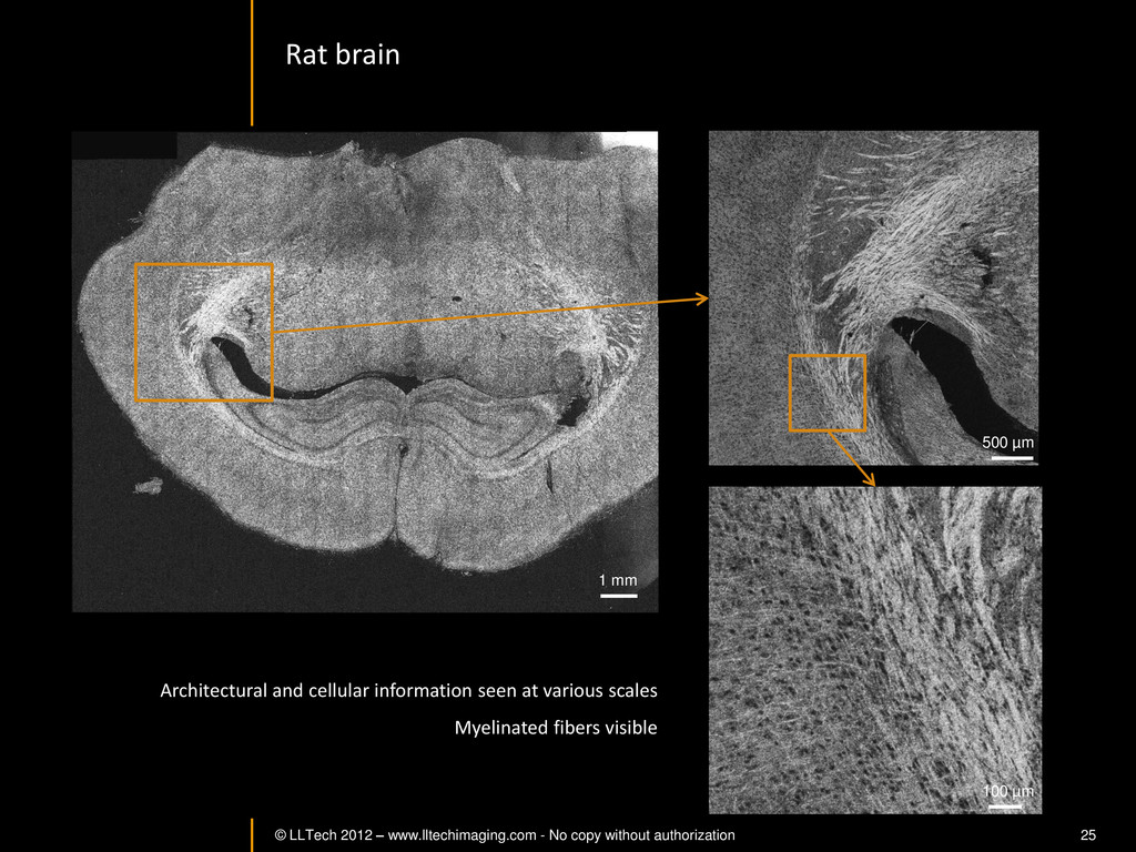
25 Rat brain Architectural and cellular information seen at various scales Myelinated fibers visible 1 mm 500 µm 100 µm -
© LLTech 2012 – www.lltechimaging.com - No copy without authorization
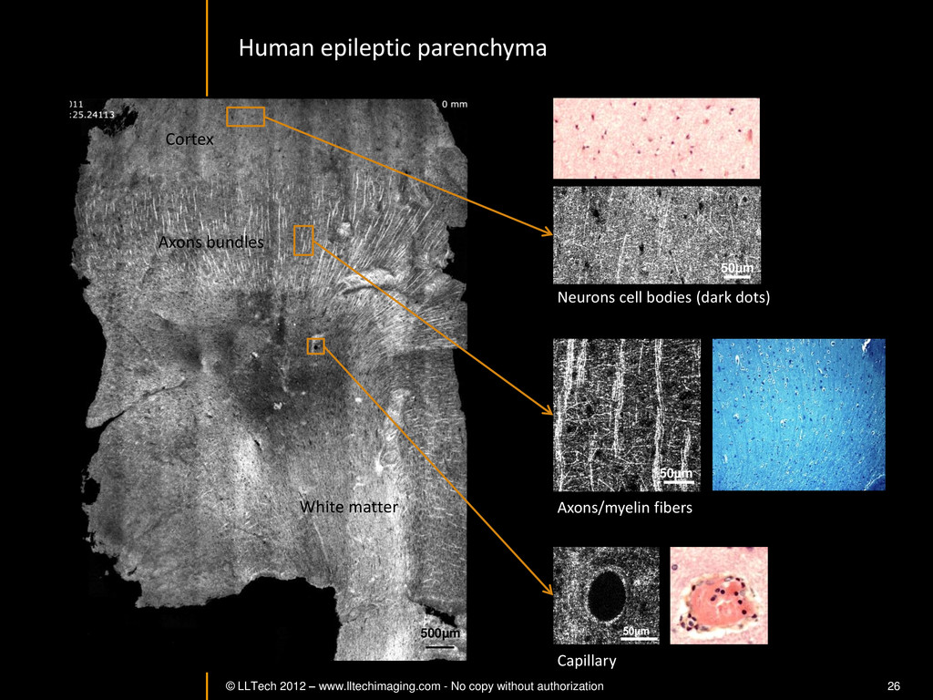
26 Neurons cell bodies (dark dots) Axons/myelin fibers Capillary 50µm 50µm 50µm 500µm Human epileptic parenchyma Cortex White matter Axons bundles -
© LLTech 2012 – www.lltechimaging.com - No copy without authorization
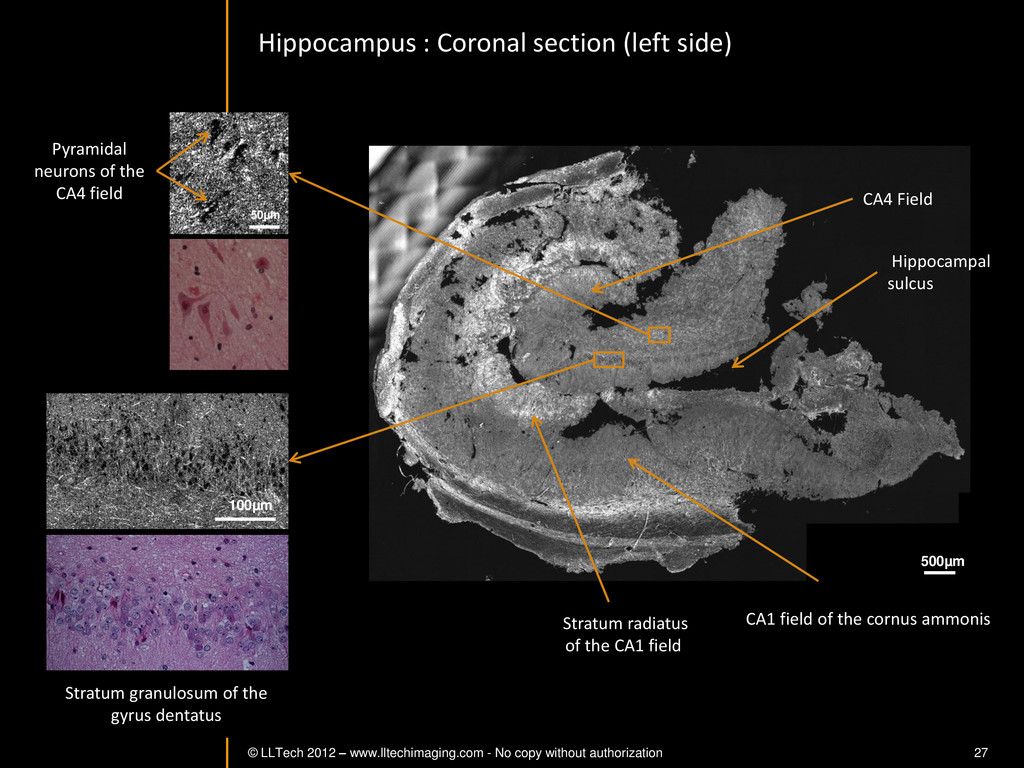
27 Stratum granulosum of the gyrus dentatus Hippocampus : Coronal section (left side) 100µm 500µm CA4 Field CA1 field of the cornus ammonis Hippocampal sulcus Stratum radiatus of the CA1 field Pyramidal neurons of the CA4 field 50µm -
© LLTech 2012 – www.lltechimaging.com - No copy without authorization
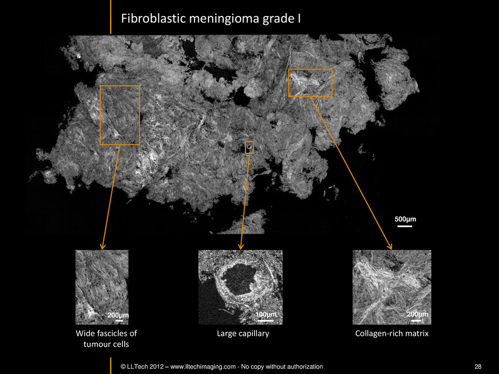
28 © LLTECH 2011 Collagen-rich matrix Wide fascicles of tumour cells Large capillary Fibroblastic meningioma grade I 500µm 100µm 200µm 200µm -
© LLTech 2012 – www.lltechimaging.com - No copy without authorization
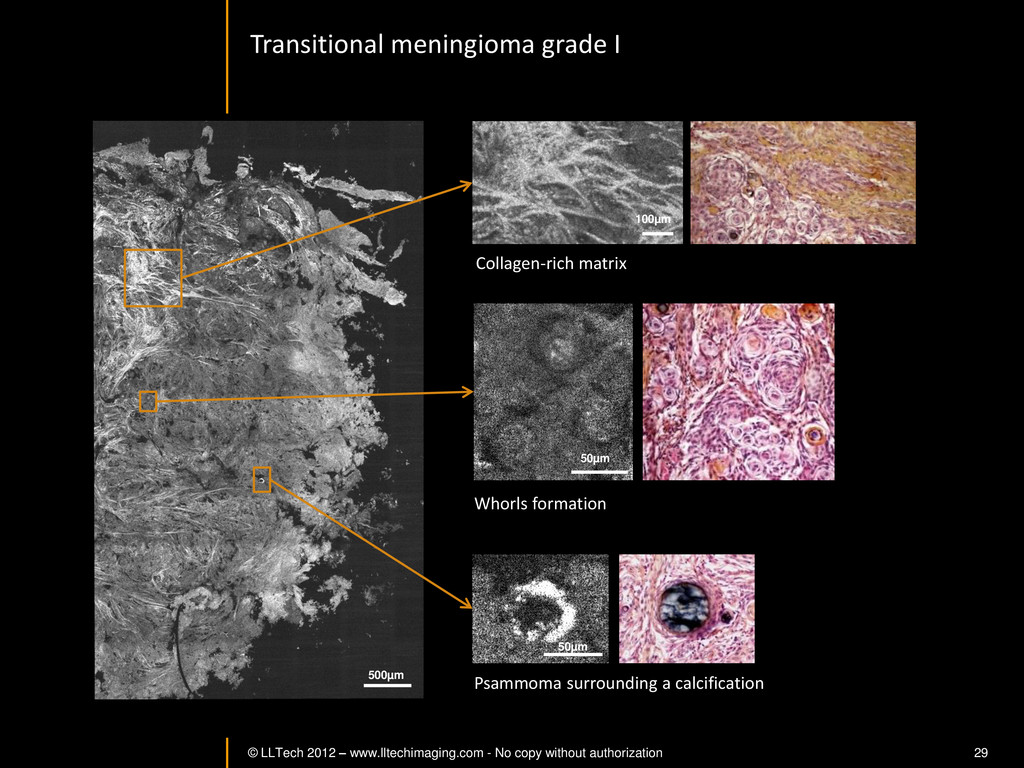
29 Transitional meningioma grade I Psammoma surrounding a calcification Whorls formation Collagen-rich matrix 500µm 100µm 50µm 50µm