project. We are creating a platform for collecting and analysis of clinical neuroscience data. • Partners: leading russian medical centers, including ◦ Burdenko Neurosurgery Institute ◦ Scientific Center of Neurology ◦ And more • Data: ◦ More than 50000 patients with MRI / CT imaging ◦ Other modalities: сlinical data, genetics, EEG data. • Algorithms: specially developed algorithms for medical data analysis • Hardware: 2 PB of storage & heavy computational power
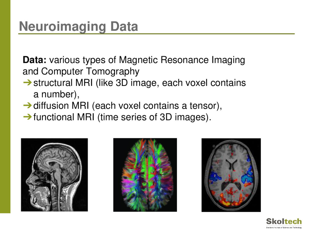
➔structural MRI (like 3D image, each voxel contains a number), ➔diffusion MRI (each voxel contains a tensor), ➔functional MRI (time series of 3D images). Neuroimaging Data
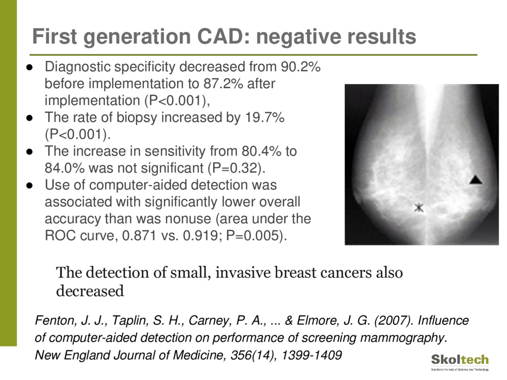
H., Carney, P. A., ... & Elmore, J. G. (2007). Influence of computer-aided detection on performance of screening mammography. New England Journal of Medicine, 356(14), 1399-1409 • Diagnostic specificity decreased from 90.2% before implementation to 87.2% after implementation (P<0.001), • The rate of biopsy increased by 19.7% (P<0.001). • The increase in sensitivity from 80.4% to 84.0% was not significant (P=0.32). • Use of computer-aided detection was associated with significantly lower overall accuracy than was nonuse (area under the ROC curve, 0.871 vs. 0.919; P=0.005). The detection of small, invasive breast cancers also decreased
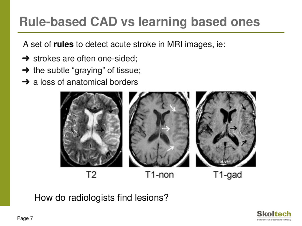
images, ie: ➜ strokes are often one-sided; ➜ the subtle “graying” of tissue; ➜ a loss of anatomical borders Page 7 Rule-based CAD vs learning based ones How do radiologists find lesions?
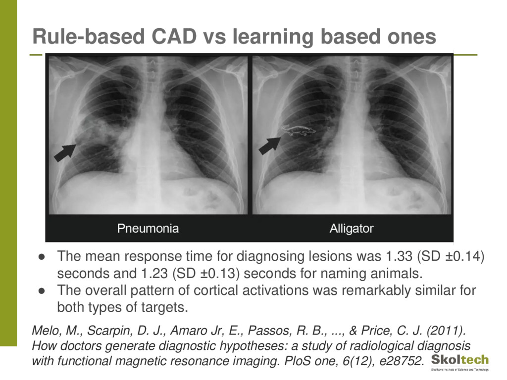
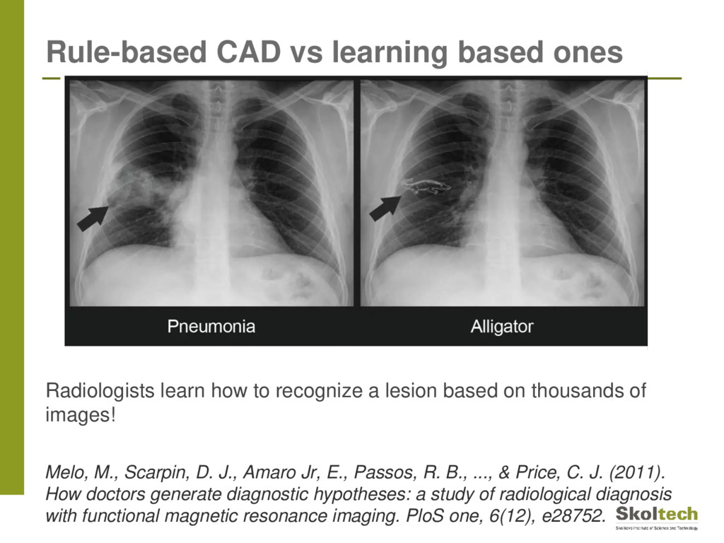
J., Amaro Jr, E., Passos, R. B., ..., & Price, C. J. (2011). How doctors generate diagnostic hypotheses: a study of radiological diagnosis with functional magnetic resonance imaging. PloS one, 6(12), e28752. • The mean response time for diagnosing lesions was 1.33 (SD ±0.14) seconds and 1.23 (SD ±0.13) seconds for naming animals. • The overall pattern of cortical activations was remarkably similar for both types of targets.
J., Amaro Jr, E., Passos, R. B., ..., & Price, C. J. (2011). How doctors generate diagnostic hypotheses: a study of radiological diagnosis with functional magnetic resonance imaging. PloS one, 6(12), e28752. Radiologists learn how to recognize a lesion based on thousands of images!
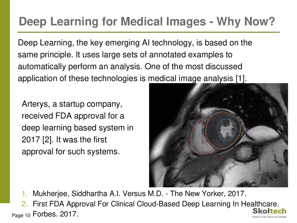
the same principle. It uses large sets of annotated examples to automatically perform an analysis. One of the most discussed application of these technologies is medical image analysis [1]. 1. Mukherjee, Siddhartha A.I. Versus M.D. - The New Yorker, 2017. 2. First FDA Approval For Clinical Cloud-Based Deep Learning In Healthcare. Forbes. 2017. Page 10 Deep Learning for Medical Images - Why Now? Arterys, a startup company, received FDA approval for a deep learning based system in 2017 [2]. It was the first approval for such systems.
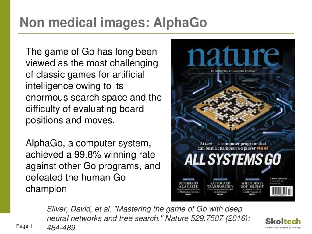
has long been viewed as the most challenging of classic games for artificial intelligence owing to its enormous search space and the difficulty of evaluating board positions and moves. AlphaGo, a computer system, achieved a 99.8% winning rate against other Go programs, and defeated the human Go champion Silver, David, et al. "Mastering the game of Go with deep neural networks and tree search." Nature 529.7587 (2016): 484-489.
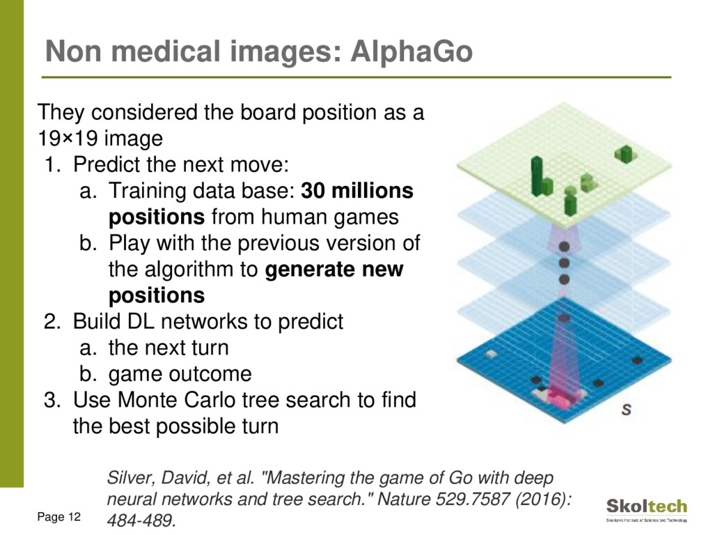
position as a 19×19 image 1. Predict the next move: a. Training data base: 30 millions positions from human games b. Play with the previous version of the algorithm to generate new positions 2. Build DL networks to predict a. the next turn b. game outcome 3. Use Monte Carlo tree search to find the best possible turn Silver, David, et al. "Mastering the game of Go with deep neural networks and tree search." Nature 529.7587 (2016): 484-489.
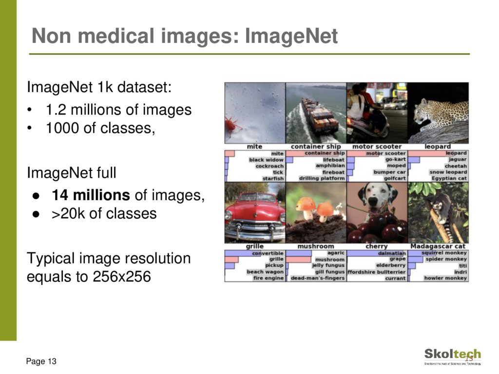
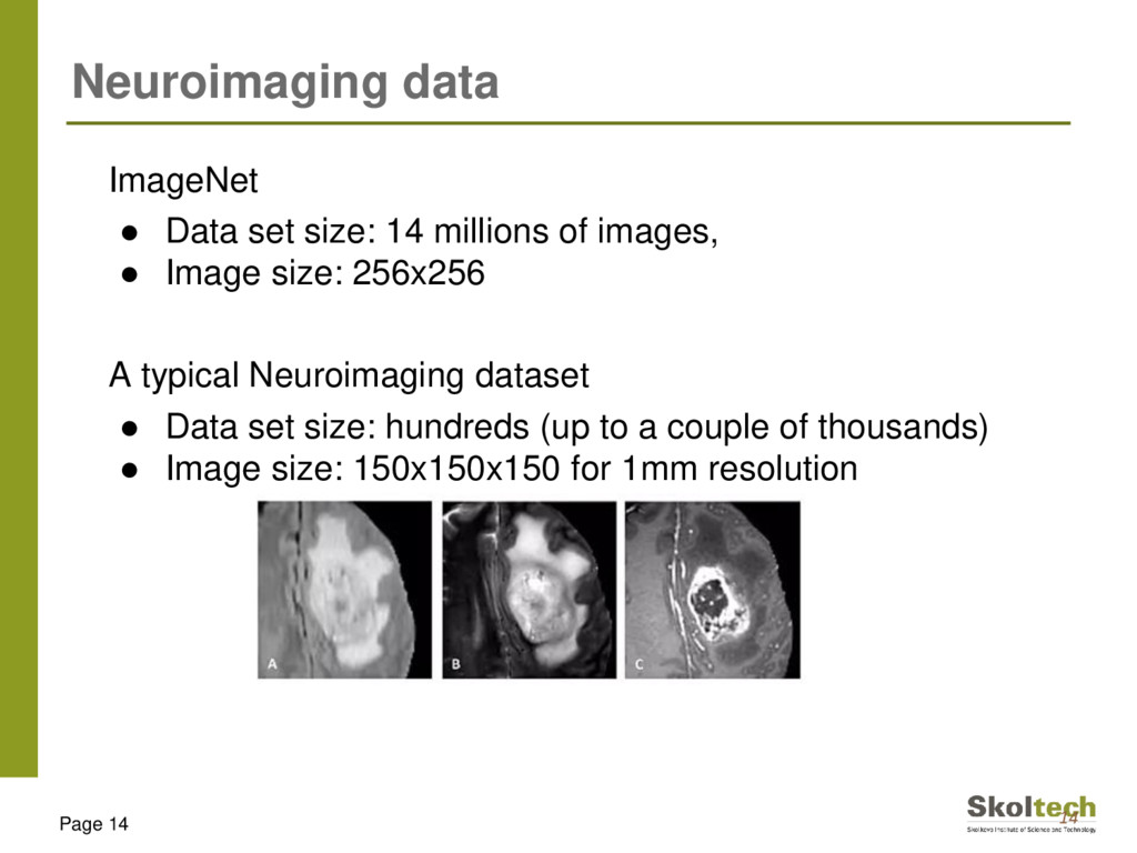
of images, • Image size: 256x256 A typical Neuroimaging dataset • Data set size: hundreds (up to a couple of thousands) • Image size: 150x150x150 for 1mm resolution Page 14
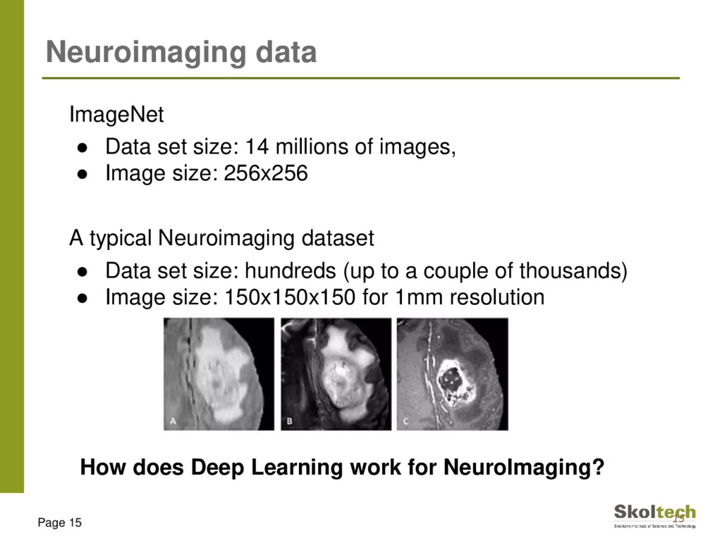
of images, • Image size: 256x256 A typical Neuroimaging dataset • Data set size: hundreds (up to a couple of thousands) • Image size: 150x150x150 for 1mm resolution Page 15 How does Deep Learning work for NeuroImaging?
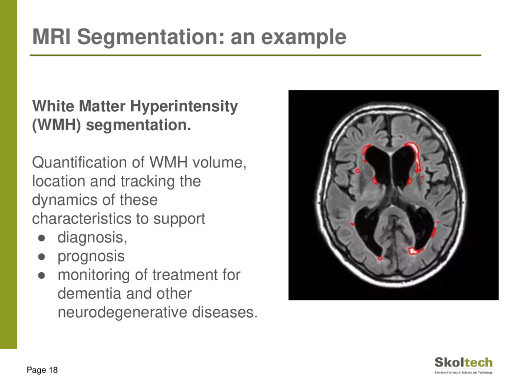
segmentation. Quantification of WMH volume, location and tracking the dynamics of these characteristics to support • diagnosis, • prognosis • monitoring of treatment for dementia and other neurodegenerative diseases.
standardized than other images: • The target is usually located in the centre • There is a predefined number of their instances in each image There are two opposite strategies to deal with data variability: • decrease variability in all data by some data preprocessing or • use extensive data augmentation, increasing data variability and the size of the training set. The medical image computing community historically prefers preprocessing.
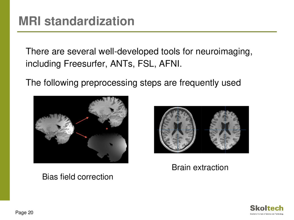
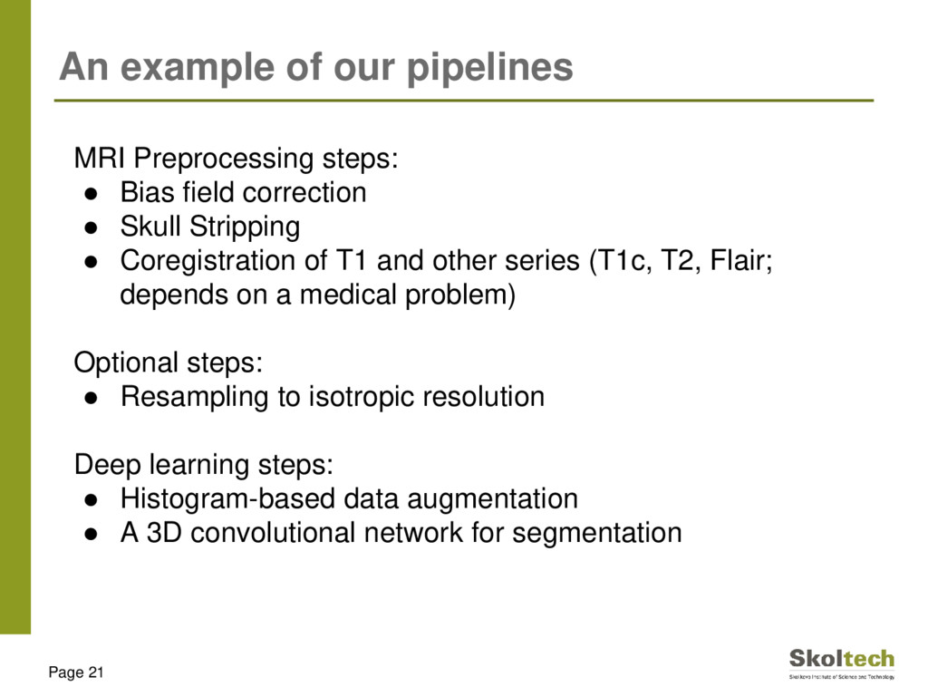
• Bias field correction • Skull Stripping • Coregistration of T1 and other series (T1c, T2, Flair; depends on a medical problem) Optional steps: • Resampling to isotropic resolution Deep learning steps: • Histogram-based data augmentation • A 3D convolutional network for segmentation
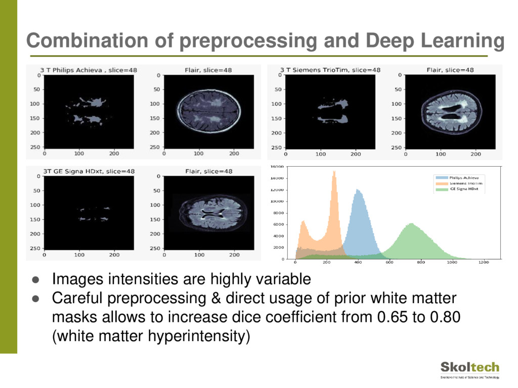
direct usage of prior white matter masks allows to increase dice coefficient from 0.65 to 0.80 (white matter hyperintensity) Combination of preprocessing and Deep Learning
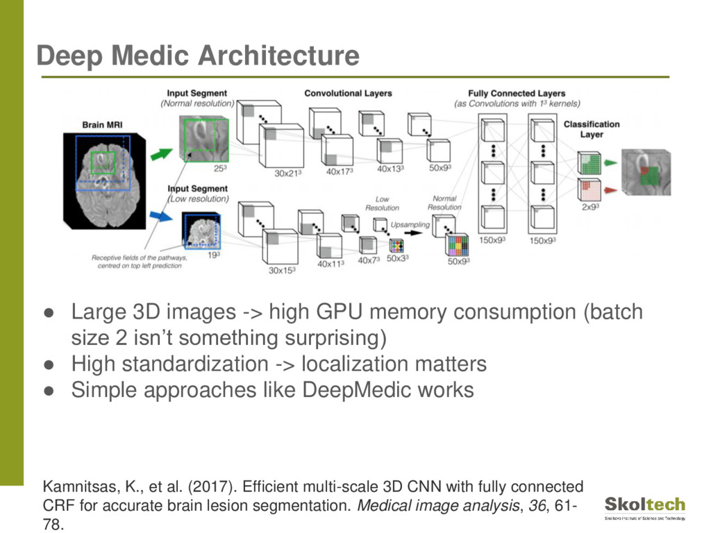
size 2 isn’t something surprising) • High standardization -> localization matters • Simple approaches like DeepMedic works Deep Medic Architecture Kamnitsas, K., et al. (2017). Efficient multi-scale 3D CNN with fully connected CRF for accurate brain lesion segmentation. Medical image analysis, 36, 61- 78.
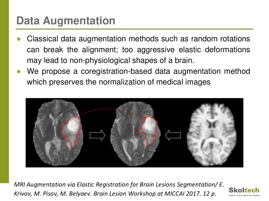
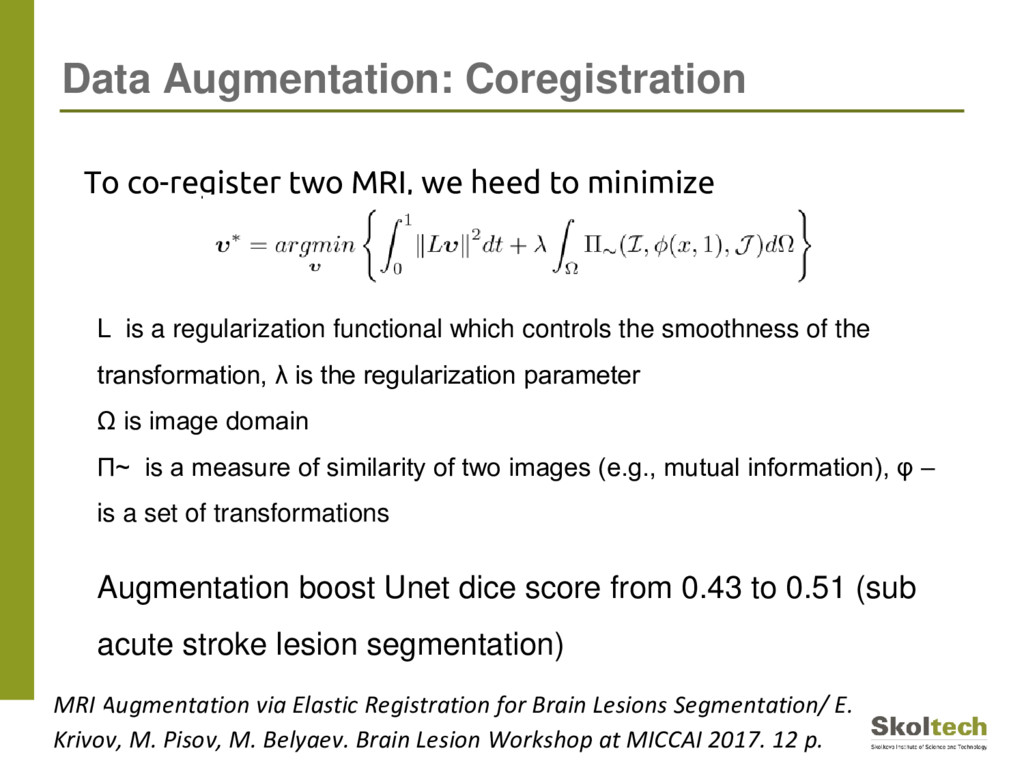
break the alignment; too aggressive elastic deformations may lead to non-physiological shapes of a brain. • We propose a coregistration-based data augmentation method which preserves the normalization of medical images Data Augmentation MRI Augmentation via Elastic Registration for Brain Lesions Segmentation/ E. Krivov, M. Pisov, M. Belyaev. Brain Lesion Workshop at MICCAI 2017. 12 p.
Lesions Segmentation/ E. Krivov, M. Pisov, M. Belyaev. Brain Lesion Workshop at MICCAI 2017. 12 p. To co-register two MRI, we heed to minimize L is a regularization functional which controls the smoothness of the transformation, λ is the regularization parameter Ω is image domain П~ is a measure of similarity of two images (e.g., mutual information), φ – is a set of transformations Augmentation boost Unet dice score from 0.43 to 0.51 (sub acute stroke lesion segmentation)
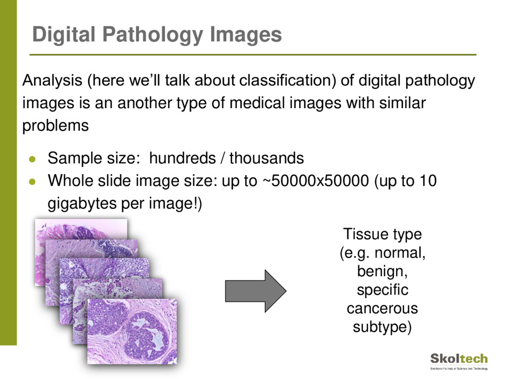
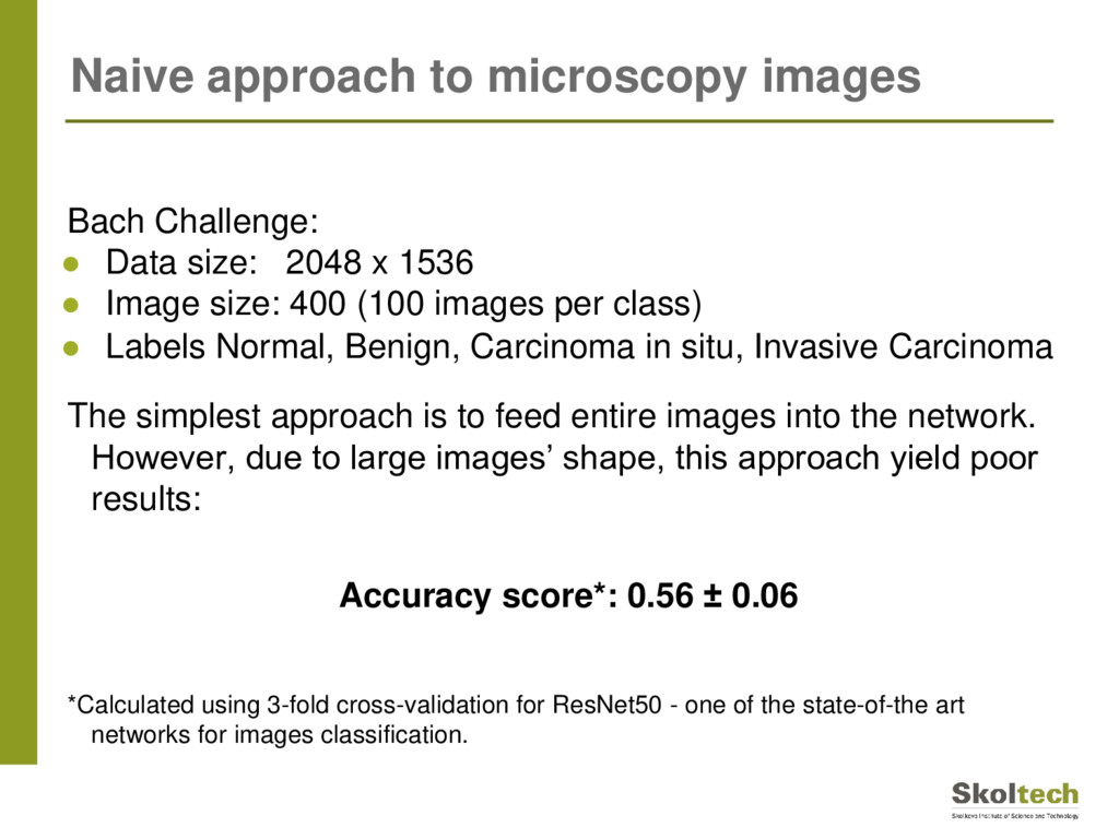
is an another type of medical images with similar problems • Sample size: hundreds / thousands • Whole slide image size: up to ~50000x50000 (up to 10 gigabytes per image!) Tissue type (e.g. normal, benign, specific cancerous subtype) Digital Pathology Images
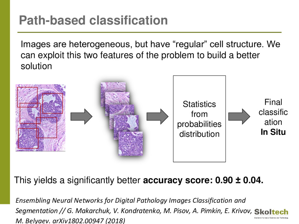
size: 400 (100 images per class) • Labels Normal, Benign, Carcinoma in situ, Invasive Carcinoma The simplest approach is to feed entire images into the network. However, due to large images’ shape, this approach yield poor results: Accuracy score*: 0.56 ± 0.06 *Calculated using 3-fold cross-validation for ResNet50 - one of the state-of-the art networks for images classification. Naive approach to microscopy images
exploit this two features of the problem to build a better solution Statistics from probabilities distribution Final classific ation In Situ This yields a significantly better accuracy score: 0.90 ± 0.04. Path-based classification Ensembling Neural Networks for Digital Pathology Images Classification and Segmentation // G. Makarchuk, V. Kondratenko, M. Pisov, A. Pimkin, E. Krivov, M. Belyaev. arXiv1802.00947 (2018)
to limited amount of data available • Deep learning still is a very powerful set of methods especially if data is carefully prepared and network architectures are carefully adopted
to limited amount of data available • Deep learning still is a very powerful set of methods especially if data is carefully prepared and network architectures are carefully adopted Cobrain-Analytics project is looking for a business partners who wants to build products and services based on brain-related data analysis.
![Thank you! Mikhail Belyaev [email protected]](https://files.speakerdeck.com/presentations/76b817ee7b1446f1941dcce72927df0c/slide_31.jpg)