Upgrade to Pro — share decks privately, control downloads, hide ads and more …

JTGGA_5_sup1_25_27.pdf

shyza82
October 11, 2011

 JTGGA_5_sup1_25_27.pdf

shyza82

October 11, 2011
Tweet

More Decks by shyza82

Other Decks in Education

Transcript

  1. 25 J TURKISH GERMAN GYNECOL ASSOC, Vol. 5, Suppl. 1,

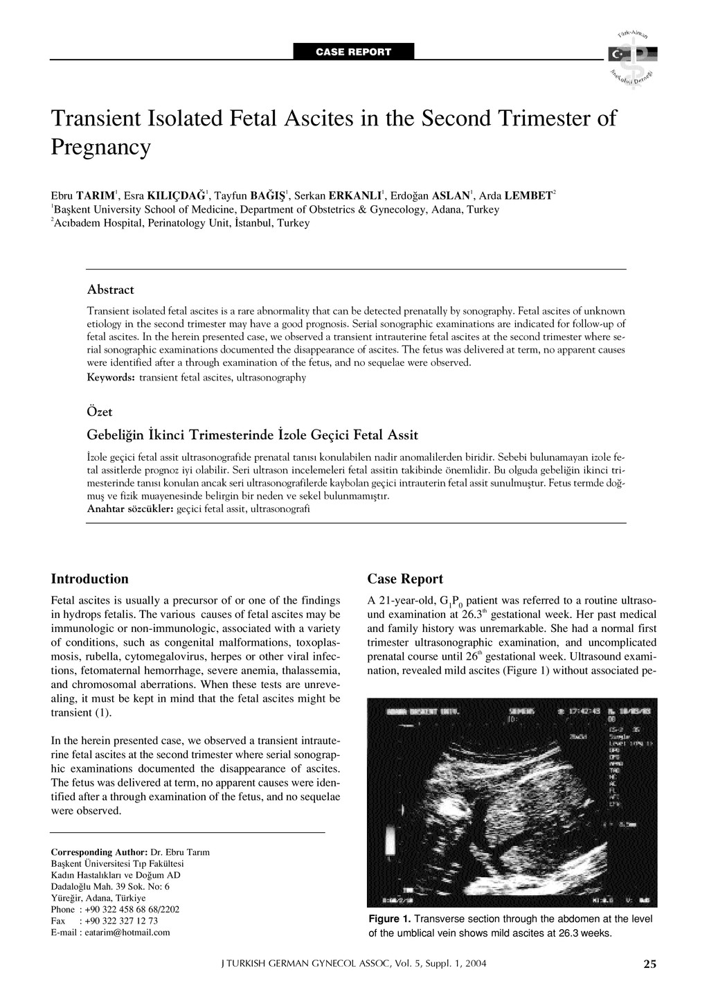
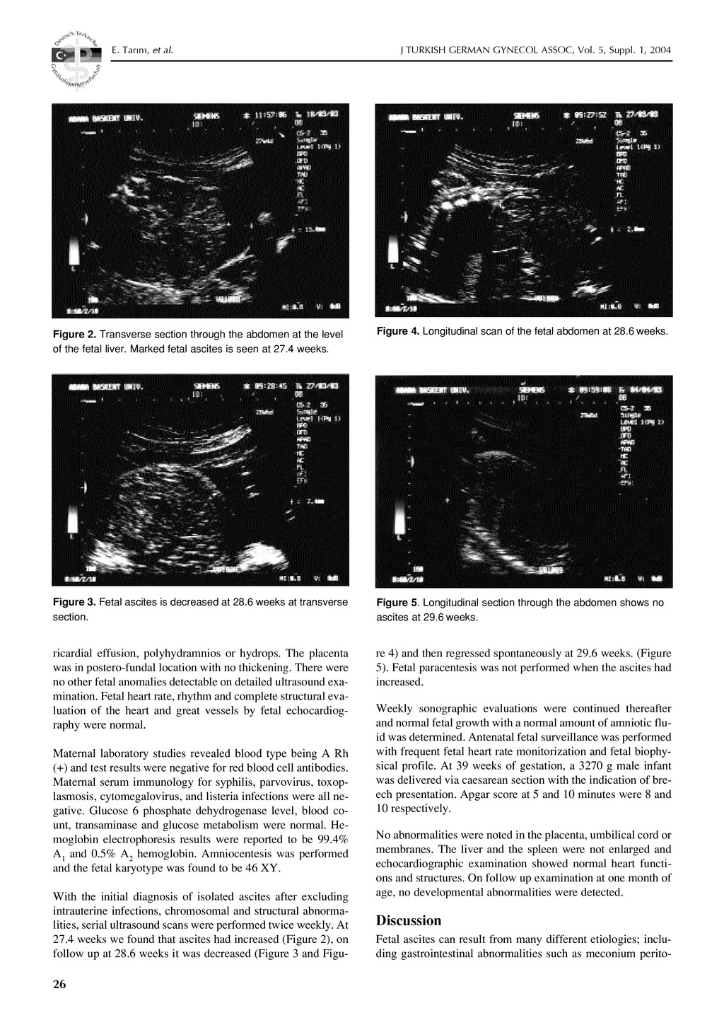
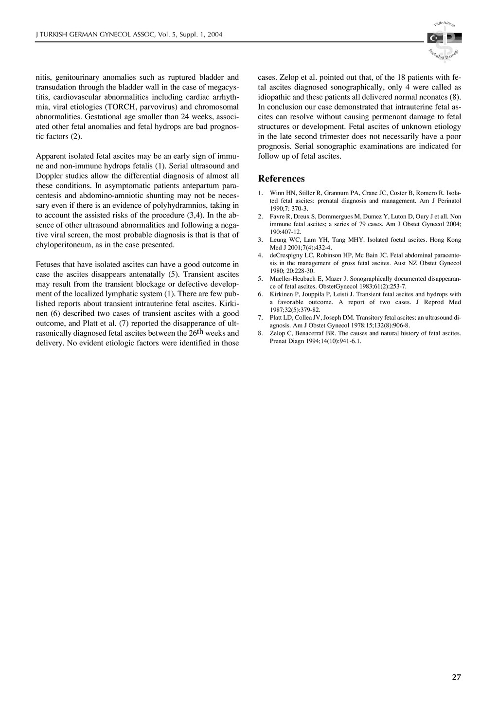
    2004 Introduction Fetal ascites is usually a precursor of or one of the findings in hydrops fetalis. The various causes of fetal ascites may be immunologic or non-immunologic, associated with a variety of conditions, such as congenital malformations, toxoplas- mosis, rubella, cytomegalovirus, herpes or other viral infec- tions, fetomaternal hemorrhage, severe anemia, thalassemia, and chromosomal aberrations. When these tests are unreve- aling, it must be kept in mind that the fetal ascites might be transient (1). In the herein presented case, we observed a transient intraute- rine fetal ascites at the second trimester where serial sonograp- hic examinations documented the disappearance of ascites. The fetus was delivered at term, no apparent causes were iden- tified after a through examination of the fetus, and no sequelae were observed. Case Report A 21-year-old, G 1 P 0 patient was referred to a routine ultraso- und examination at 26.3th gestational week. Her past medical and family history was unremarkable. She had a normal first trimester ultrasonographic examination, and uncomplicated prenatal course until 26th gestational week. Ultrasound exami- nation, revealed mild ascites (Figure 1) without associated pe- Transient Isolated Fetal Ascites in the Second Trimester of Pregnancy Ebru TARIM1, Esra KILIÇDA⁄1, Tayfun BA⁄Ifi1, Serkan ERKANLI1, Erdo¤an ASLAN1, Arda LEMBET2 1Baflkent University School of Medicine, Department of Obstetrics & Gynecology, Adana, Turkey 2Ac›badem Hospital, Perinatology Unit, ‹stanbul, Turkey Abstract Transient isolated fetal ascites is a rare abnormality that can be detected prenatally by sonography. Fetal ascites of unknown etiology in the second trimester may have a good prognosis. Serial sonographic examinations are indicated for follow-up of fetal ascites. In the herein presented case, we observed a transient intrauterine fetal ascites at the second trimester where se- rial sonographic examinations documented the disappearance of ascites. The fetus was delivered at term, no apparent causes were identified after a through examination of the fetus, and no sequelae were observed. Keywords: transient fetal ascites, ultrasonography Özet Gebeli¤in ‹kinci Trimesterinde ‹zole Geçici Fetal Assit ‹zole geçici fetal assit ultrasonografide prenatal tan›s› konulabilen nadir anomalilerden biridir. Sebebi bulunamayan izole fe- tal assitlerde prognoz iyi olabilir. Seri ultrason incelemeleri fetal assitin takibinde önemlidir. Bu olguda gebeli¤in ikinci tri- mesterinde tan›s› konulan ancak seri ultrasonografilerde kaybolan geçici intrauterin fetal assit sunulmufltur. Fetus termde do¤- mufl ve fizik muayenesinde belirgin bir neden ve sekel bulunmam›flt›r. Anahtar sözcükler: geçici fetal assit, ultrasonografi Corresponding Author: Dr. Ebru Tar›m Baflkent Üniversitesi T›p Fakültesi Kad›n Hastal›klar› ve Do¤um AD Dadalo¤lu Mah. 39 Sok. No: 6 Yüre¤ir, Adana, Türkiye Phone : +90 322 458 68 68/2202 Fax : +90 322 327 12 73 E-mail : [email protected] Figure 1. Transverse section through the abdomen at the level of the umblical vein shows mild ascites at 26.3 weeks. CASE REPORT
  2. 26 J TURKISH GERMAN GYNECOL ASSOC, Vol. 5, Suppl. 1,

    2004 E. Tar›m, et al. ricardial effusion, polyhydramnios or hydrops. The placenta was in postero-fundal location with no thickening. There were no other fetal anomalies detectable on detailed ultrasound exa- mination. Fetal heart rate, rhythm and complete structural eva- luation of the heart and great vessels by fetal echocardiog- raphy were normal. Maternal laboratory studies revealed blood type being A Rh (+) and test results were negative for red blood cell antibodies. Maternal serum immunology for syphilis, parvovirus, toxop- lasmosis, cytomegalovirus, and listeria infections were all ne- gative. Glucose 6 phosphate dehydrogenase level, blood co- unt, transaminase and glucose metabolism were normal. He- moglobin electrophoresis results were reported to be 99.4% A 1 and 0.5% A 2 hemoglobin. Amniocentesis was performed and the fetal karyotype was found to be 46 XY. With the initial diagnosis of isolated ascites after excluding intrauterine infections, chromosomal and structural abnorma- lities, serial ultrasound scans were performed twice weekly. At 27.4 weeks we found that ascites had increased (Figure 2), on follow up at 28.6 weeks it was decreased (Figure 3 and Figu- re 4) and then regressed spontaneously at 29.6 weeks. (Figure 5). Fetal paracentesis was not performed when the ascites had increased. Weekly sonographic evaluations were continued thereafter and normal fetal growth with a normal amount of amniotic flu- id was determined. Antenatal fetal surveillance was performed with frequent fetal heart rate monitorization and fetal biophy- sical profile. At 39 weeks of gestation, a 3270 g male infant was delivered via caesarean section with the indication of bre- ech presentation. Apgar score at 5 and 10 minutes were 8 and 10 respectively. No abnormalities were noted in the placenta, umbilical cord or membranes. The liver and the spleen were not enlarged and echocardiographic examination showed normal heart functi- ons and structures. On follow up examination at one month of age, no developmental abnormalities were detected. Discussion Fetal ascites can result from many different etiologies; inclu- ding gastrointestinal abnormalities such as meconium perito- Figure 2. Transverse section through the abdomen at the level of the fetal liver. Marked fetal ascites is seen at 27.4 weeks. Figure 5. Longitudinal section through the abdomen shows no ascites at 29.6 weeks. Figure 4. Longitudinal scan of the fetal abdomen at 28.6 weeks. Figure 3. Fetal ascites is decreased at 28.6 weeks at transverse section.
  3. 27 J TURKISH GERMAN GYNECOL ASSOC, Vol. 5, Suppl. 1,

    2004 nitis, genitourinary anomalies such as ruptured bladder and transudation through the bladder wall in the case of megacys- titis, cardiovascular abnormalities including cardiac arrhyth- mia, viral etiologies (TORCH, parvovirus) and chromosomal abnormalities. Gestational age smaller than 24 weeks, associ- ated other fetal anomalies and fetal hydrops are bad prognos- tic factors (2). Apparent isolated fetal ascites may be an early sign of immu- ne and non-immune hydrops fetalis (1). Serial ultrasound and Doppler studies allow the differential diagnosis of almost all these conditions. In asymptomatic patients antepartum para- centesis and abdomino-amniotic shunting may not be neces- sary even if there is an evidence of polyhydramnios, taking in to account the assisted risks of the procedure (3,4). In the ab- sence of other ultrasound abnormalities and following a nega- tive viral screen, the most probable diagnosis is that is that of chyloperitoneum, as in the case presented. Fetuses that have isolated ascites can have a good outcome in case the ascites disappears antenatally (5). Transient ascites may result from the transient blockage or defective develop- ment of the localized lymphatic system (1). There are few pub- lished reports about transient intrauterine fetal ascites. Kirki- nen (6) described two cases of transient ascites with a good outcome, and Platt et al. (7) reported the disapperance of ult- rasonically diagnosed fetal ascites between the 26th weeks and delivery. No evident etiologic factors were identified in those cases. Zelop et al. pointed out that, of the 18 patients with fe- tal ascites diagnosed sonographically, only 4 were called as idiopathic and these patients all delivered normal neonates (8). In conclusion our case demonstrated that intrauterine fetal as- cites can resolve without causing permenant damage to fetal structures or development. Fetal ascites of unknown etiology in the late second trimester does not necessarily have a poor prognosis. Serial sonographic examinations are indicated for follow up of fetal ascites. References 1. Winn HN, Stiller R, Grannum PA, Crane JC, Coster B, Romero R. Isola- ted fetal ascites: prenatal diagnosis and management. Am J Perinatol 1990;7: 370-3. 2. Favre R, Dreux S, Dommergues M, Dumez Y, Luton D, Oury J et all. Non immune fetal ascites; a series of 79 cases. Am J Obstet Gynecol 2004; 190:407-12. 3. Leung WC, Lam YH, Tang MHY. Isolated foetal ascites. Hong Kong Med J 2001;7(4):432-4. 4. deCrespigny LC, Robinson HP, Mc Bain JC. Fetal abdominal paracente- sis in the management of gross fetal ascites. Aust NZ Obstet Gynecol 1980; 20:228-30. 5. Mueller-Heubach E, Mazer J. Sonographically documented disappearan- ce of fetal ascites. ObstetGynecol 1983;61(2):253-7. 6. Kirkinen P, Jouppila P, Leisti J. Transient fetal ascites and hydrops with a favorable outcome. A report of two cases. J Reprod Med 1987;32(5):379-82. 7. Platt LD, Collea JV, Joseph DM. Transitory fetal ascites: an ultrasound di- agnosis. Am J Obstet Gynecol 1978:15;132(8):906-8. 8. Zelop C, Benacerraf BR. The causes and natural history of fetal ascites. Prenat Diagn 1994;14(10):941-6.1.