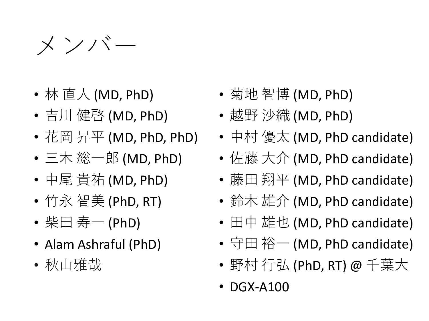
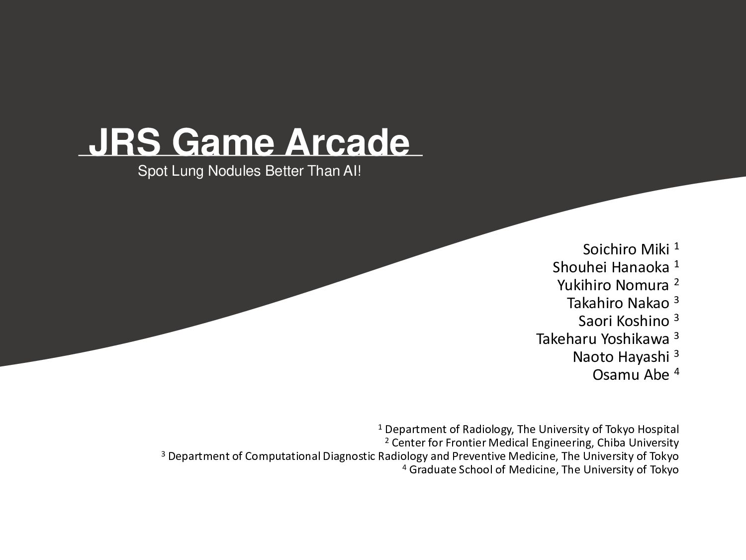
Miki 1 Shouhei Hanaoka 1 Yukihiro Nomura 2 Takahiro Nakao 3 Saori Koshino 3 Takeharu Yoshikawa 3 Naoto Hayashi 3 Osamu Abe 4 1 Department of Radiology, The University of Tokyo Hospital 2 Center for Frontier Medical Engineering, Chiba University 3 Department of Computational Diagnostic Radiology and Preventive Medicine, The University of Tokyo 4 Graduate School of Medicine, The University of Tokyo
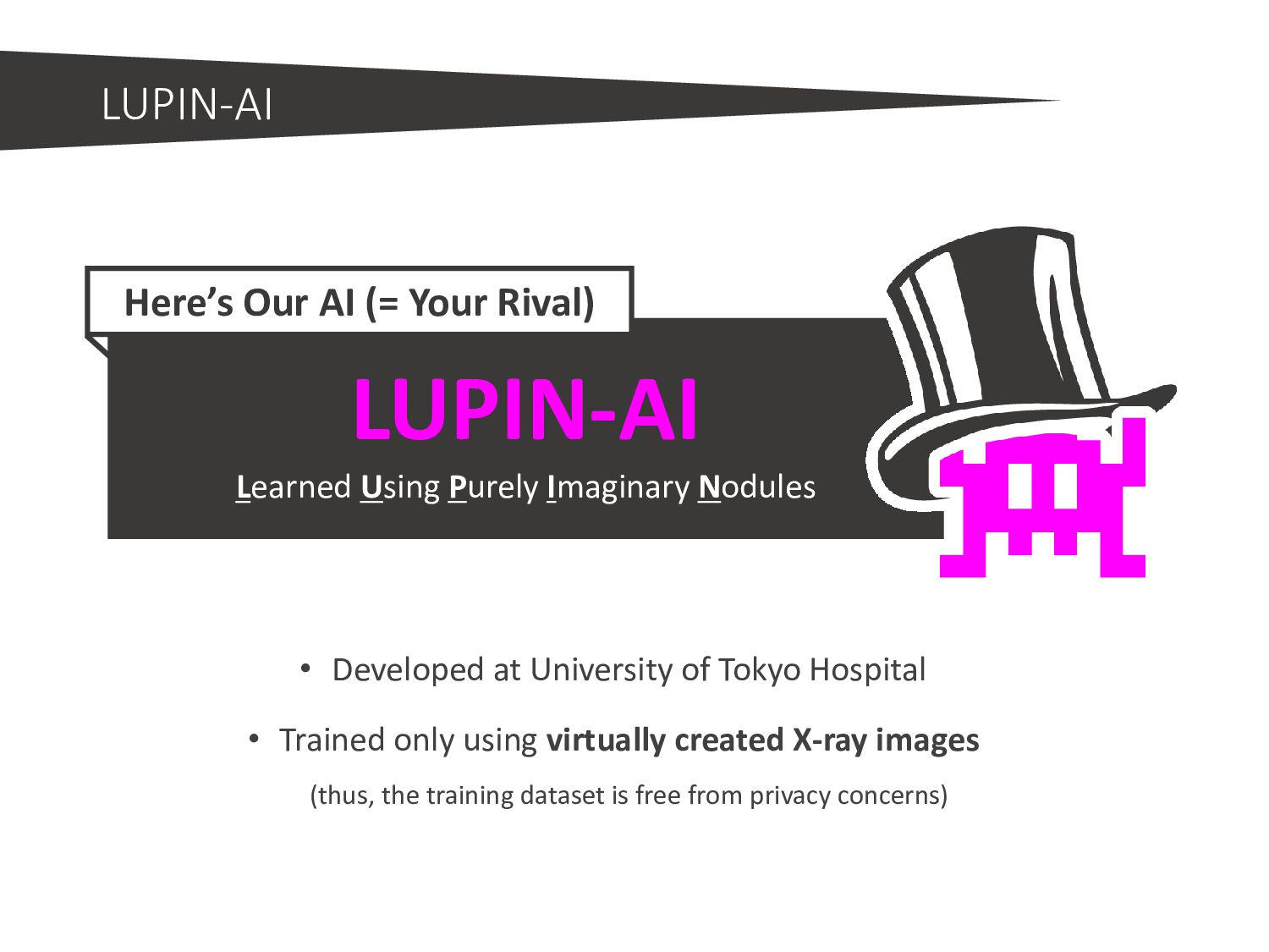
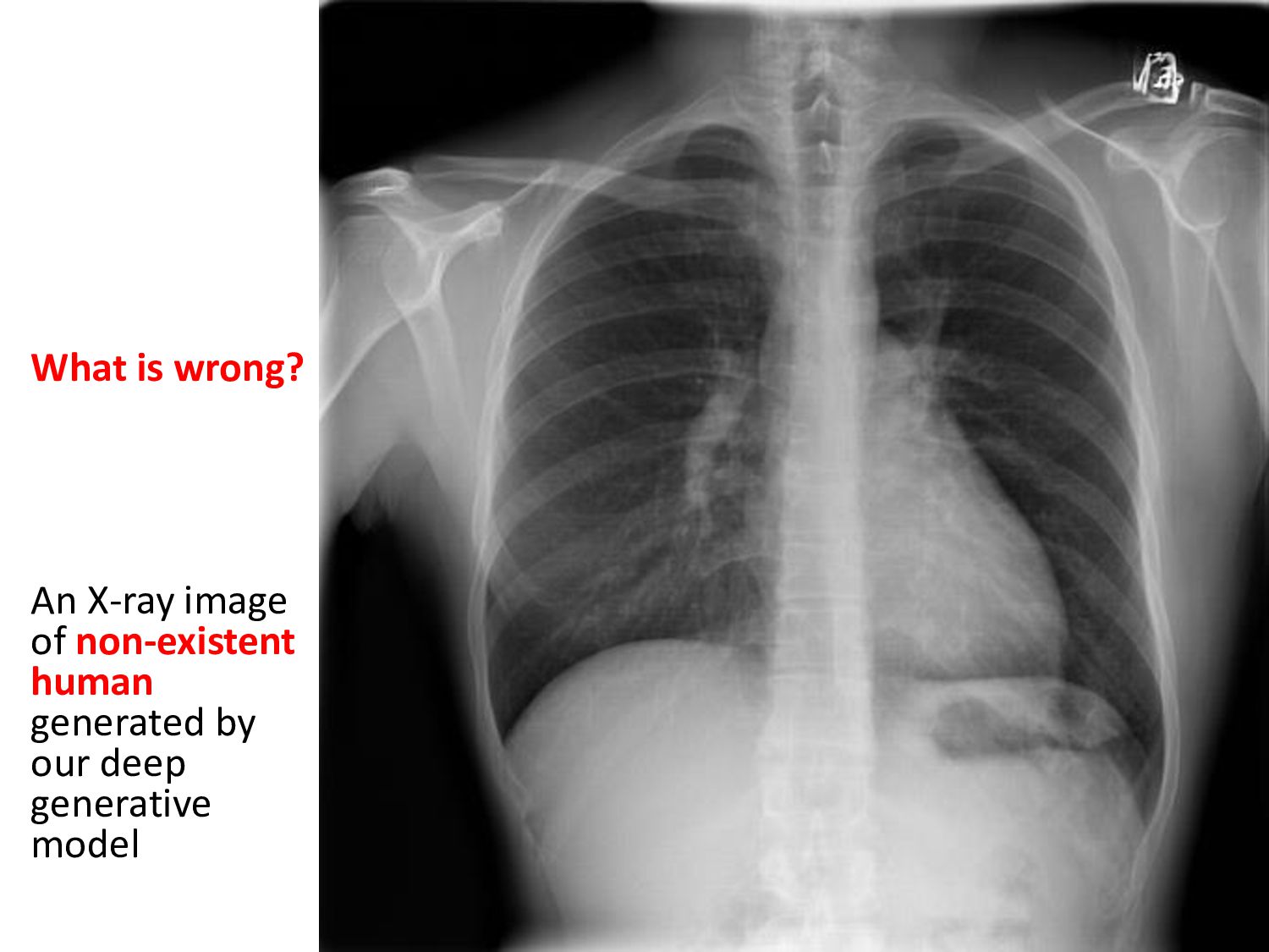
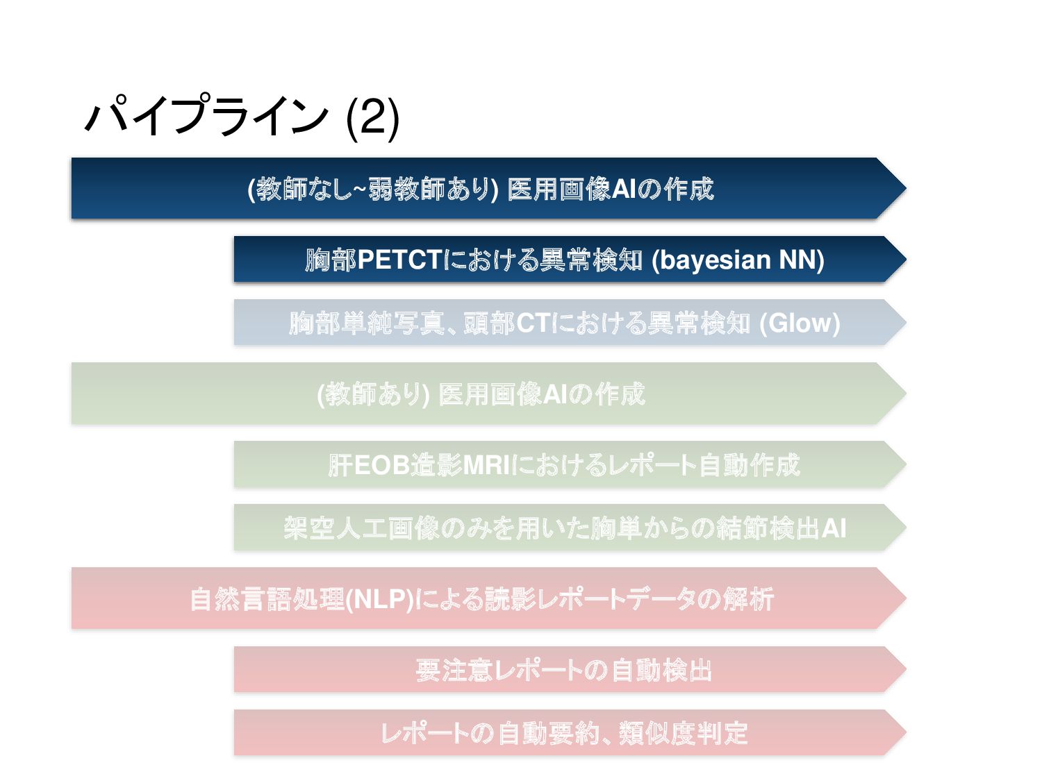
only using virtually created X-ray images (thus, the training dataset is free from privacy concerns) LUPIN-AI Learned Using Purely Imaginary Nodules Here’s Our AI (= Your Rival)
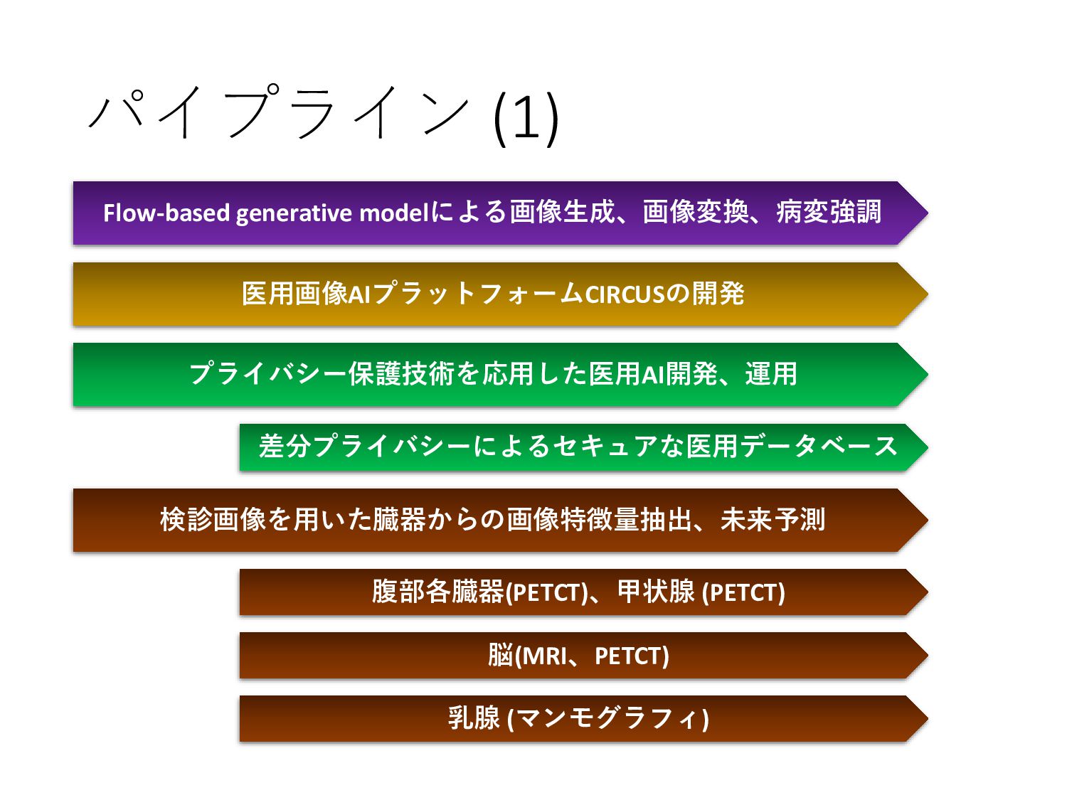
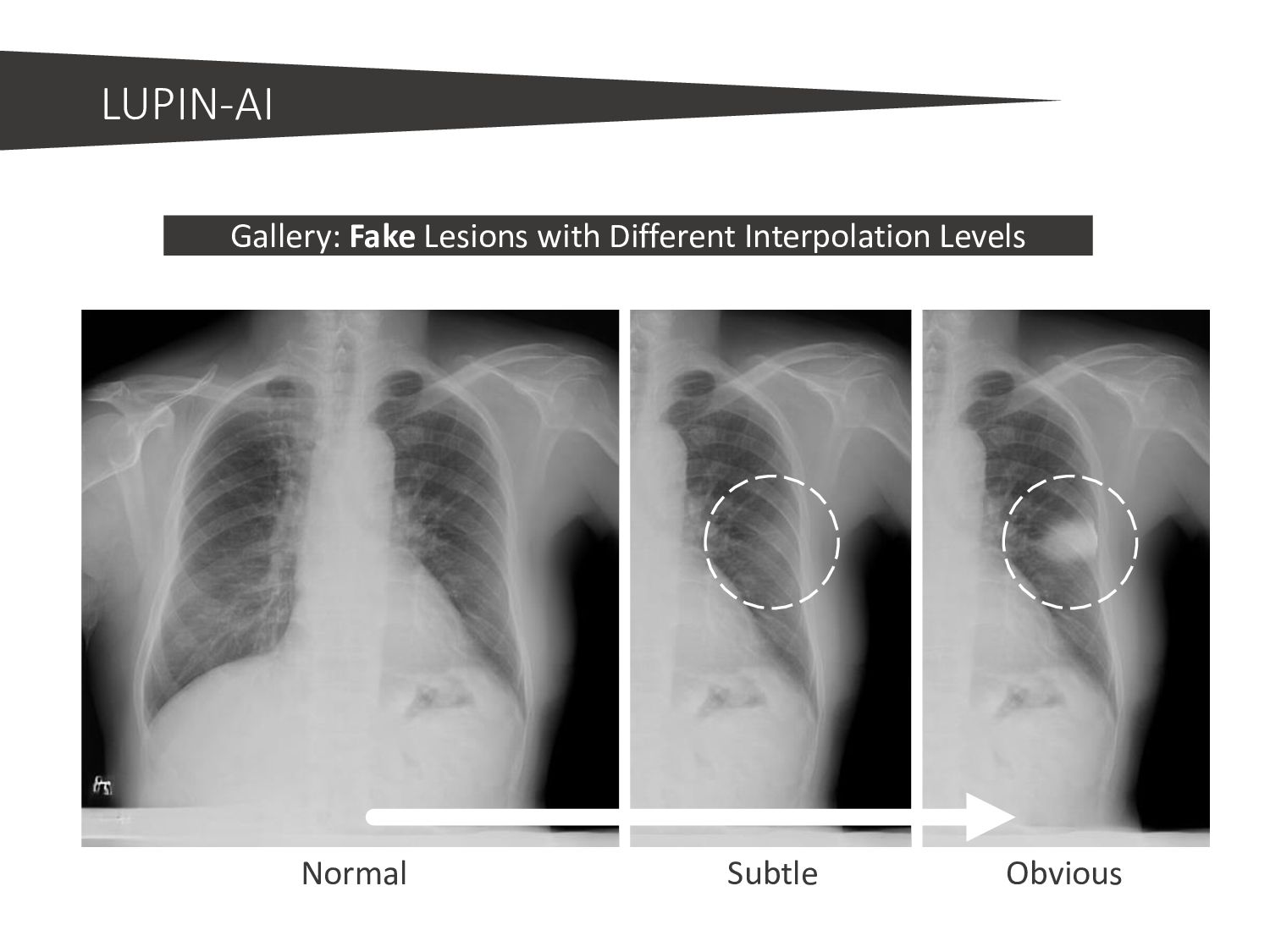
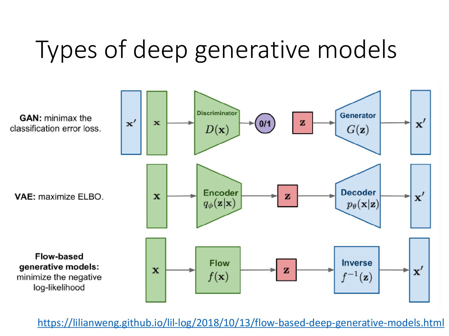
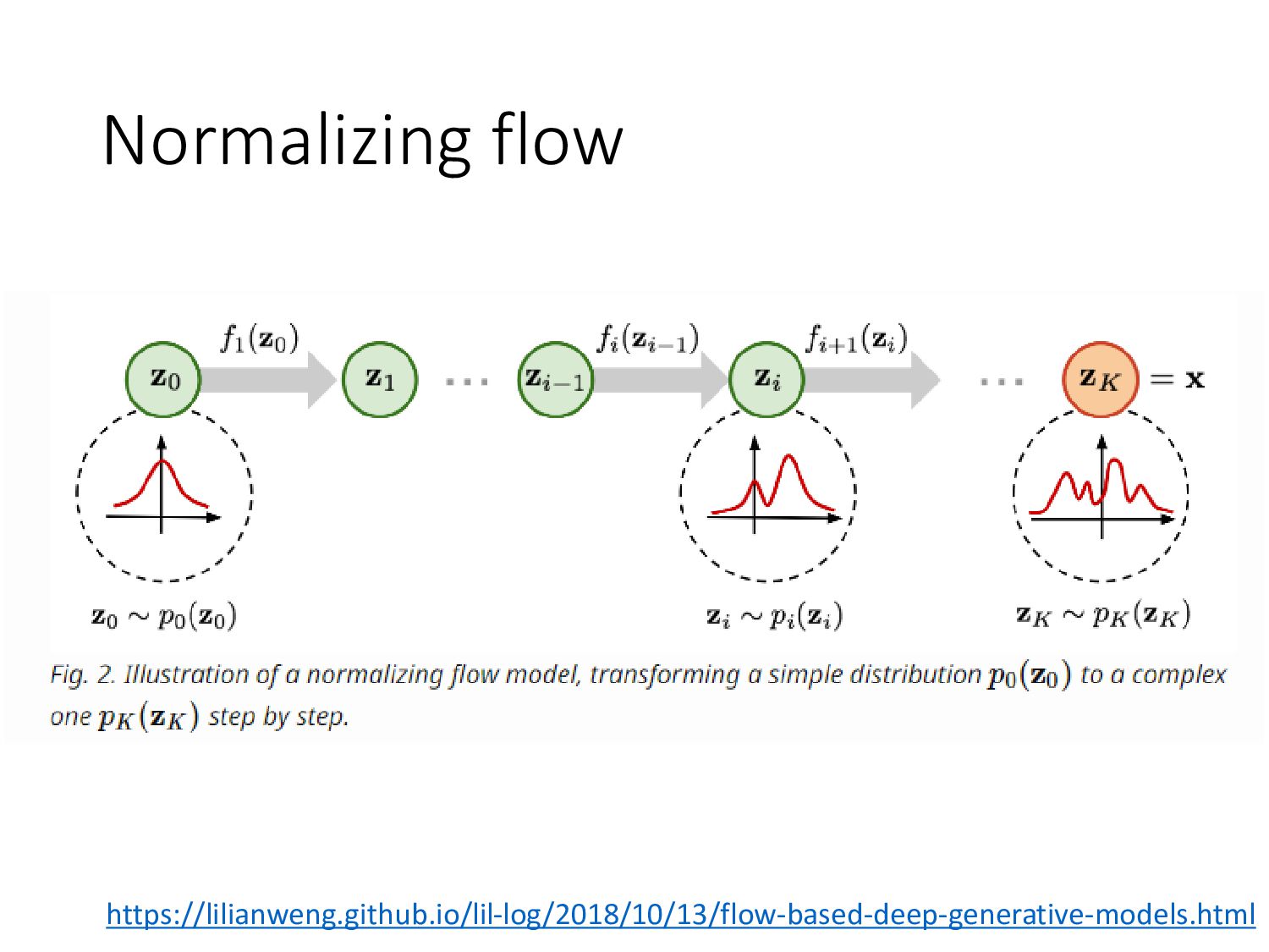
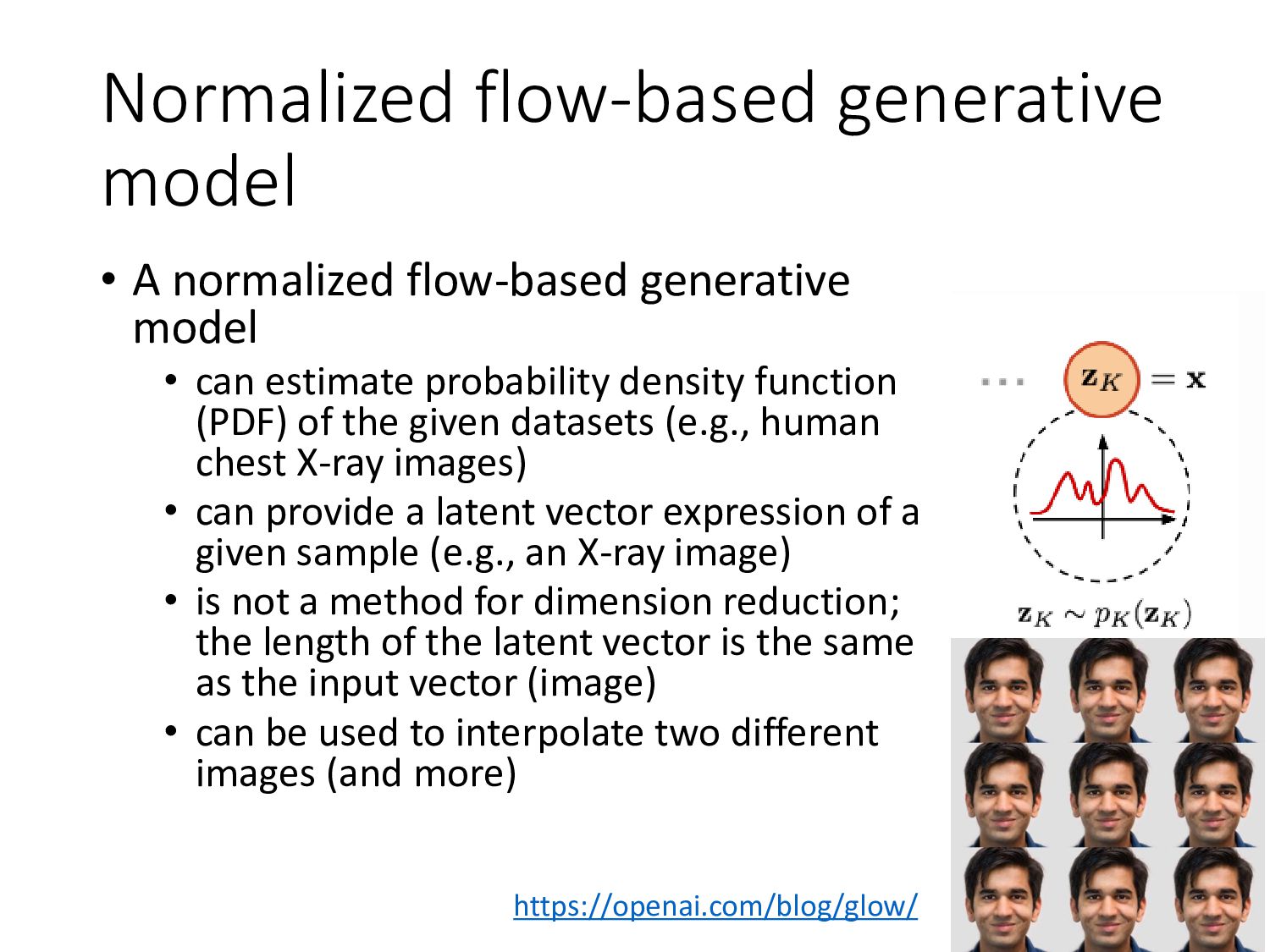
• can estimate probability density function (PDF) of the given datasets (e.g., human chest X-ray images) • can provide a latent vector expression of a given sample (e.g., an X-ray image) • is not a method for dimension reduction; the length of the latent vector is the same as the input vector (image) • can be used to interpolate two different images (and more) https://openai.com/blog/glow/
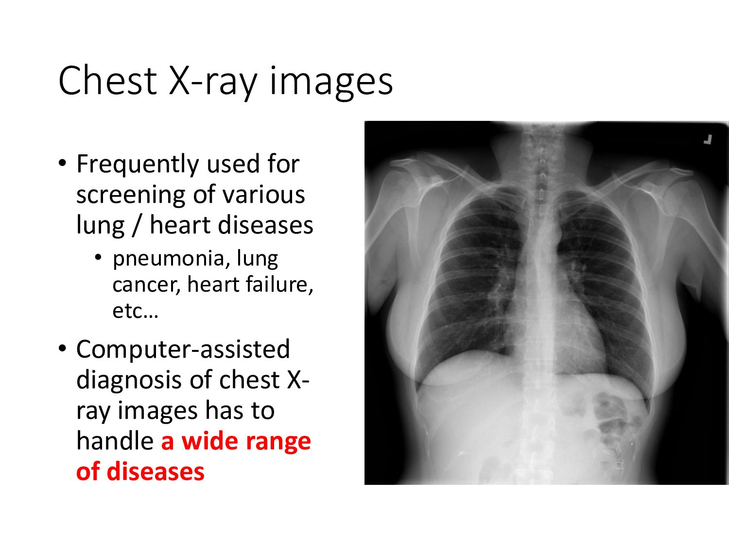
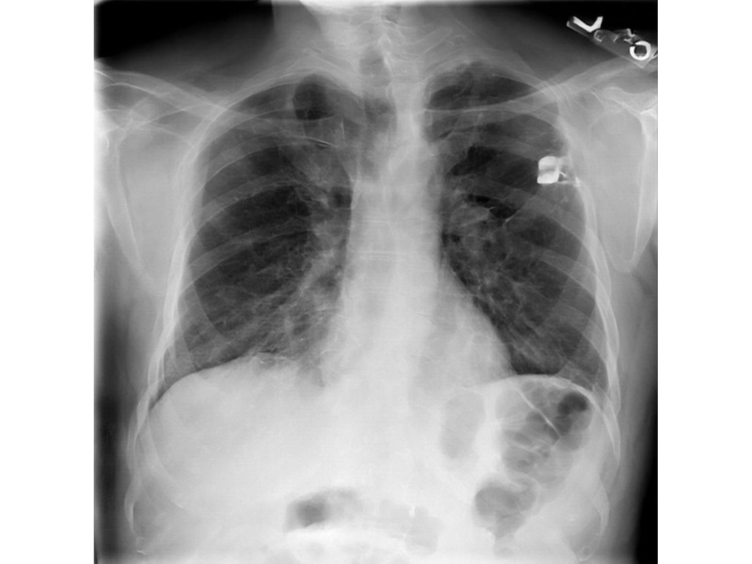
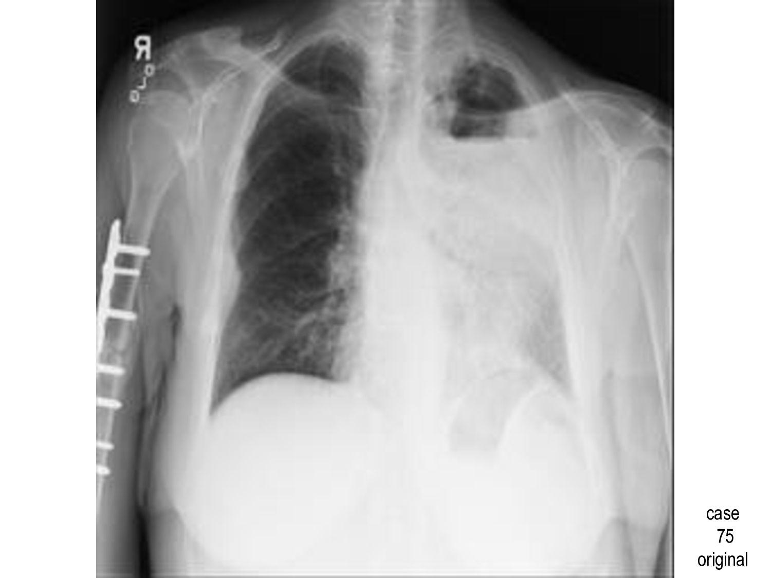
lung / heart diseases • pneumonia, lung cancer, heart failure, etc… • Computer-assisted diagnosis of chest X- ray images has to handle a wide range of diseases
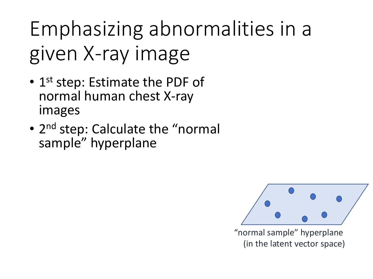
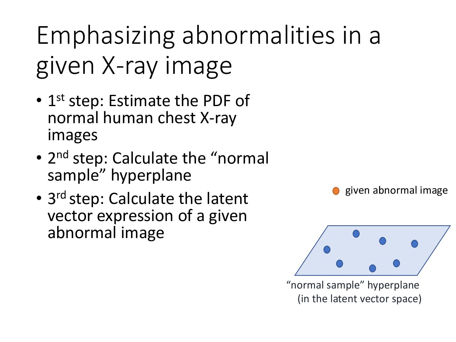
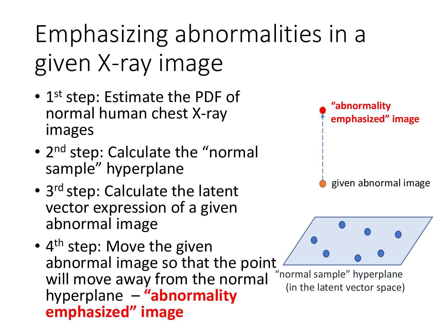
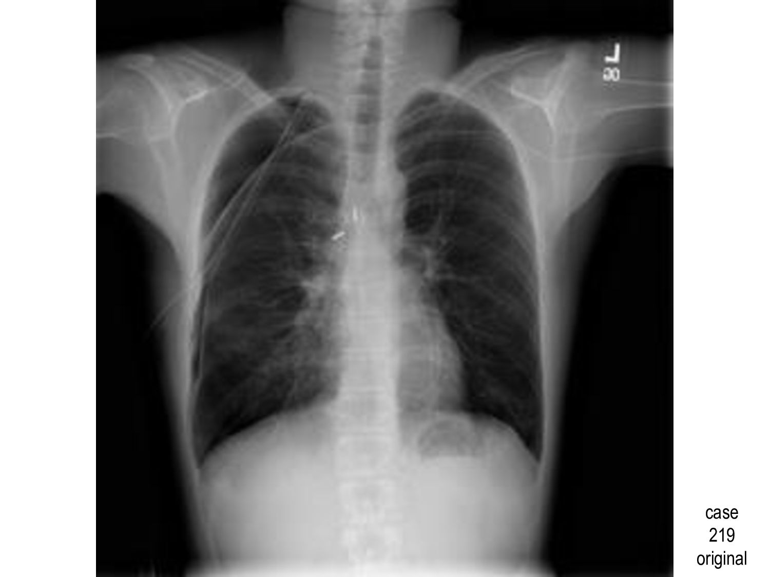
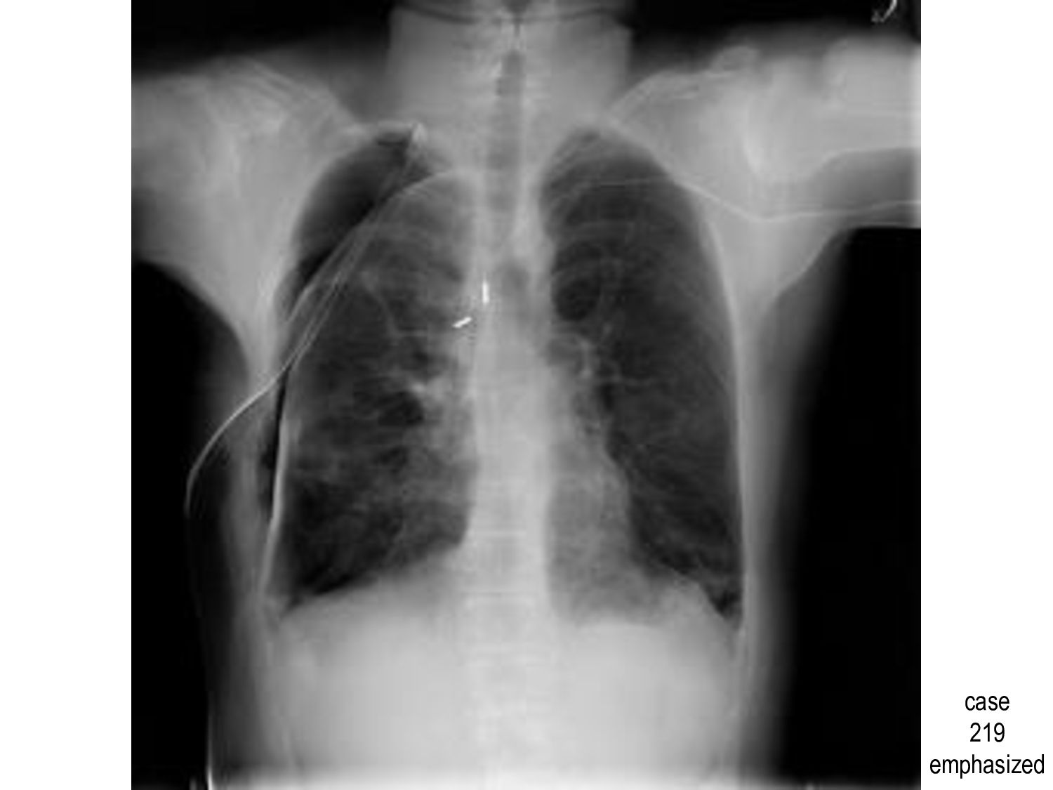
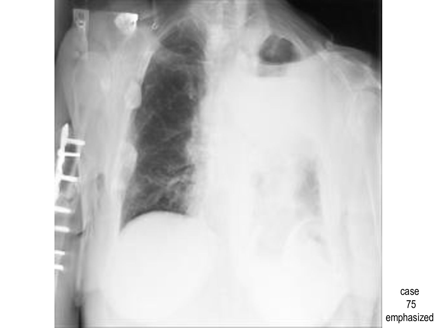
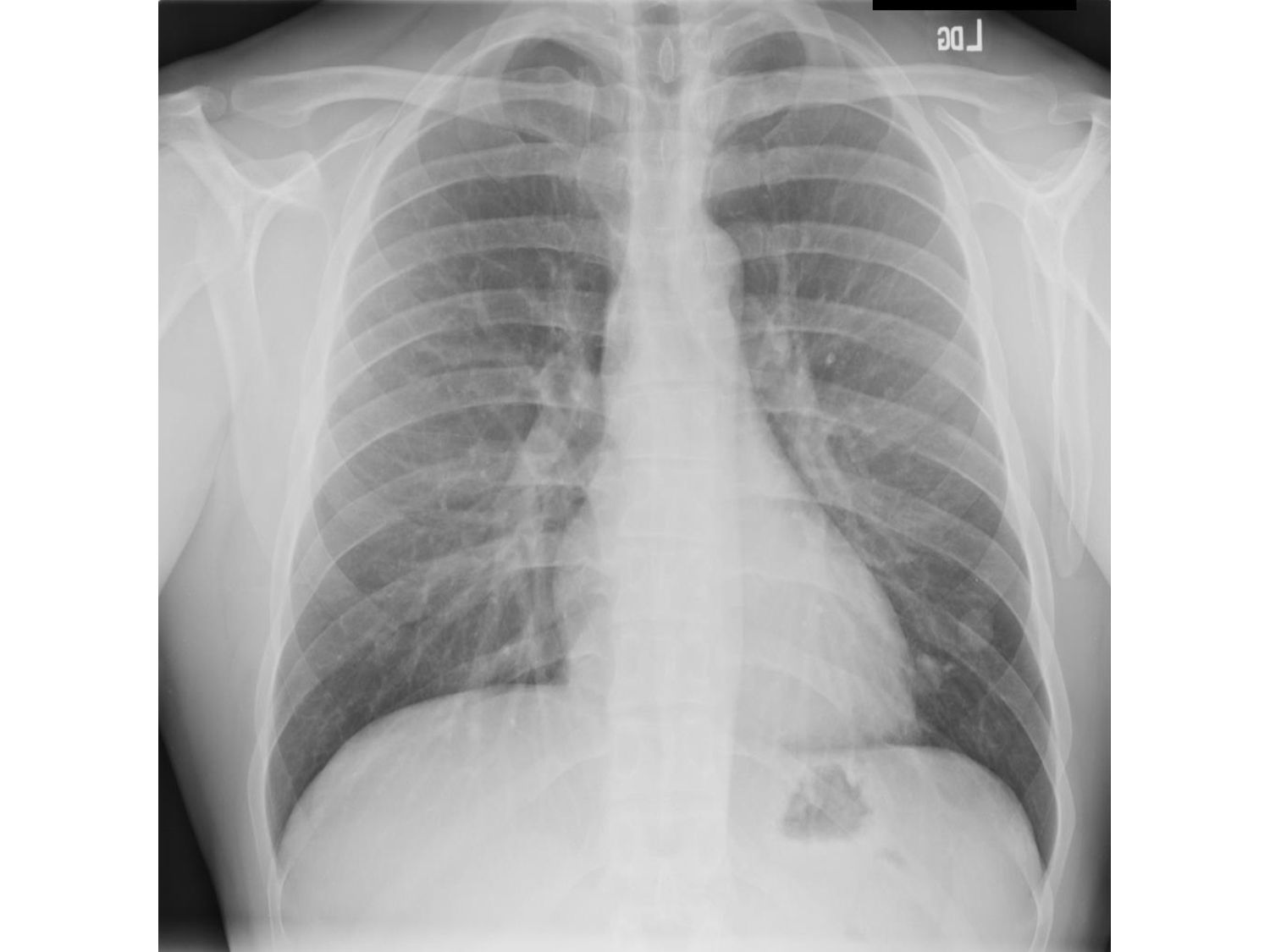
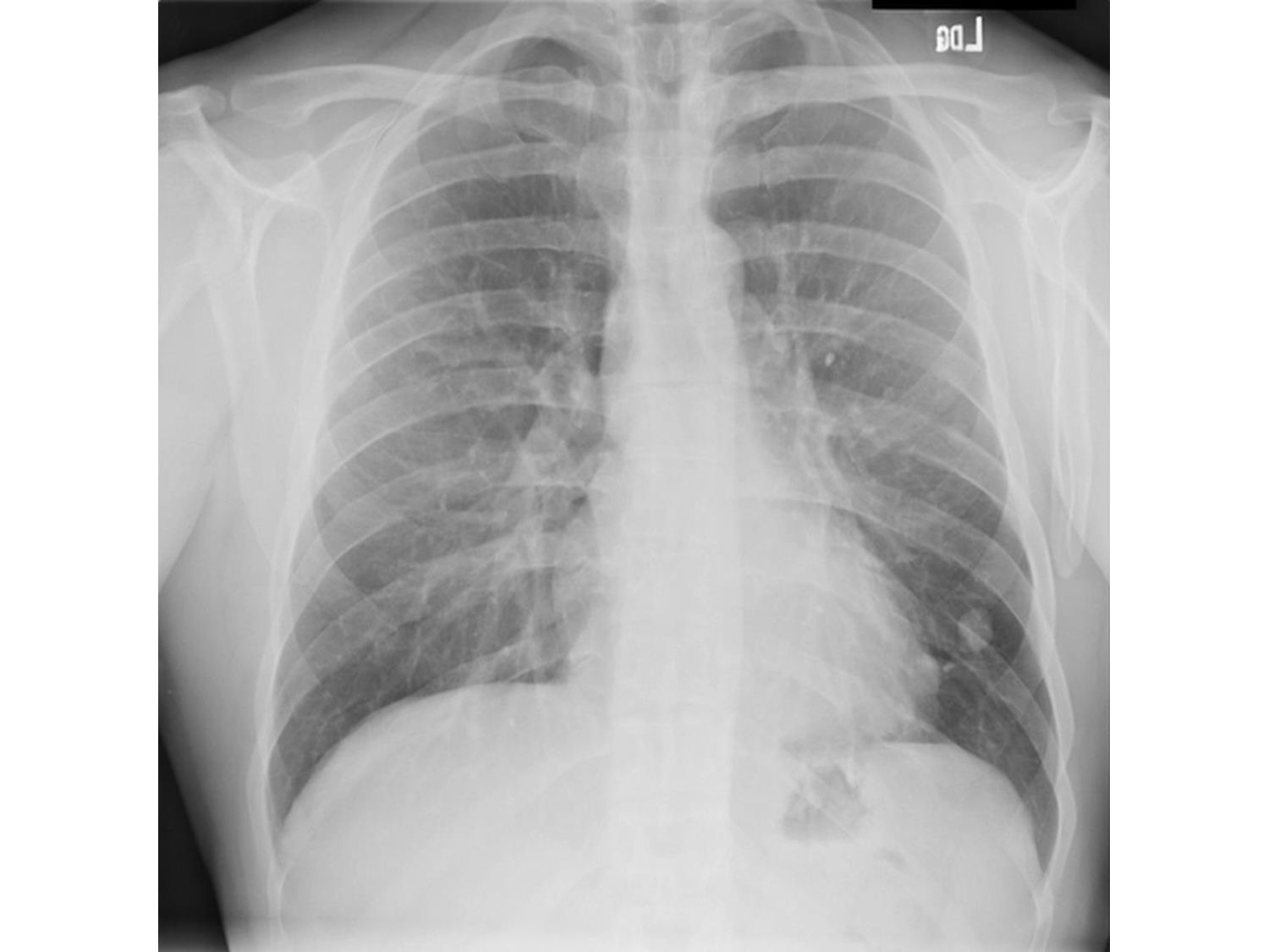
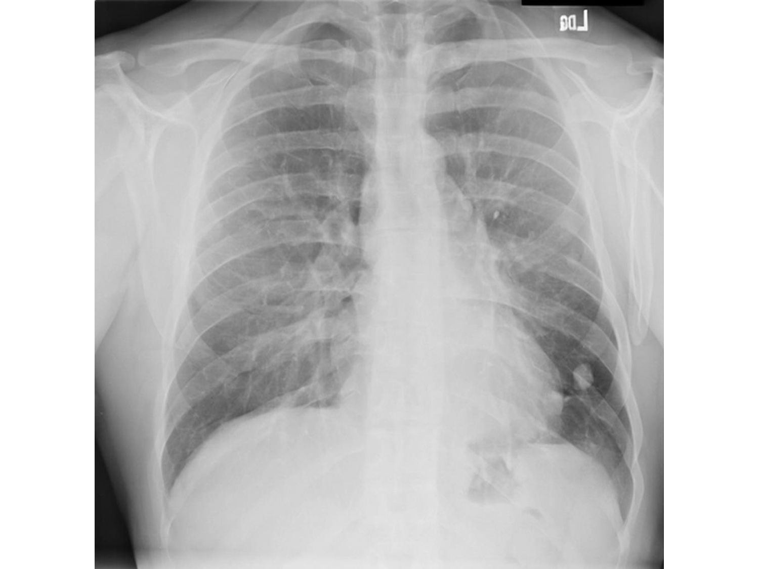
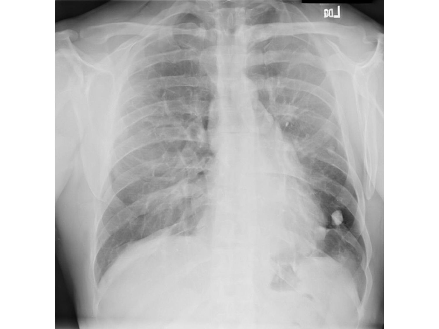
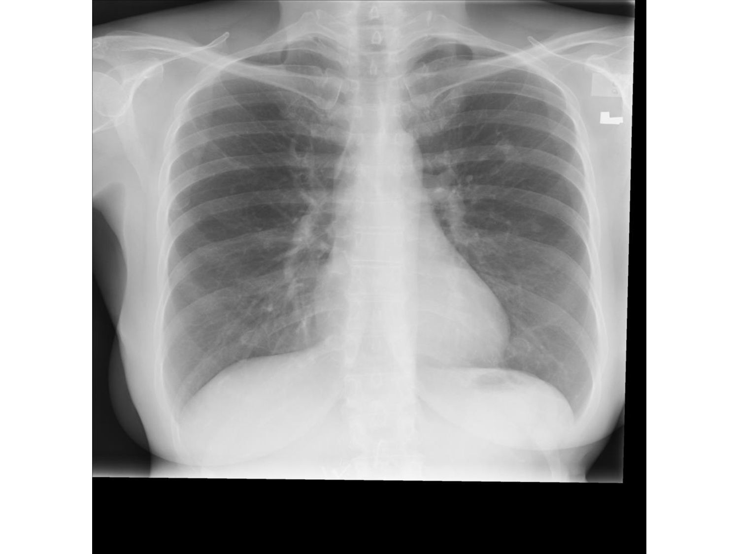
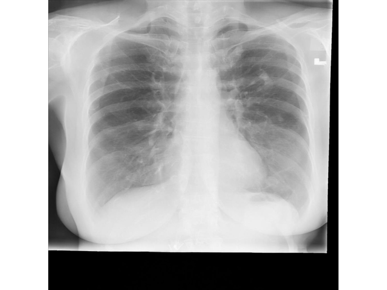
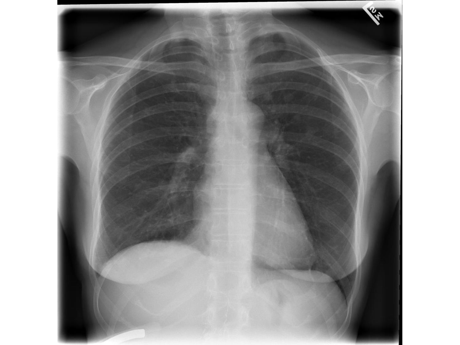
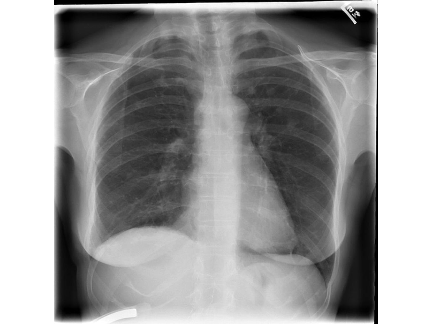
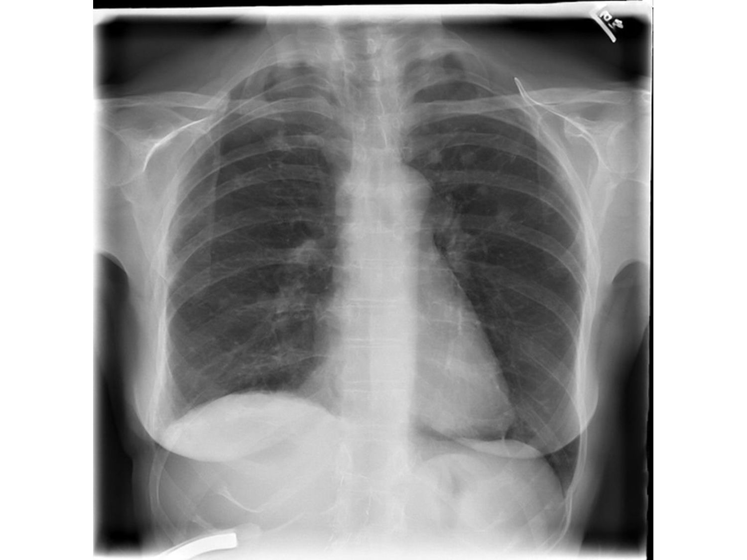
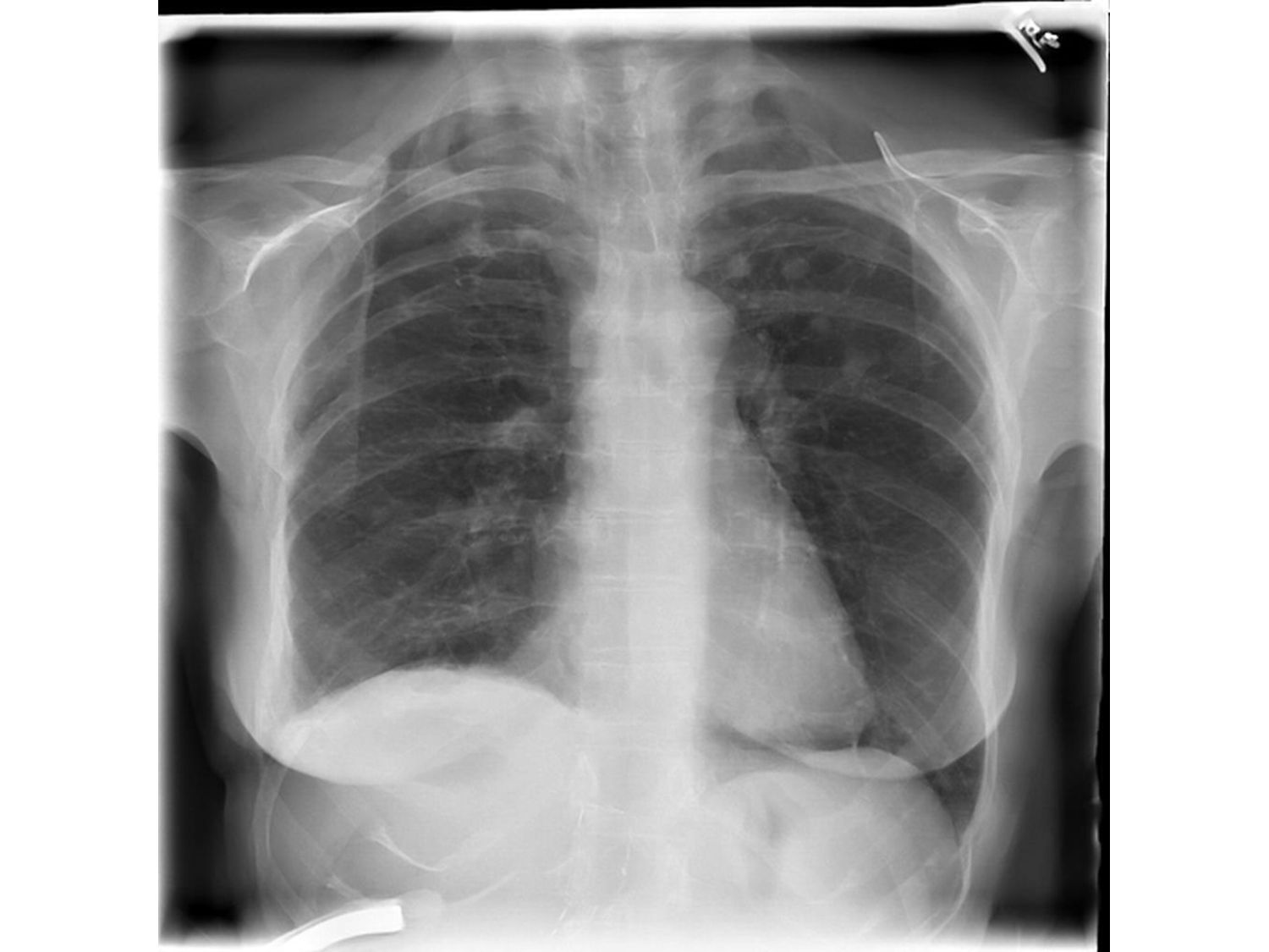
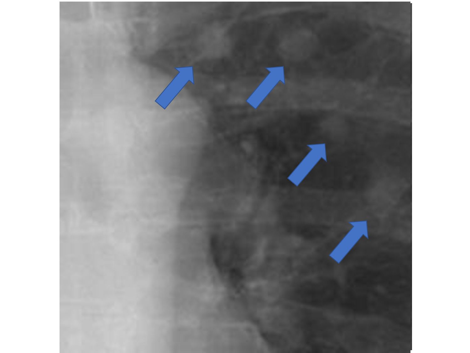
Estimate the PDF of normal human chest X-ray images • 2nd step: Calculate the “normal sample” hyperplane • 3rd step: Calculate the latent vector expression of a given abnormal image • 4th step: Move the given abnormal image so that the point will go away from the original point – “abnormality emphasized image” “normal sample” hyperplane (in the latent vector space)
“normal sample” hyperplane (in the latent vector space) • 1st step: Estimate the PDF of normal human chest X-ray images • 2nd step: Calculate the “normal sample” hyperplane • 3rd step: Calculate the latent vector expression of a given abnormal image • 4th step: Move the given abnormal image so that the point will go away from the original point – “abnormality emphasized image”
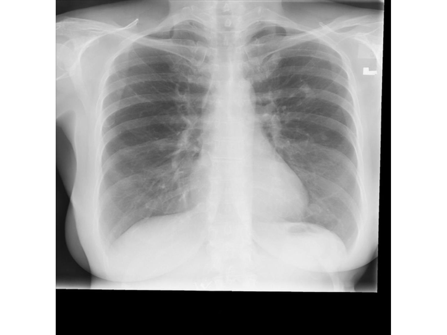
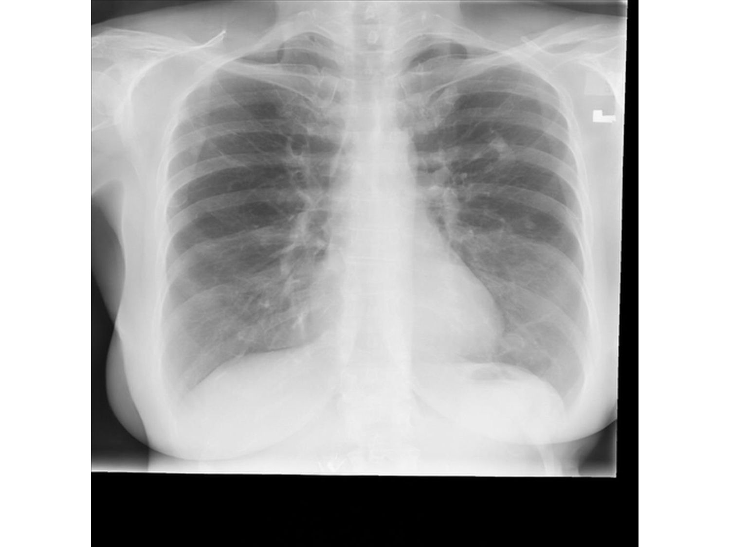
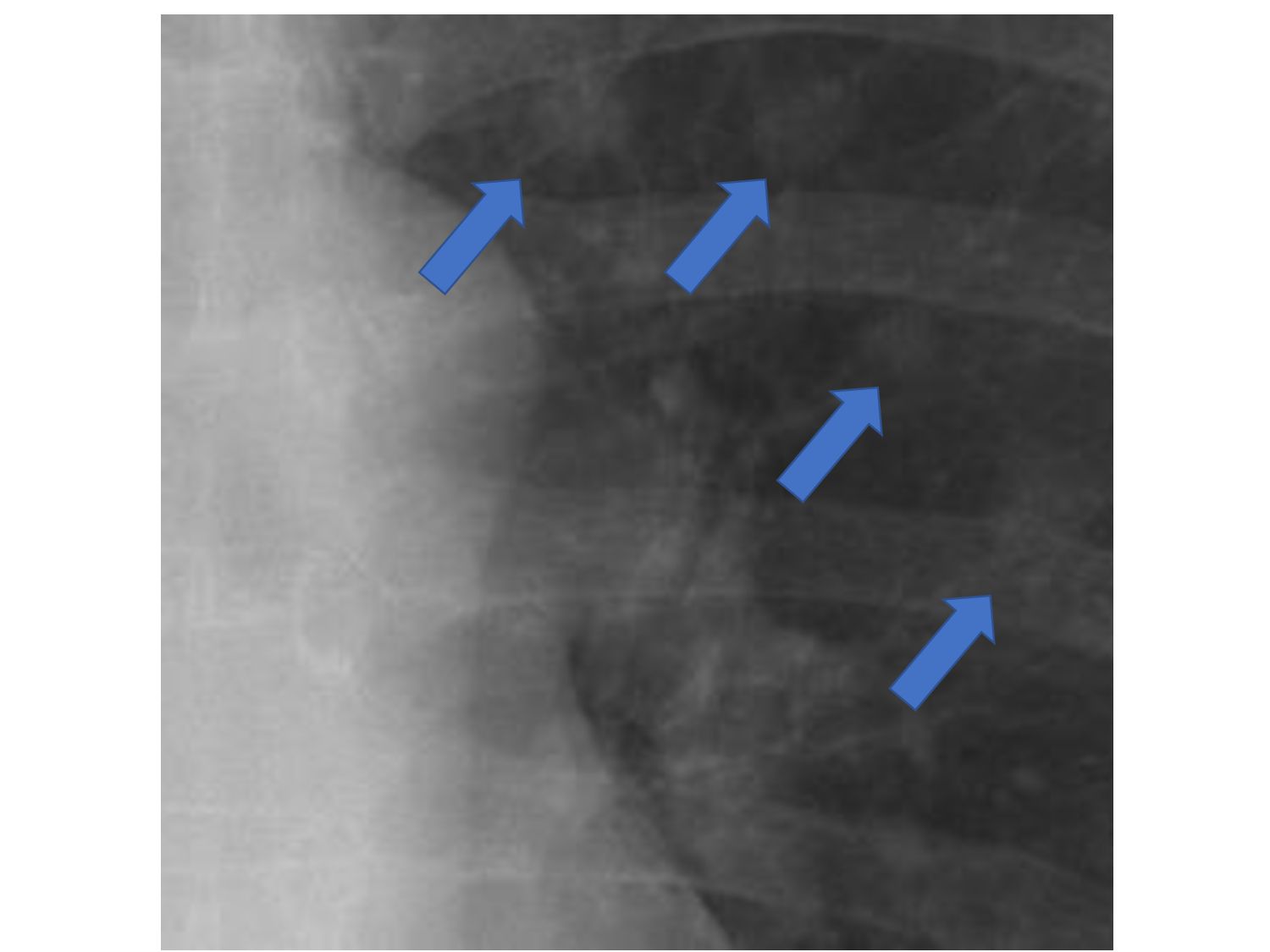
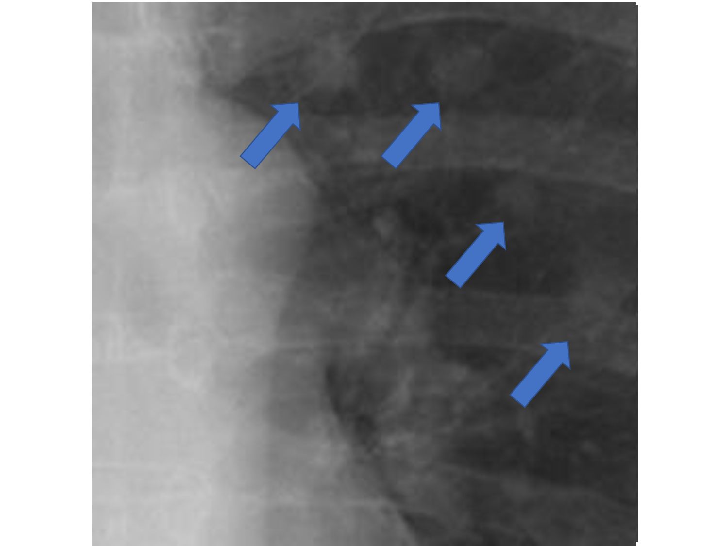
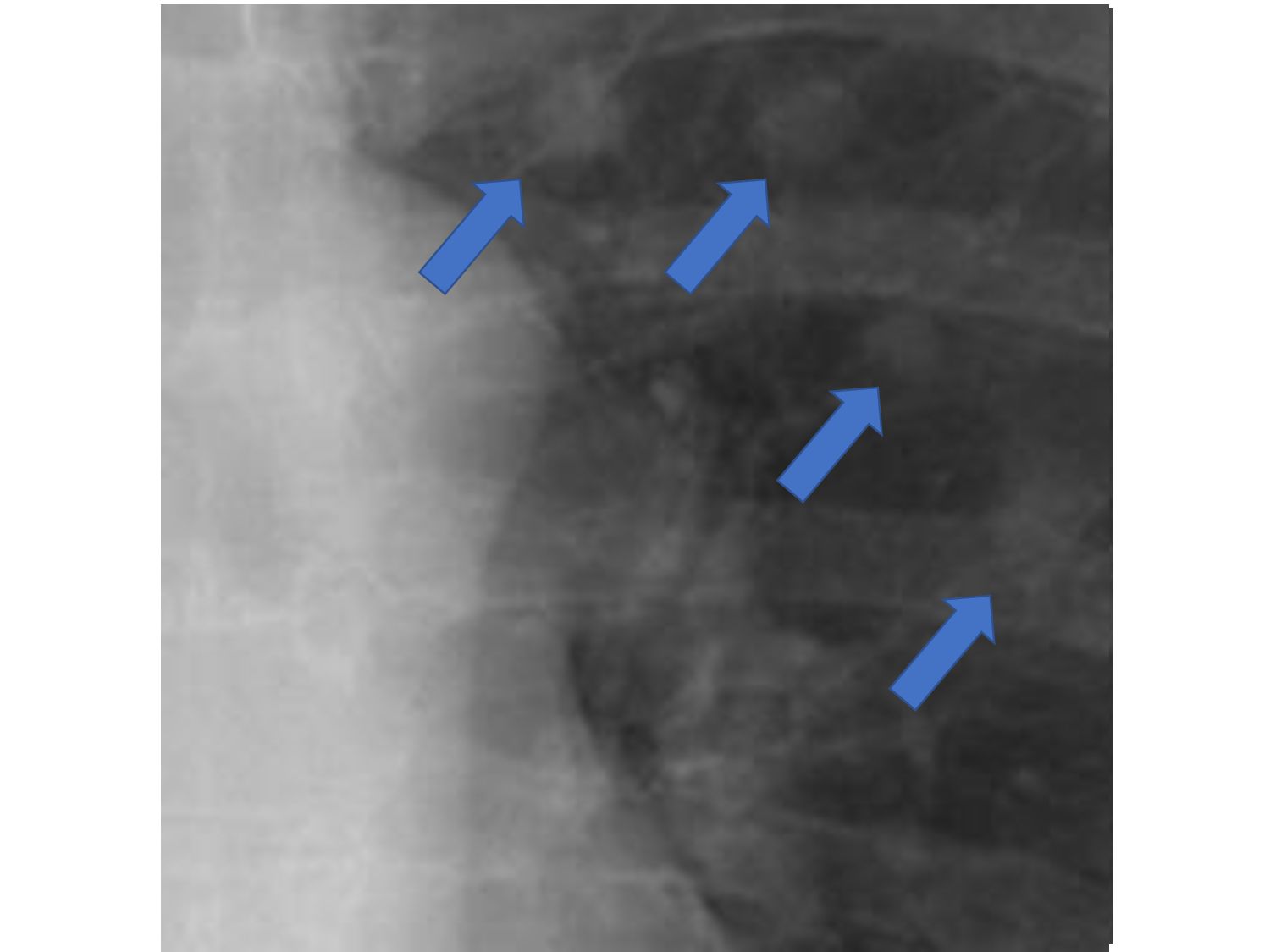
given abnormal image “normal sample” hyperplane (in the latent vector space) • 1st step: Estimate the PDF of normal human chest X-ray images • 2nd step: Calculate the “normal sample” hyperplane • 3rd step: Calculate the latent vector expression of a given abnormal image • 4th step: Move the given abnormal image so that the point will move away from the normal hyperplane – “abnormality emphasized” image
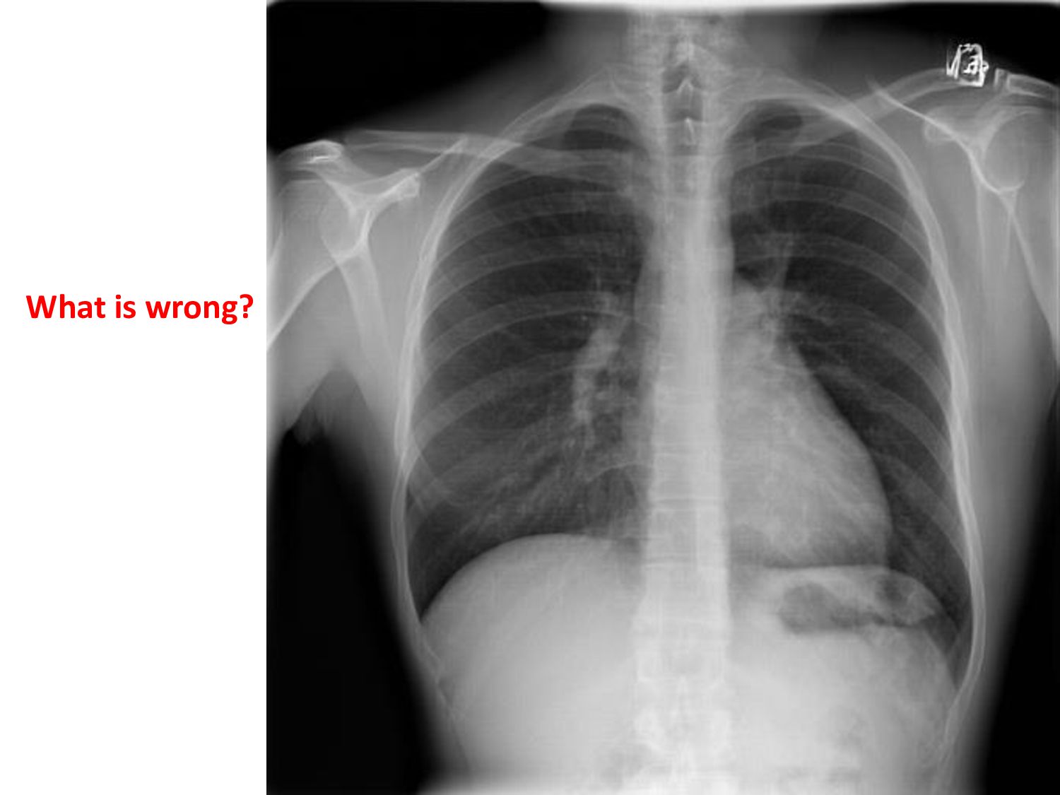
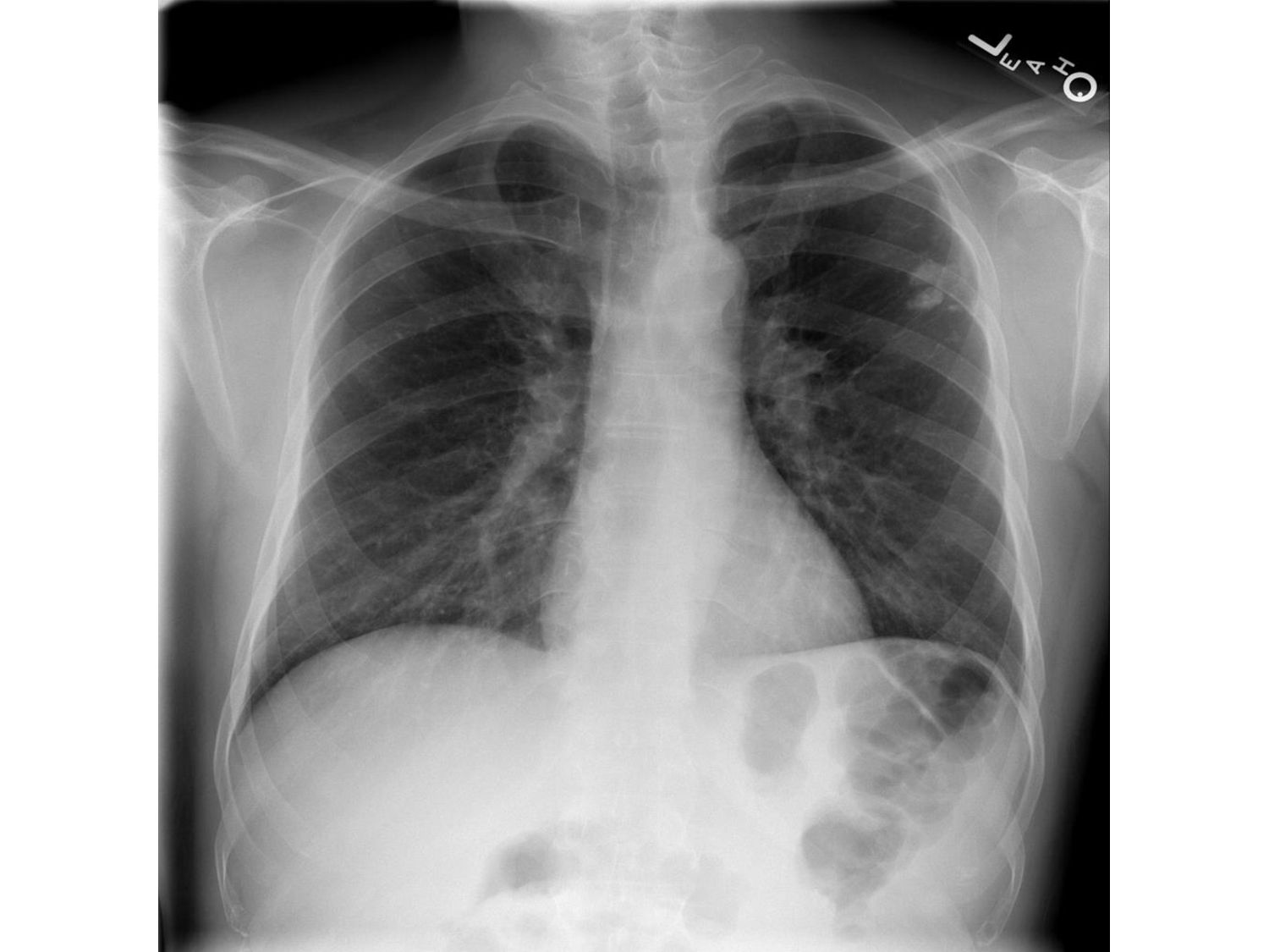
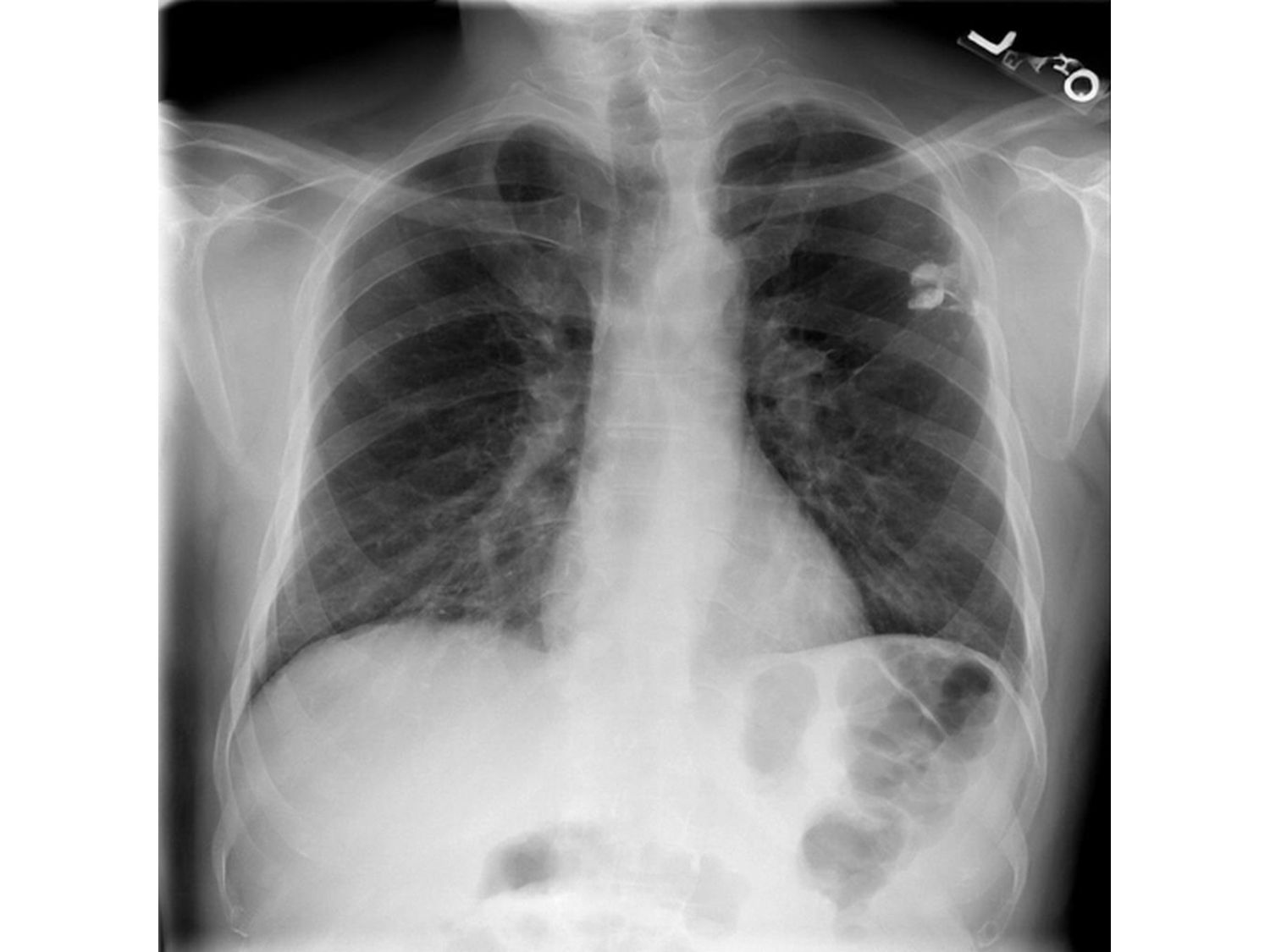
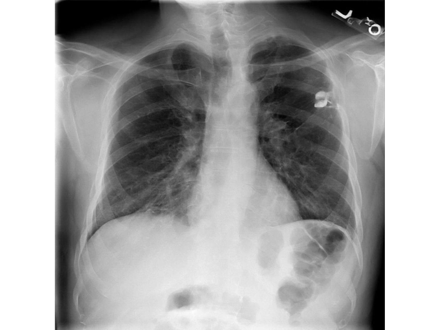
given X-ray image is presented. • It can emphasize arbitrary types of abnormalities, thus we expect that it can be used in daily routine X-ray image diagnosis tasks.
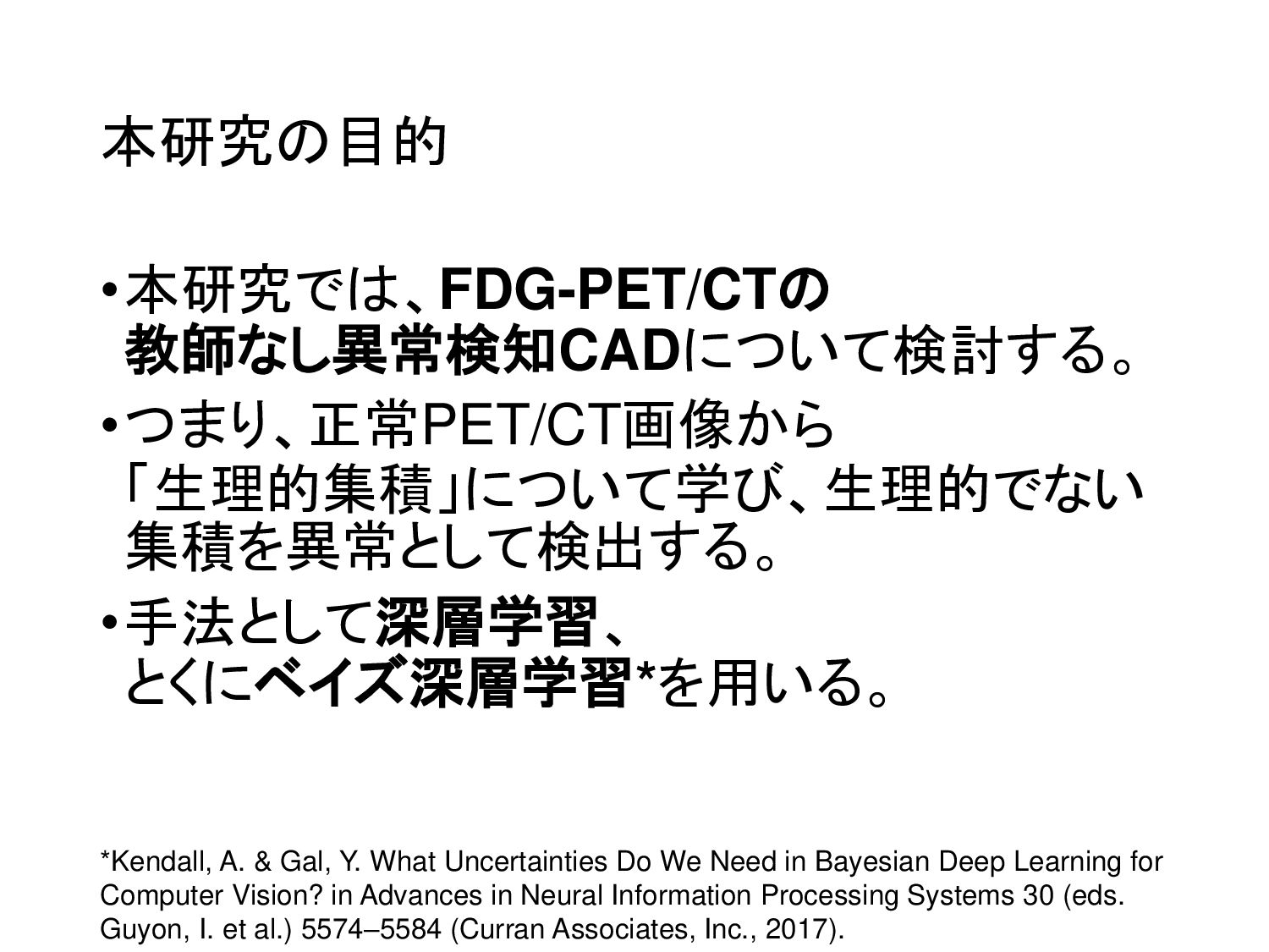
& Gal, Y. What Uncertainties Do We Need in Bayesian Deep Learning for Computer Vision? in Advances in Neural Information Processing Systems 30 (eds. Guyon, I. et al.) 5574–5584 (Curran Associates, Inc., 2017).


 hanaokalog
 juanmartin2026
 jdbedics
 robintux
 signer
 naochim2026
 bvr7352
 3l4l5
 spring_aki
 frievea
 bunnchinn3
 soudai
 vanstee
 3n
 kimpetersen
 yeseniaperezcruz
 uxyall
 marcelosomers
 carmenintech
 honzajavorek
 andyhume
 aleyda
 reverentgeek
 nonsquared