Upgrade to Pro — share decks privately, control downloads, hide ads and more …

Cardiac image analysis in Python: Learning from medical images

Cardiac image analysis in Python: Learning from medical images

Presented at PyDays Vienna 2017

Can Python help us to make cardiology more efficient?
Become a part of our start-up journey at KardioMe. We will see how Python and deep learning can be used for analysis of medical images.
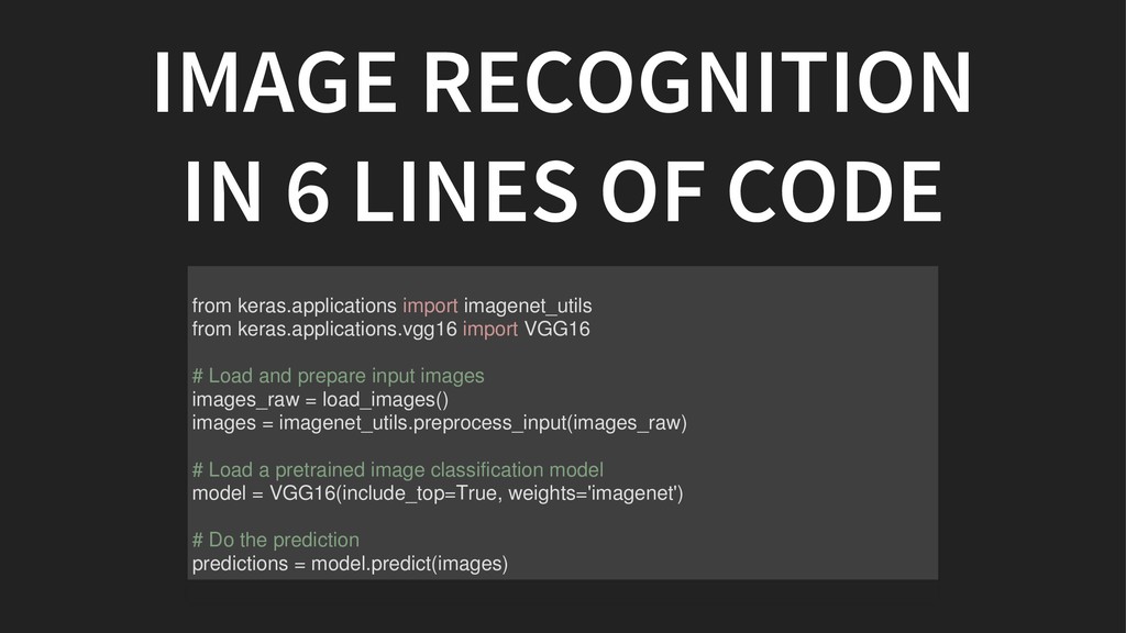
You will see how simple it is to start analysing your images too.

Artificial intelligence is booming and it is up to all of us to make tools that will improve our lives.
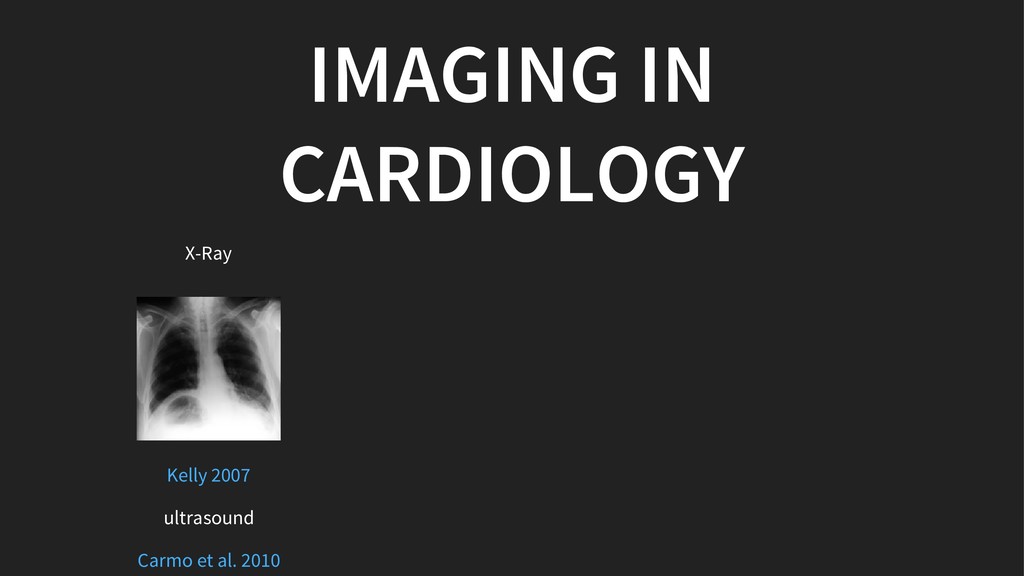
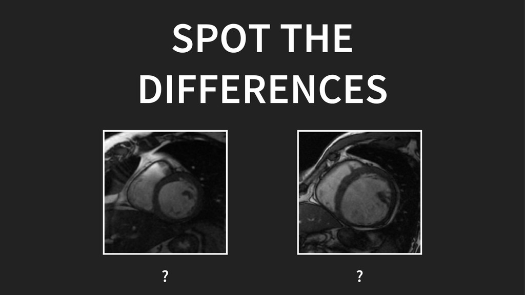
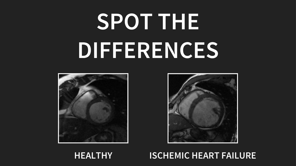
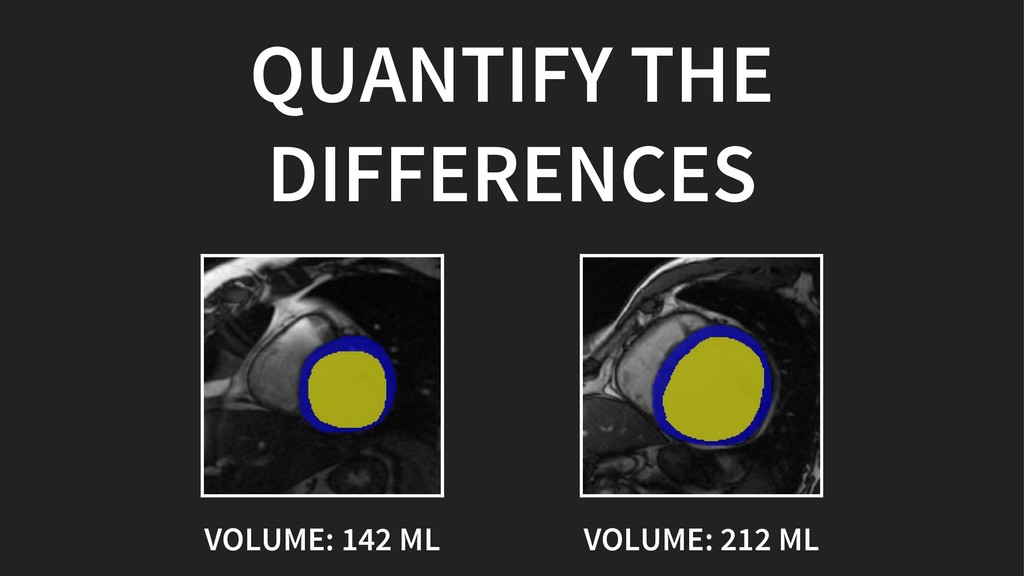
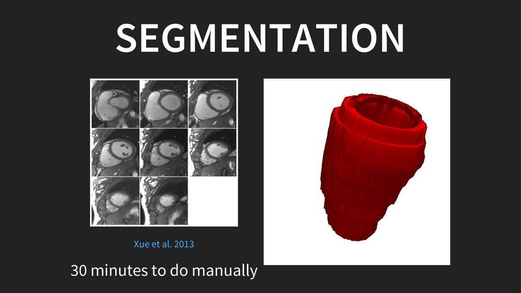
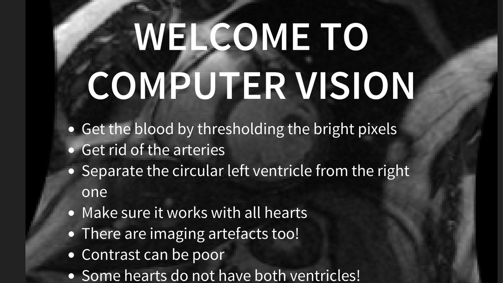
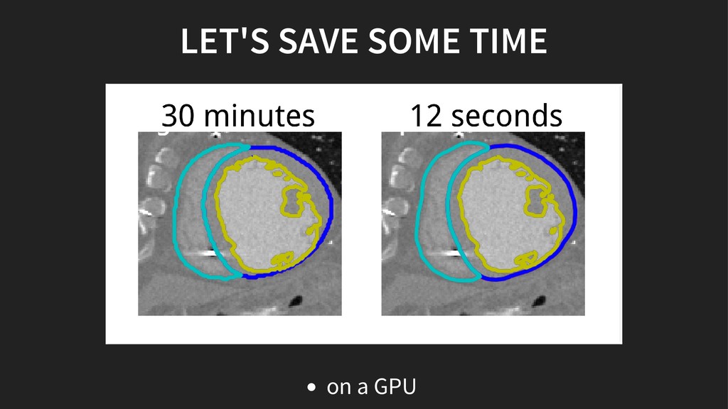
In this talk, we will see tools that can help radiologists be more efficient when analysing our hearts.

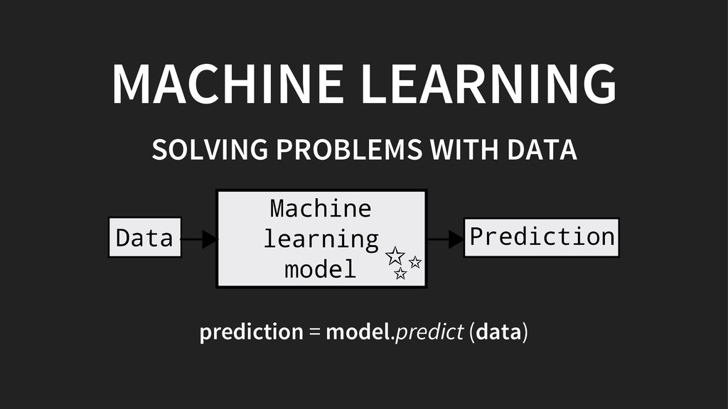
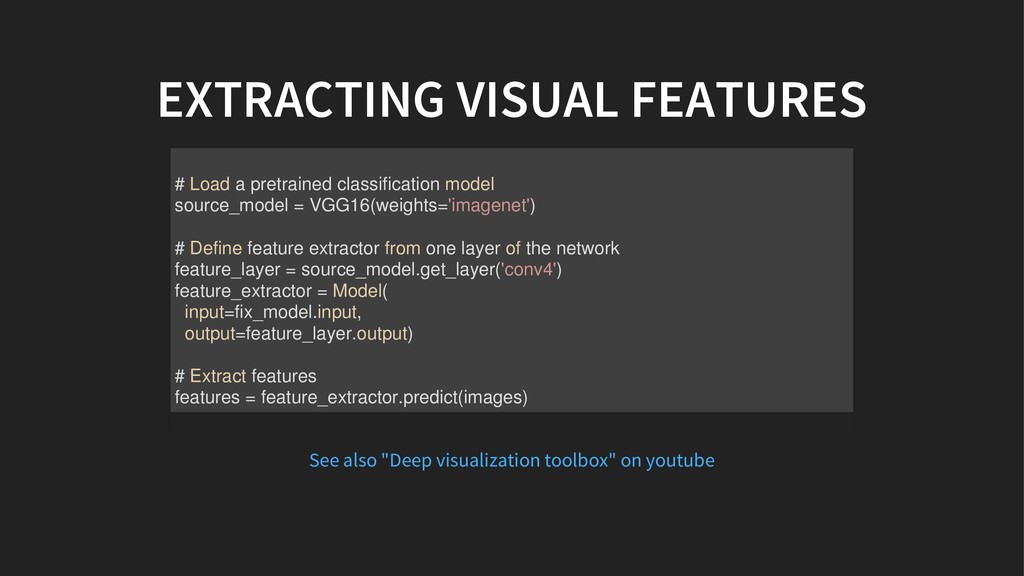
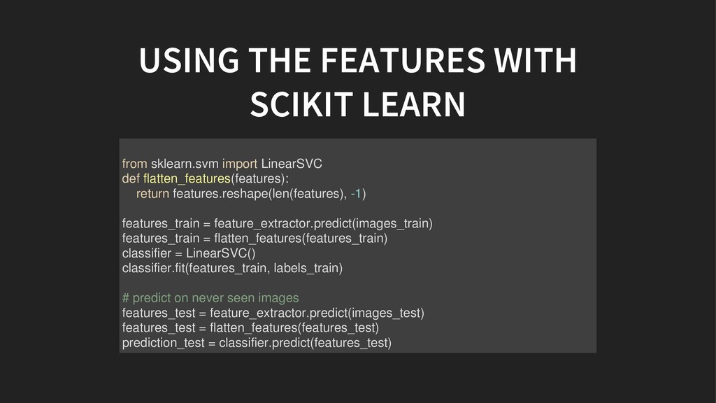
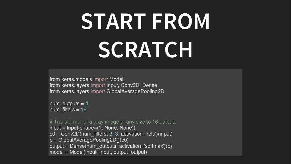
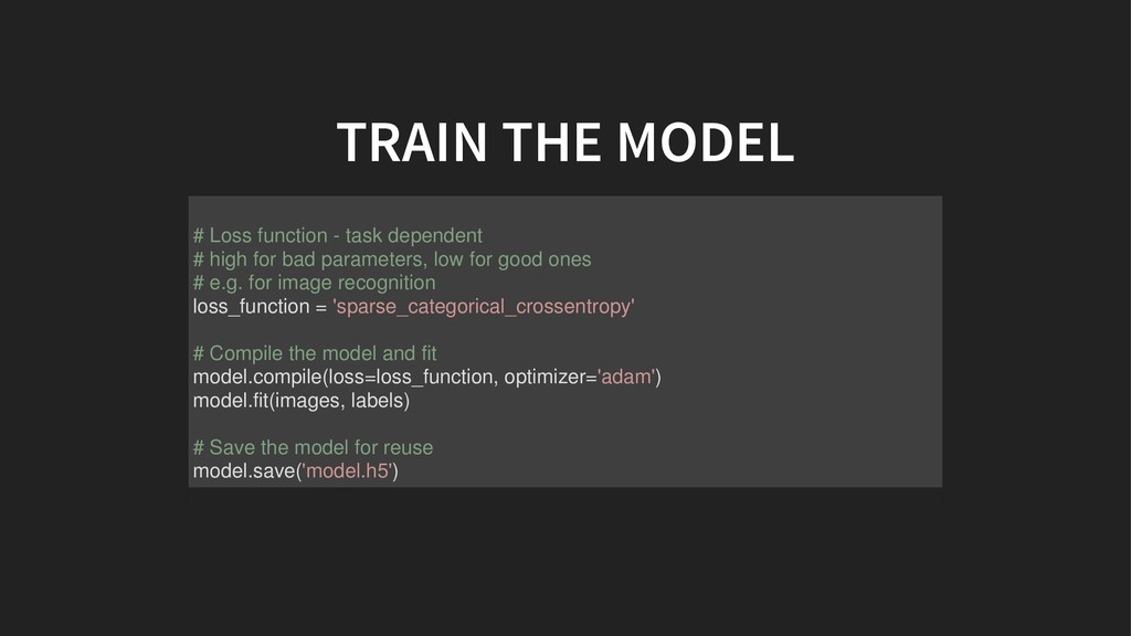
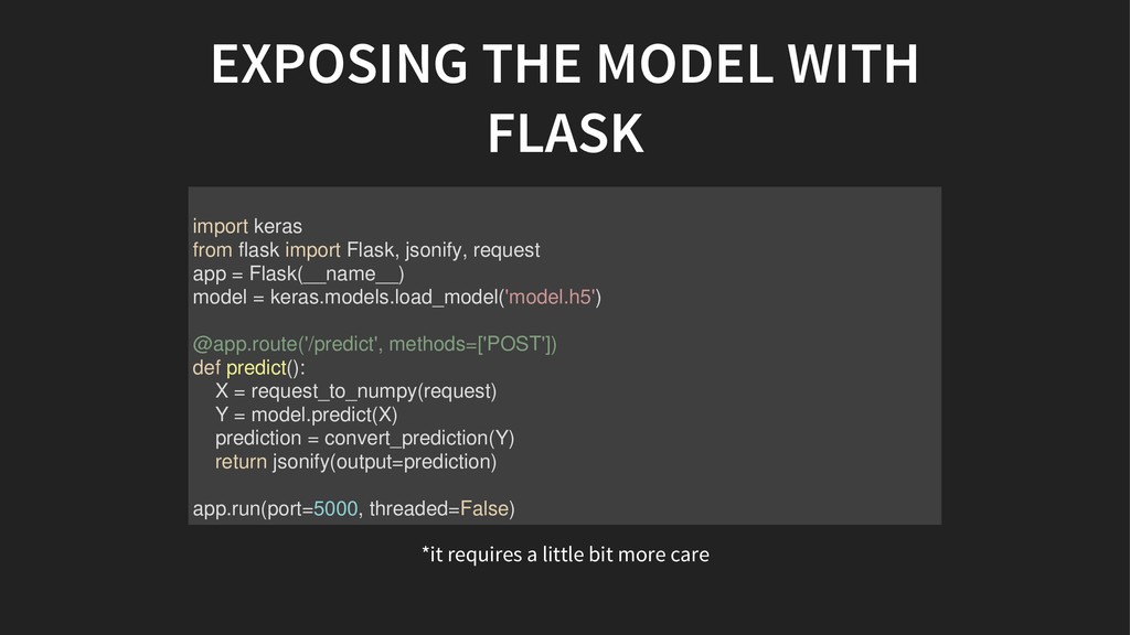
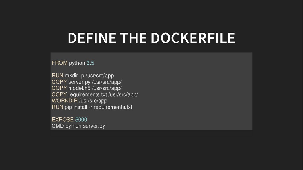
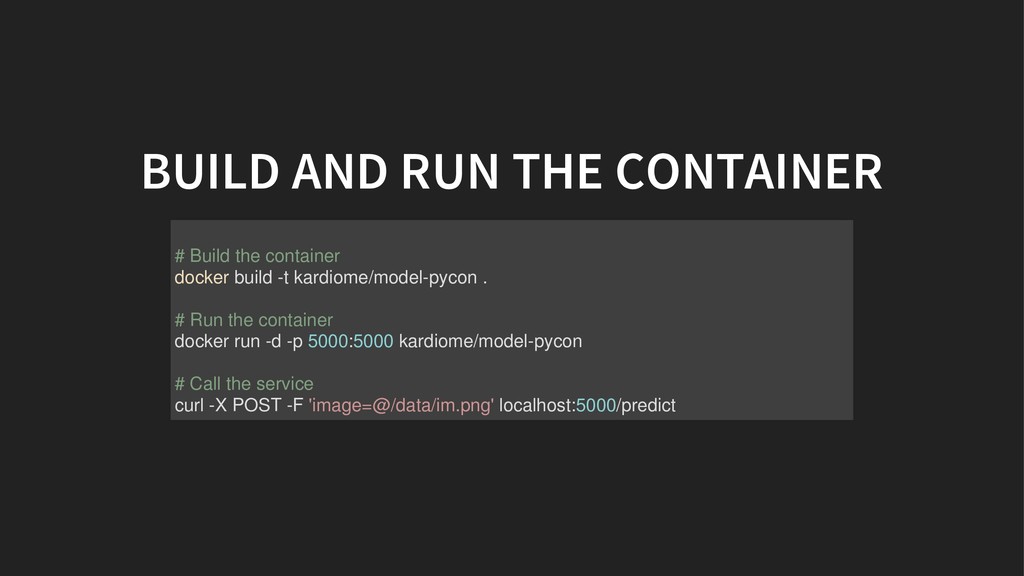
I will share tips for training machine learning systems in Python, and how to bring them from experimentation to production.
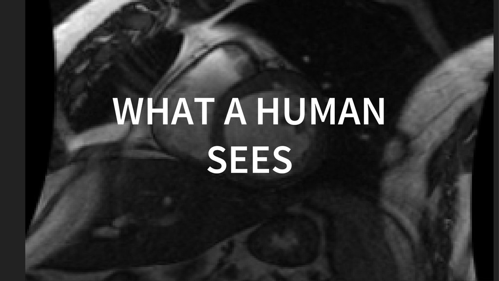
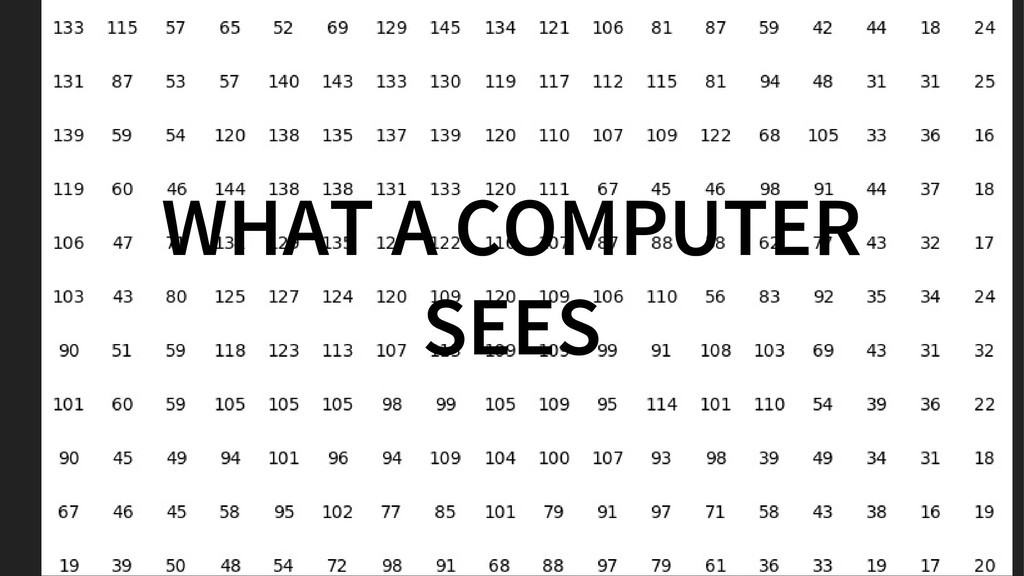
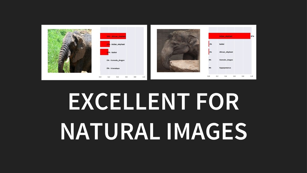
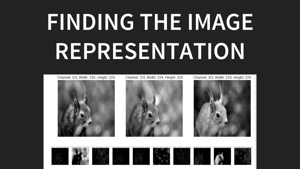
We will discuss how convolutional neural networks see the world and how we can tame them.

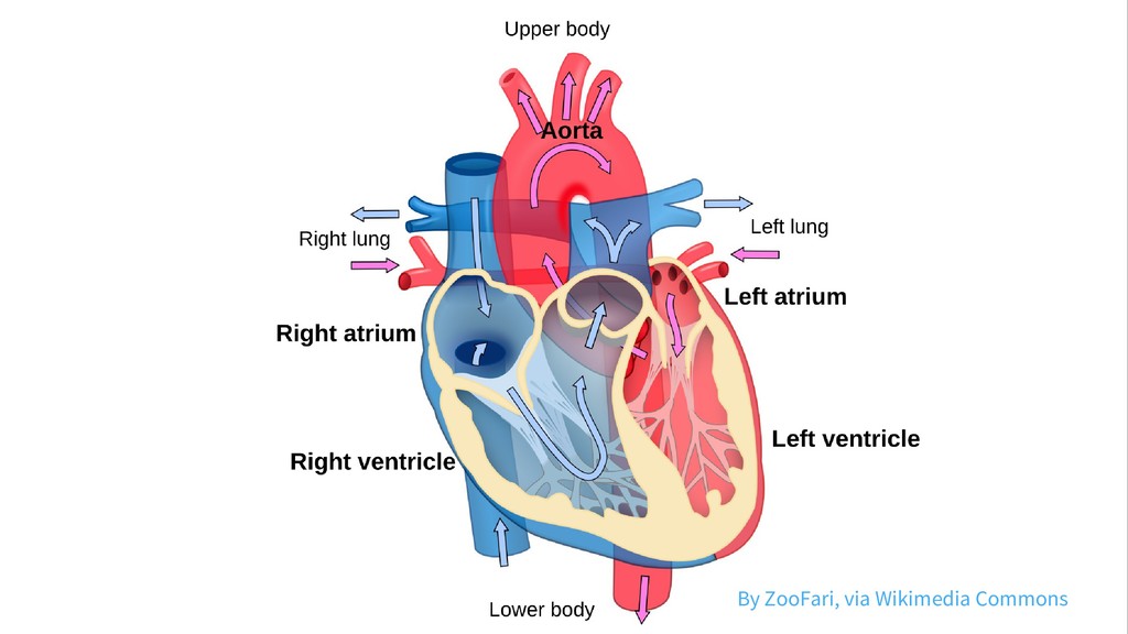
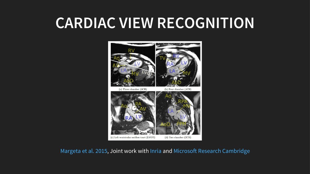
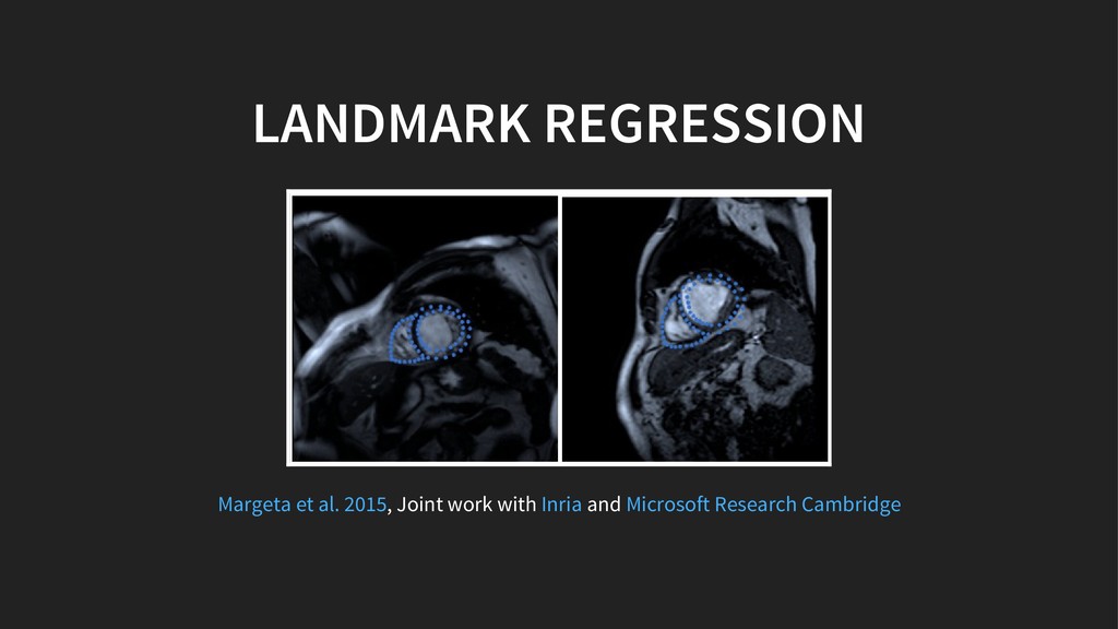
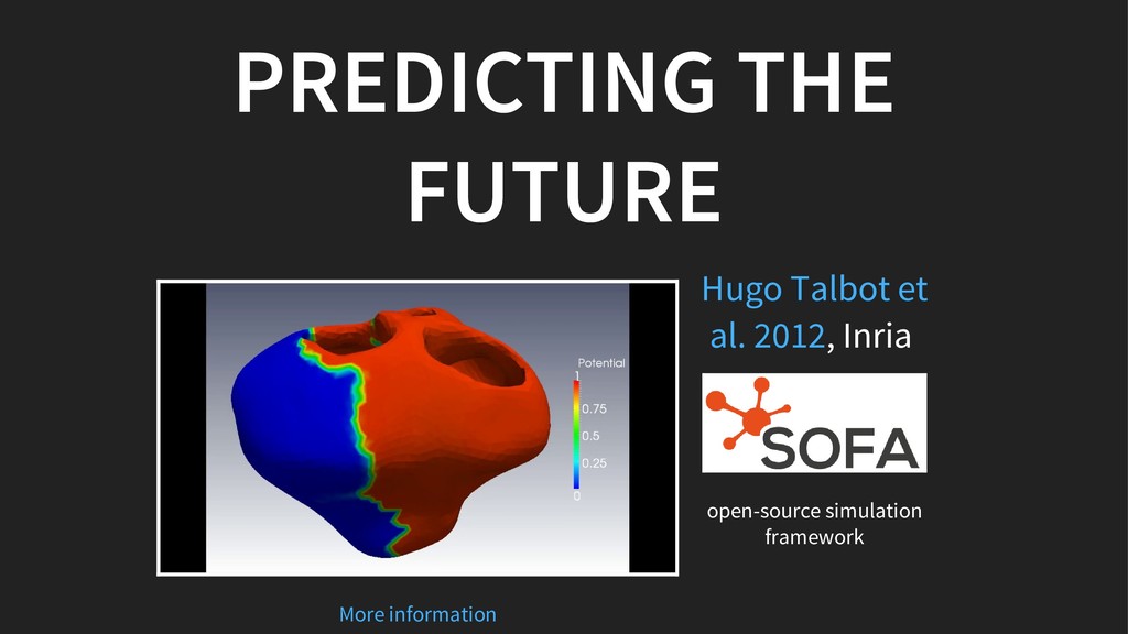
Our main application is cardiac imaging. That includes images from computed tomography and magnetic resonance.
But similar principles can be applied to many other computer vision challenges beyond medical imaging.

Jan Margeta

May 05, 2018
Tweet

More Decks by Jan Margeta

Other Decks in Programming

Transcript

  1. WELCOME TO COMPUTER VISION Get the blood by thresholding the

    bright pixels Get rid of the arteries Separate the circular left ventricle from the right one Make sure it works with all hearts There are imaging artefacts too! Contrast can be poor Some hearts do not have both ventricles!
  2. IMAGE RECOGNITION IN 6 LINES OF CODE from keras.applications import

    imagenet_utils from keras.applications.vgg16 import VGG16 # Load and prepare input images images_raw = load_images() images = imagenet_utils.preprocess_input(images_raw) # Load a pretrained image classification model model = VGG16(include_top=True, weights='imagenet') # Do the prediction predictions = model.predict(images)
  3. EXTRACTING VISUAL FEATURES # Load a pretrained classification model source_model

    = VGG16(weights='imagenet') # Define feature extractor from one layer of the network feature_layer = source_model.get_layer('conv4') feature_extractor = Model( input=fix_model.input, output=feature_layer.output) # Extract features features = feature_extractor.predict(images) See also "Deep visualization toolbox" on youtube
  4. USING THE FEATURES WITH SCIKIT LEARN from sklearn.svm import LinearSVC

    def flatten_features(features): return features.reshape(len(features), -1) features_train = feature_extractor.predict(images_train) features_train = flatten_features(features_train) classifier = LinearSVC() classifier.fit(features_train, labels_train) # predict on never seen images features_test = feature_extractor.predict(images_test) features_test = flatten_features(features_test) prediction_test = classifier.predict(features_test)
  5. CARDIAC VIEW RECOGNITION , Joint work with and Margeta et

    al. 2015 Inria Microsoft Research Cambridge
  6. LANDMARK REGRESSION , Joint work with and Margeta et al.

    2015 Inria Microsoft Research Cambridge
  7. START FROM SCRATCH from keras.models import Model from keras.layers import

    Input, Conv2D, Dense from keras.layers import GlobalAveragePooling2D num_outputs = 4 num_filters = 16 # Transformer of a gray image of any size to 16 outputs input = Input(shape=(1, None, None)) c0 = Conv2D(num_filters, 3, 3, activation='relu')(input) p = GlobalAveragePooling2D()(c0) output = Dense(num_outputs, activation='softmax')(p) model = Model(input=input, output=output)
  8. TRAIN THE MODEL # Loss function - task dependent #

    high for bad parameters, low for good ones # e.g. for image recognition loss_function = 'sparse_categorical_crossentropy' # Compile the model and fit model.compile(loss=loss_function, optimizer='adam') model.fit(images, labels) # Save the model for reuse model.save('model.h5')
  9. EXPOSING THE MODEL WITH FLASK *it requires a little bit

    more care import keras from flask import Flask, jsonify, request app = Flask(__name__) model = keras.models.load_model('model.h5') @app.route('/predict', methods=['POST']) def predict(): X = request_to_numpy(request) Y = model.predict(X) prediction = convert_prediction(Y) return jsonify(output=prediction) app.run(port=5000, threaded=False)
  10. DEFINE THE DOCKERFILE FROM python:3.5 RUN mkdir -p /usr/src/app COPY

    server.py /usr/src/app/ COPY model.h5 /usr/src/app/ COPY requirements.txt /usr/src/app/ WORKDIR /usr/src/app RUN pip install -r requirements.txt EXPOSE 5000 CMD python server.py
  11. BUILD AND RUN THE CONTAINER # Build the container docker

    build -t kardiome/model-pycon . # Run the container docker run -d -p 5000:5000 kardiome/model-pycon # Call the service curl -X POST -F 'image=@/data/im.png' localhost:5000/predict
  12. PROGRESS WITH CONFIDENCE AND REPEATABLE PIPELINES Gitlab's continuous integration is

    excellent start for simple pipelines (see also Airflow, Luigi, Flink)
  13. GOT UNLABELED DATA? DON'T BE LAZY, JUST ANNOTATE IT IF

    YOU CAN, THERE ARE TOOLS TO HELP YOU , Joint work with and Check out also Scikit learn example on or Sloth Margeta et al. 2015 Inria Microsoft Research Cambridge Label Propagation digits active learning
  14. PYTHON + AI + MEDICINE = A⚕ by Michael F.

    Mehnert, , via Wikimedia Commons Original file CC BY-SA 3.0
  15. THANKS , , , , , , , , ,

    , , CONNECT WITH ME Jan Margeta | | Krissy Hubert Hugo Karol Loïc Maxime Rocío Asclepios IHU Liryc Microsoft Research Cambridge NumFOCUS PyData [email protected] @jmargeta
  16. CREDITS By original file by Michael F. Mehnert , via

    Wikimedia Commons Radau P, Lu Y, Connelly K, Paul G, Dick AJ, Wright GA. “Evaluation Framework for Algorithms Segmenting Short Axis Cardiac MRI.” The MIDAS Journal – Cardiac MR Left Ventricle Segmentation Challenge, Statue of Asklepius, exhibited in the Museum of Epidaurus Theatre. CC BY-SA 3.0 http://hdl.handle.net/10380/3070
  17. Some results come from my PhD thesis funded by through

    its PhD Scholarship Programme and by the Microsoft Research ERC Advanced Grant MedYMA
  18. RESOURCES PhD thesis - Jan Margeta PhD thesis - Rocío

    Cabrera Lozoya PhD thesis - Hugo Talbot Book From Andrew Ng Fast AI Notebooks and course Visualizing convnets Conv filter visualization
  19. Transfer learning with MNIST Label propagation with scikit learn Keras

    and pretrained models Staying organized - Templates for data science Cardiac atlas project Sunnybrook cardiac dataset UK biobank
  20. Mimesis team @ Inria Asclepios @ Inria SOFA - Opensource

    simulation framework Cardiovascular death stats Detecting cancer with deep learning Dermatologist-level classification of skin cancer with deep neural networks
  21. REFERENCES Kelly, M. D. (2007). Laparoscopic repair of strangulated Morgagni

    hernia. World Journal of Emergency Surgery. Springer Nature. Carmo, P., Andrade, M. J., Aguiar, C., Rodrigues, R., Gouveia, R., & Silva, J. A. (2010, December). Mitral annular disjunction in myxomatous mitral valve disease: a relevant abnormality recognizable by transthoracic echocardiography. Cardiovascular Ultrasound. Springer Nature. Arnold, J. R., West, N. E., van Gaal, W. J., Karamitsos, T. D., & Banning, A. P. (2008, May 31). The role of Intravascular Ultrasound in the management of spontaneous coronary artery dissection. Cardiovascular Ultrasound. Springer Nature. https://doi.org/10.1186/1749-7922-2-27 https://doi.org/10.1186/1476-7120-8-53 https://doi.org/10.1186/1476- 7120-6-24
  22. Foley, P. W., Hamaad, A., El-Gendi, H., & Leyva, F.

    (2010). Incidental cardiac findings on computed tomography imaging of the thorax. BMC Research Notes. Springer Nature. Vanezis, A. P., Baig, M. K., Mitchel, I. M., Shajar, M., Naik, S. K., Henderson, R. A., & Mathew, T. (2011). Pseudoaneurysm of the left ventricle following apical approach TAVI. Journal of Cardiovascular Magnetic Resonance. Springer Nature. Xue, H., Kellman, P., LaRocca, G., Arai, A. E., & Hansen, M. S. (2013). High spatial and temporal resolution retrospective cine cardiovascular magnetic resonance from shortened free breathing real-time acquisitions. Journal of Cardiovascular Magnetic Resonance. Springer Nature. Margeta, J., McLeod, K., Criminisi, A., & Ayache, N. (2014). Decision Forests for Segmentation of the Left Atrium from 3D MRI. Statistical Atlases and Computational Models of the Heart. Imaging and Modelling Challenges. Springer Berlin Heidelberg. https://doi.org/10.1186/1756-0500-3-326 https://doi.org/10.1186/1532-429x- 13-79 https://doi.org/10.1186/1532-429x-15-102 https://doi.org/10.1007/978-3-642-54268-8_6
  23. Tobon-Gomez, C., Geers, A. J., Peters, J., Weese, J., Pinto,

    K., Karim, R., … Rhode, K. S. (2015, July). Benchmark for Algorithms Segmenting the Left Atrium From 3D CT and MRI Datasets. IEEE Transactions on Medical Imaging. Institute of Electrical and Electronics Engineers (IEEE). Margeta, J., Criminisi, A., Cabrera Lozoya, R., Lee, D. C., & Ayache, N. (2015, August 13). Fine-tuned convolutional neural nets for cardiac MRI acquisition plane recognition. Computer Methods in Biomechanics and Biomedical Engineering: Imaging & Visualization. Informa UK Limited. McLeod, K., Wall, S., Leren, I. S., Saberniak, J., & Haugaa, K. H. (2016, October 14). Ventricular structure in ARVC: going beyond volumes as a measure of risk. Journal of Cardiovascular Magnetic Resonance. Springer Nature. https://doi.org/10.1109/tmi.2015.2398818 https://doi.org/10.1080/21681163.2015.1061448 https://doi.org/10.1186/s12968- 016-0291-9
  24. Talbot, H., Marchesseau, S., Duriez, C., Sermesant, M., Cotin, S.,

    & Delingette, H. (2013, February 21). Towards an interactive electromechanical model of the heart. Interface Focus. The Royal Society. Demarcy, T., Vandersteen, C., Raffaelli, C., Gnansia, D., Guevara, N., Ayache, N., & Delingette, H. (2016). Uncertainty Quantification of Cochlear Implant Insertion from CT Images. Clinical Image-Based Procedures. Translational Research in Medical Imaging. Springer International Publishing. Montuoro, A., Waldstein, S. M., Gerendas, B. S., Schmidt-Erfurth, U., & Bogunović, H. (2017, February 27). Joint retinal layer and fluid segmentation in OCT scans of eyes with severe macular edema using unsupervised representation and auto-context. Biomedical Optics Express. The Optical Society. Demarcy, T., Vandersteen, C., Raffaelli, C., Gnansia, D., Guevara, N., Ayache, N., & Delingette, H. (2017). Automated Analysis of Human Cochlea Shape Variability from segmented μCT images. Computerized Medical Imaging and Graphics (to appear). https://doi.org/10.1098/rsfs.2012.0091 https://doi.org/10.1007/978-3-319-46472-5_4 https://doi.org/10.1364/boe.8.001874
  25. Radau, P., Lu, Y., Connelly, K., Paul, G., Dick, AJ.,

    Wright, GA. (2009). Evaluation Framework for Algorithms Segmenting Short Axis Cardiac MRI. The MIDAS Journal – Cardiac MR Left Ventricle Segmentation Challenge. Kadish, A. H., Bello, D., Finn, J. P., Bonow, R. O., Schaechter, A., Subacius, H., … Goldberger, J. J. (2009, September). Rationale and Design for the Defibrillators to Reduce Risk by Magnetic Resonance Imaging Evaluation (DETERMINE) Trial. Journal of Cardiovascular Electrophysiology. Wiley-Blackwell. Fonseca, C. G., Backhaus, M., Do Chung, J., Tao, W., Medrano-Gracia, P., Cowan, B. R., … Young, A. A. (2010). The Cardiac Atlas Project: Rationale, Design and Procedures. Statistical Atlases and Computational Models of the Heart. Springer Berlin Heidelberg. http://hdl.handle.net/10380/3070 https://doi.org/10.1111/j.1540- 8167.2009.01503.x https://doi.org/10.1007/978-3-642-15835-3_4