Upgrade to Pro — share decks privately, control downloads, hide ads and more …

Bringing Medical imaging data to life

Bringing Medical imaging data to life

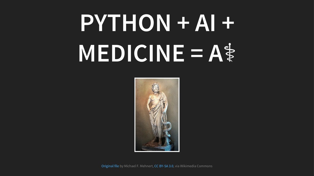
Presented at PyData Bratislava Meetup

Event description: Join us on a journey into our own hearts through the images and data. Come to discuss artificial intelligence, healthcare, computer vision, and Python. This meetup is a sequel of my PyCon SK 2017 talk from two weeks ago.

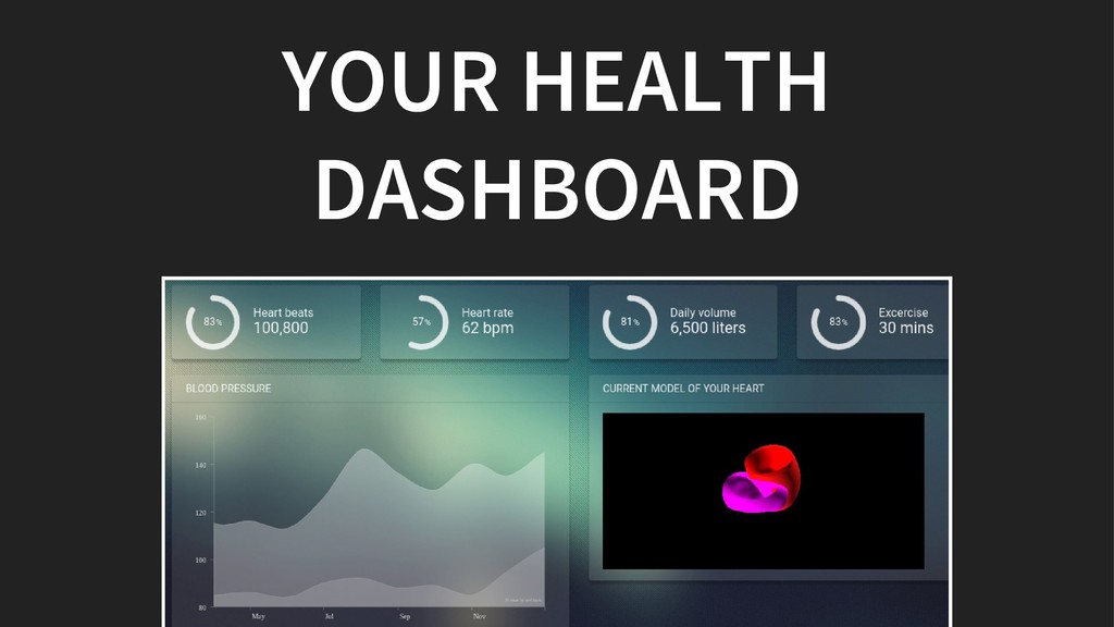
Today, we are acquiring more health data about ourselves than ever before. Many of us are already using apps and devices that record our physical activity, weight, diet, mood, heart rate, blood pressure, or sleep.

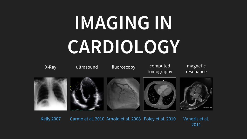
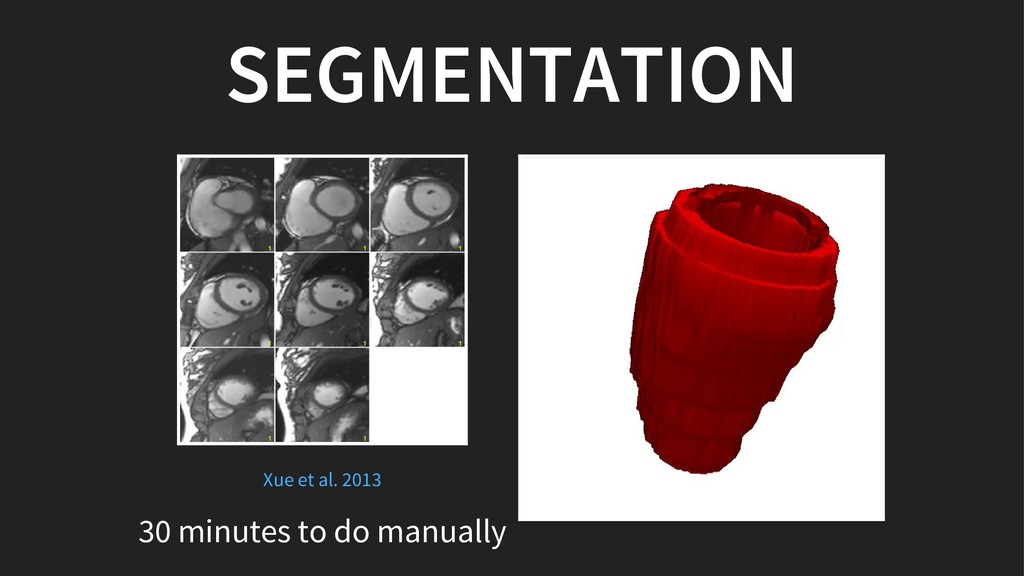
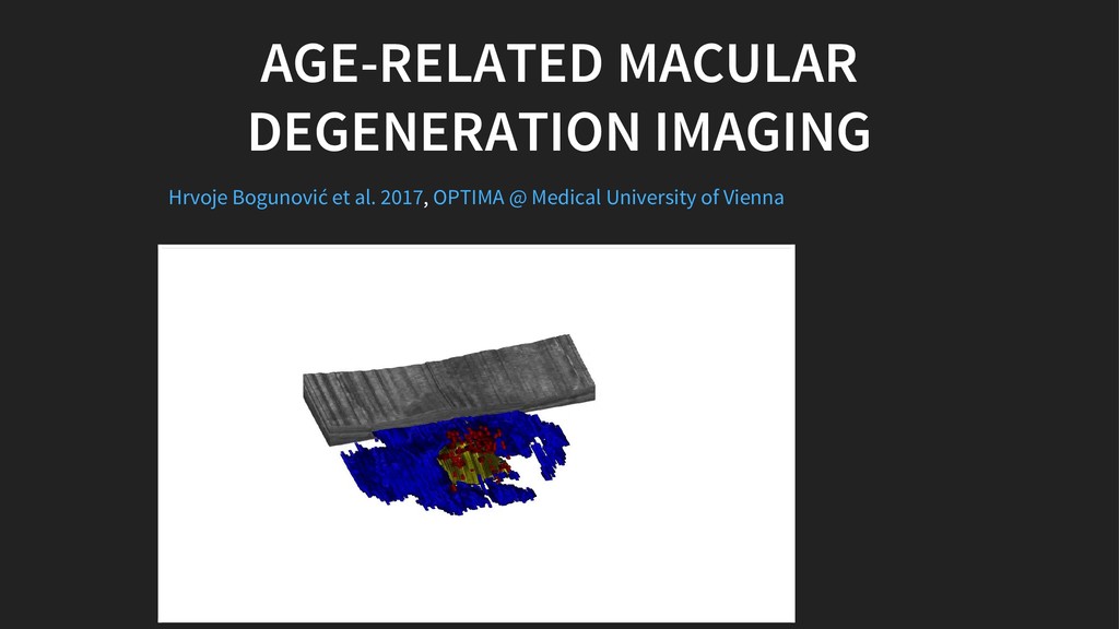
In addition to that, medical imaging techniques, such as magnetic resonance imaging (MRI) or computed tomography (CT), are giving us insights into our bodies with unprecedented detail. Yet, only few of us can interpret them (Remember the last time you saw your X-Ray?)

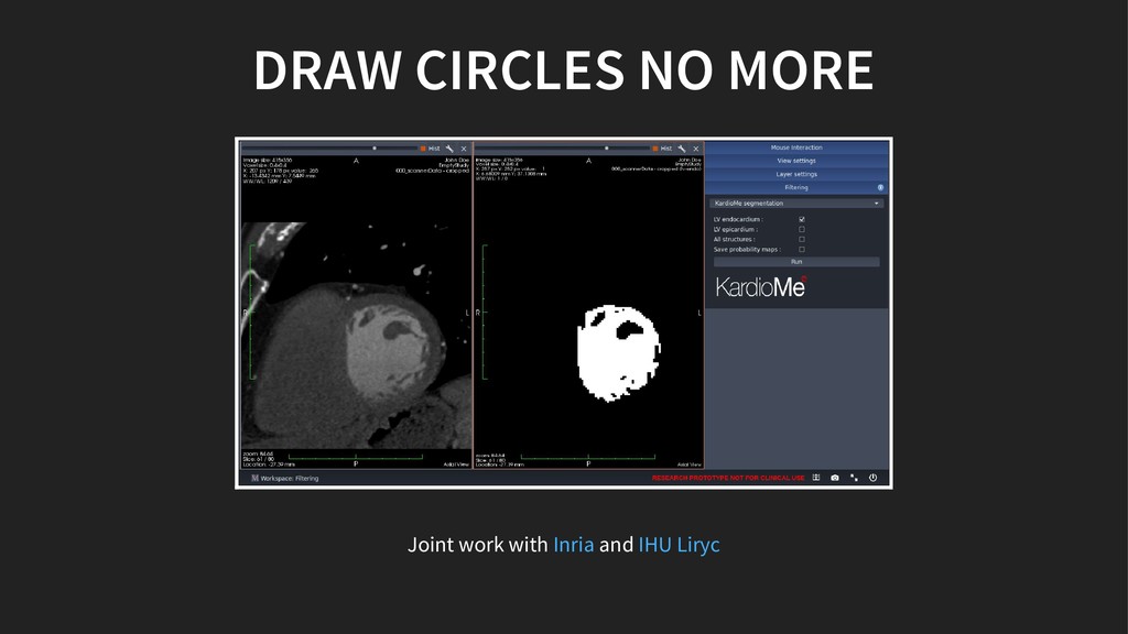
At KardioMe we build tools assisting radiologists and cardiologists to be more efficient with image analysis, tools that will empower all of us to better understand our health data and take better control of it.

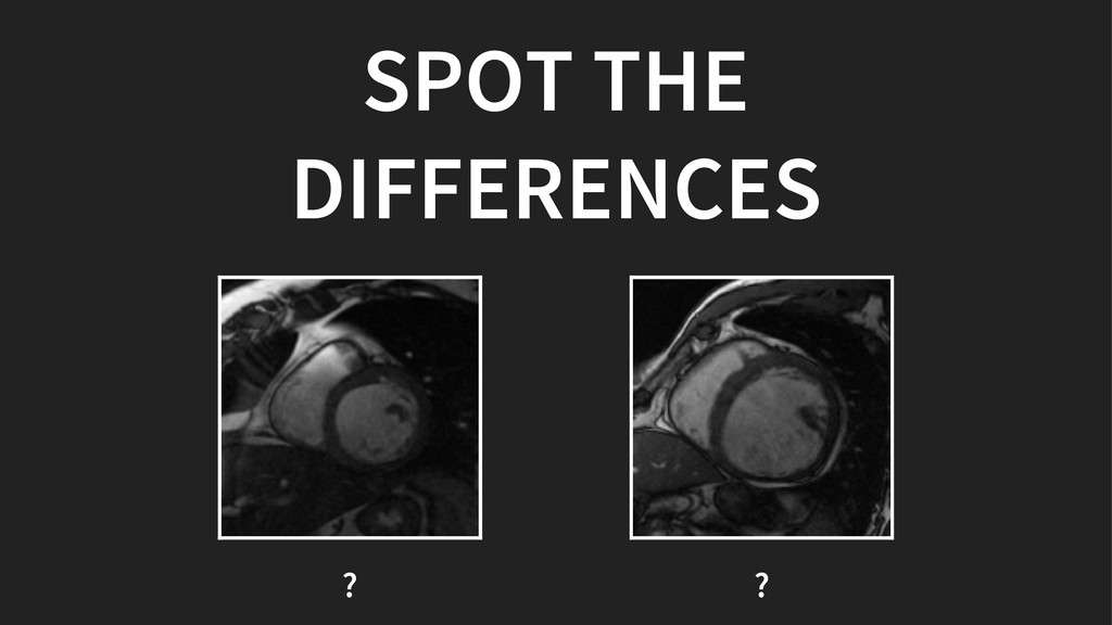
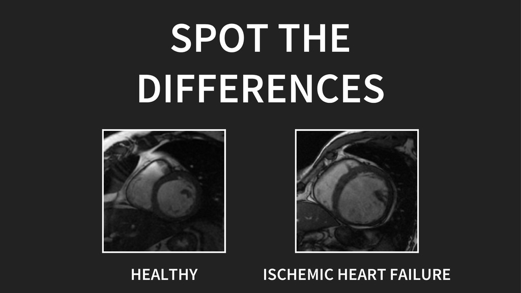
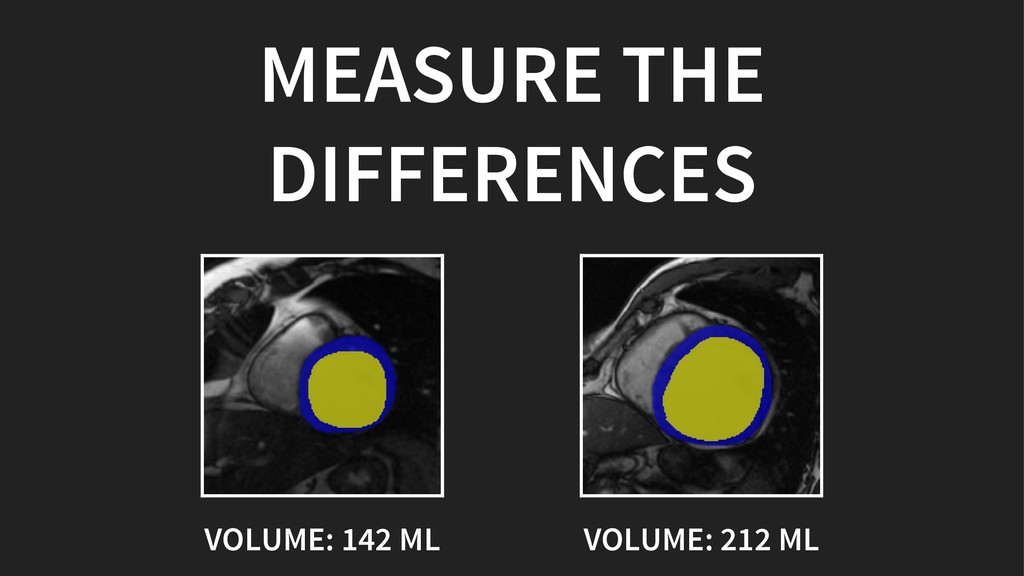
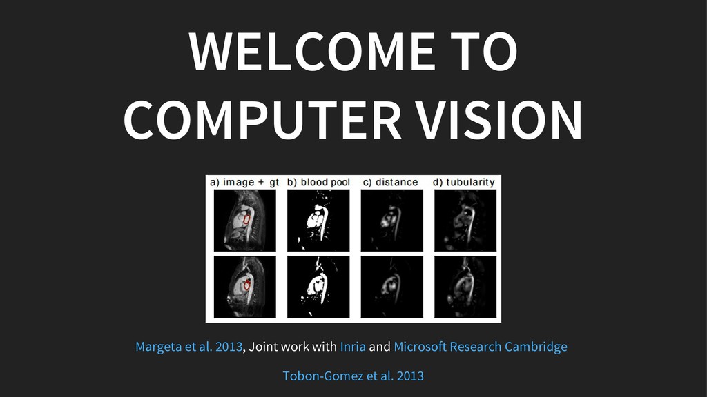
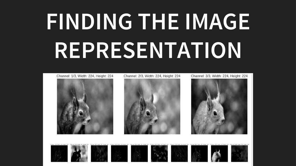
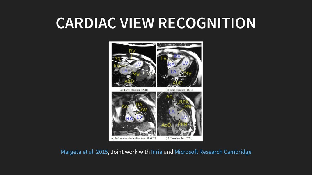
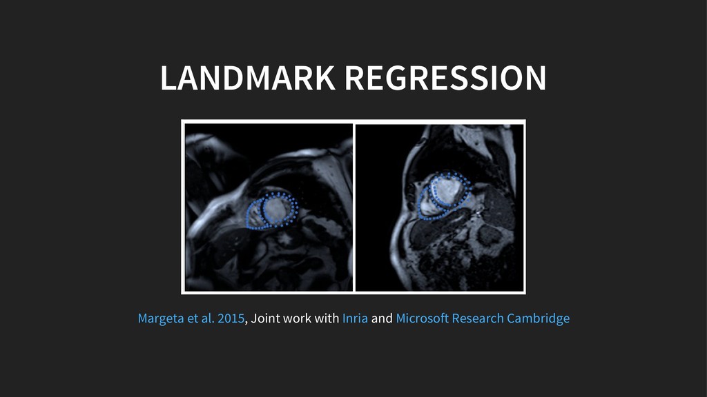
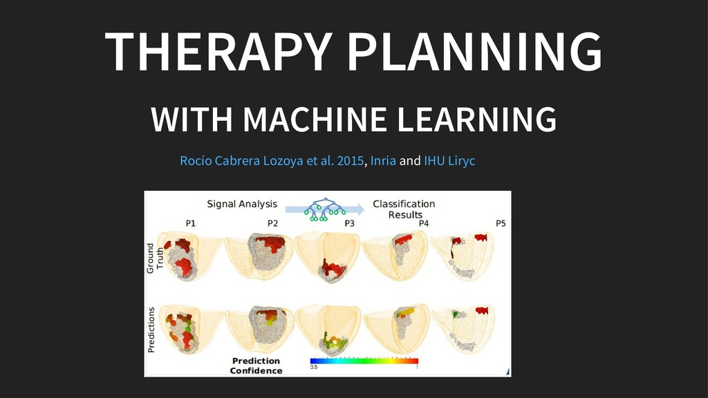
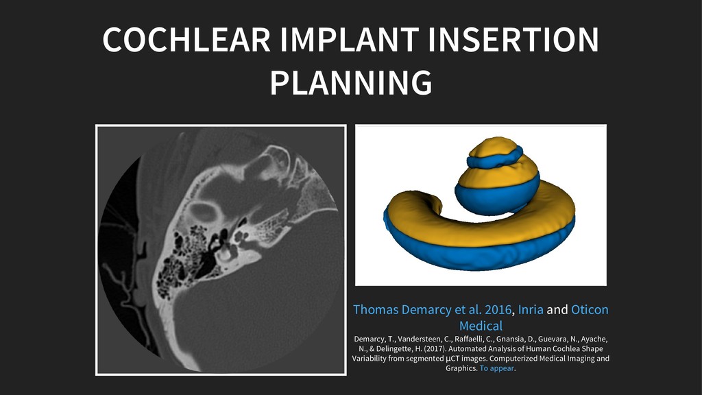
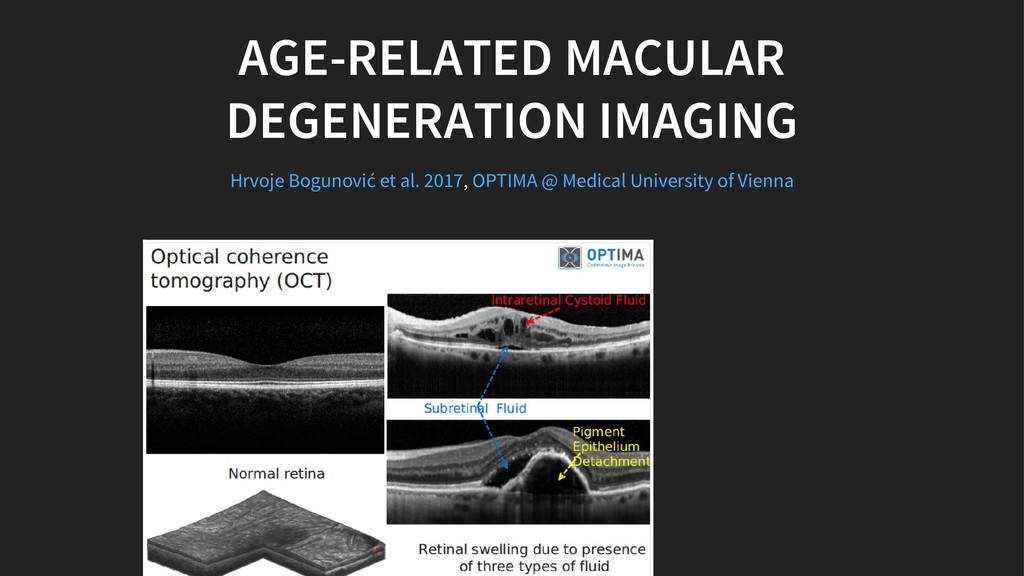
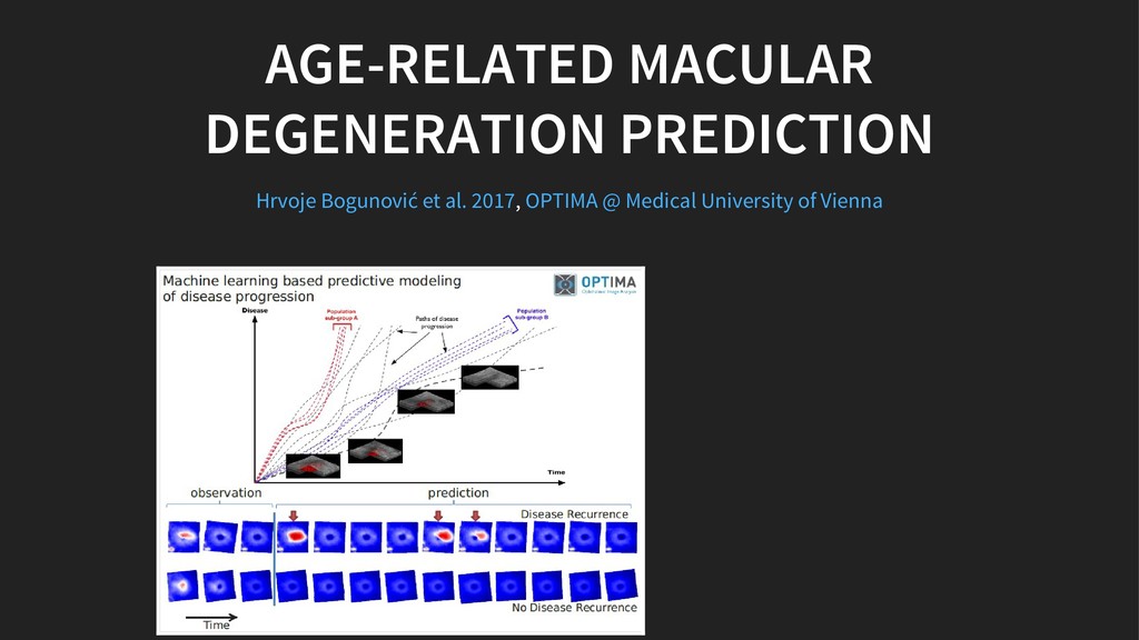
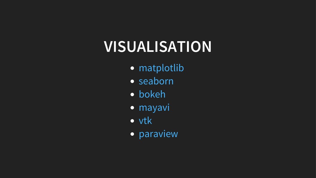
In the talk we will explore how we use machine learning and image processing to extract meaningful image descriptions of our hearts. How machine learning helps us to organise and navigate through large scale (medical) image collections for visualisation.

Do not hesitate to leave your suggestions in the comments for discussion.

Jan Margeta

March 27, 2017
Tweet

More Decks by Jan Margeta

Other Decks in Research

Transcript

  1. 45%

  2. IMAGING IN CARDIOLOGY X-Ray ultrasound fluoroscopy computed tomography magnetic resonance

    Kelly 2007 Carmo et al. 2010 Arnold et al. 2008 Foley et al. 2010 Vanezis et al. 2011
  3. WELCOME TO COMPUTER VISION , Joint work with and Margeta

    et al. 2013 Inria Microsoft Research Cambridge Tobon-Gomez et al. 2013
  4. IMAGE RECOGNITION IN 6 LINES OF CODE from keras.applications import

    imagenet_utils from keras.applications.vgg16 import VGG16 # Load and prepare input images images_raw = load_images() images = imagenet_utils.preprocess_input(images_raw) # Load a pretrained image classification model model = VGG16(include_top=True, weights='imagenet') # Do the prediction predictions = model.predict(images)
  5. CARDIAC VIEW RECOGNITION , Joint work with and Margeta et

    al. 2015 Inria Microsoft Research Cambridge
  6. LANDMARK REGRESSION , Joint work with and Margeta et al.

    2015 Inria Microsoft Research Cambridge
  7. FROM 30 MINUTES TO 12 SECONDS* *on a GPU Joint

    work with and Inria IHU Liryc
  8. , Inria ELECTROMECHANICAL COUPLING More information Hugo Talbot et al.

    2012 CHECK OUT open-source simulation framework:
  9. , and Demarcy, T., Vandersteen, C., Raffaelli, C., Gnansia, D.,

    Guevara, N., Ayache, N., & Delingette, H. (2017). Automated Analysis of Human Cochlea Shape Variability from segmented μCT images. Computerized Medical Imaging and Graphics. . COCHLEAR IMPLANT INSERTION PLANNING Thomas Demarcy et al. 2016 Inria Oticon Medical To appear
  10. GOT UNLABELED DATA? DON'T BE LAZY, JUST ANNOTATE IT IF

    YOU CAN, THERE ARE TOOLS TO HELP YOU , Joint work with and Check out also Scikit learn example on Margeta et al. 2015 Inria Microsoft Research Cambridge Label Propagation digits active learning
  11. PYTHON + AI + MEDICINE = A⚕ by Michael F.

    Mehnert, , via Wikimedia Commons Original file CC BY-SA 3.0
  12. THANKS , , , , , , , , ,

    , , , , , , , , , , CONNECT WITH ME Jan Margeta | | Krissy Hrvoje Hubert Hugo Karol Loïc Maxime Rado Rocío Thomas Maggie Asclepios GapData Institute IHU Liryc Microsoft Research Cambridge NumFOCUS PyData OPTIMA Oticon Medical Simula research laboratory [email protected] @jmargeta
  13. CREDITS By original file by Michael F. Mehnert , via

    Wikimedia Commons Radau P, Lu Y, Connelly K, Paul G, Dick AJ, Wright GA. “Evaluation Framework for Algorithms Segmenting Short Axis Cardiac MRI.” The MIDAS Journal – Cardiac MR Left Ventricle Segmentation Challenge, Statue of Asklepius, exhibited in the Museum of Epidaurus Theatre. CC BY-SA 3.0 http://hdl.handle.net/10380/3070
  14. Some results come from my PhD thesis funded by through

    its PhD Scholarship Programme and by the Microsoft Research ERC Advanced Grant MedYMA
  15. RESOURCES PhD thesis - Jan Margeta PhD thesis - Rocío

    Cabrera Lozoya PhD thesis - Hugo Talbot Book From Andrew Ng Fast AI Notebooks and course Visualizing convnets Conv filter visualization
  16. Transfer learning with MNIST Label propagation with scikit learn Keras

    and pretrained models Staying organized - Templates for data science Cardiac atlas project Sunnybrook cardiac dataset UK biobank
  17. Mimesis team @ Inria Asclepios @ Inria SOFA - Opensource

    simulation framework Cardiovascular death stats Detecting cancer with deep learning Dermatologist-level classification of skin cancer with deep neural networks
  18. REFERENCES Kelly, M. D. (2007). Laparoscopic repair of strangulated Morgagni

    hernia. World Journal of Emergency Surgery. Springer Nature. Carmo, P., Andrade, M. J., Aguiar, C., Rodrigues, R., Gouveia, R., & Silva, J. A. (2010, December). Mitral annular disjunction in myxomatous mitral valve disease: a relevant abnormality recognizable by transthoracic echocardiography. Cardiovascular Ultrasound. Springer Nature. Arnold, J. R., West, N. E., van Gaal, W. J., Karamitsos, T. D., & Banning, A. P. (2008, May 31). The role of Intravascular Ultrasound in the management of spontaneous coronary artery dissection. Cardiovascular Ultrasound. Springer Nature. https://doi.org/10.1186/1749-7922-2-27 https://doi.org/10.1186/1476-7120-8-53 https://doi.org/10.1186/1476- 7120-6-24
  19. Foley, P. W., Hamaad, A., El-Gendi, H., & Leyva, F.

    (2010). Incidental cardiac findings on computed tomography imaging of the thorax. BMC Research Notes. Springer Nature. Vanezis, A. P., Baig, M. K., Mitchel, I. M., Shajar, M., Naik, S. K., Henderson, R. A., & Mathew, T. (2011). Pseudoaneurysm of the left ventricle following apical approach TAVI. Journal of Cardiovascular Magnetic Resonance. Springer Nature. Xue, H., Kellman, P., LaRocca, G., Arai, A. E., & Hansen, M. S. (2013). High spatial and temporal resolution retrospective cine cardiovascular magnetic resonance from shortened free breathing real-time acquisitions. Journal of Cardiovascular Magnetic Resonance. Springer Nature. Margeta, J., McLeod, K., Criminisi, A., & Ayache, N. (2014). Decision Forests for Segmentation of the Left Atrium from 3D MRI. Statistical Atlases and Computational Models of the Heart. Imaging and Modelling Challenges. Springer Berlin Heidelberg. https://doi.org/10.1186/1756-0500-3-326 https://doi.org/10.1186/1532-429x- 13-79 https://doi.org/10.1186/1532-429x-15-102 https://doi.org/10.1007/978-3-642-54268-8_6
  20. Tobon-Gomez, C., Geers, A. J., Peters, J., Weese, J., Pinto,

    K., Karim, R., … Rhode, K. S. (2015, July). Benchmark for Algorithms Segmenting the Left Atrium From 3D CT and MRI Datasets. IEEE Transactions on Medical Imaging. Institute of Electrical and Electronics Engineers (IEEE). Margeta, J., Criminisi, A., Cabrera Lozoya, R., Lee, D. C., & Ayache, N. (2015, August 13). Fine-tuned convolutional neural nets for cardiac MRI acquisition plane recognition. Computer Methods in Biomechanics and Biomedical Engineering: Imaging & Visualization. Informa UK Limited. McLeod, K., Wall, S., Leren, I. S., Saberniak, J., & Haugaa, K. H. (2016, October 14). Ventricular structure in ARVC: going beyond volumes as a measure of risk. Journal of Cardiovascular Magnetic Resonance. Springer Nature. https://doi.org/10.1109/tmi.2015.2398818 https://doi.org/10.1080/21681163.2015.1061448 https://doi.org/10.1186/s12968- 016-0291-9
  21. Talbot, H., Marchesseau, S., Duriez, C., Sermesant, M., Cotin, S.,

    & Delingette, H. (2013, February 21). Towards an interactive electromechanical model of the heart. Interface Focus. The Royal Society. Demarcy, T., Vandersteen, C., Raffaelli, C., Gnansia, D., Guevara, N., Ayache, N., & Delingette, H. (2016). Uncertainty Quantification of Cochlear Implant Insertion from CT Images. Clinical Image-Based Procedures. Translational Research in Medical Imaging. Springer International Publishing. Montuoro, A., Waldstein, S. M., Gerendas, B. S., Schmidt-Erfurth, U., & Bogunović, H. (2017, February 27). Joint retinal layer and fluid segmentation in OCT scans of eyes with severe macular edema using unsupervised representation and auto-context. Biomedical Optics Express. The Optical Society. Demarcy, T., Vandersteen, C., Raffaelli, C., Gnansia, D., Guevara, N., Ayache, N., & Delingette, H. (2017). Automated Analysis of Human Cochlea Shape Variability from segmented μCT images. Computerized Medical Imaging and Graphics (to appear). https://doi.org/10.1098/rsfs.2012.0091 https://doi.org/10.1007/978-3-319-46472-5_4 https://doi.org/10.1364/boe.8.001874
  22. Radau, P., Lu, Y., Connelly, K., Paul, G., Dick, AJ.,

    Wright, GA. (2009). Evaluation Framework for Algorithms Segmenting Short Axis Cardiac MRI. The MIDAS Journal – Cardiac MR Left Ventricle Segmentation Challenge. Kadish, A. H., Bello, D., Finn, J. P., Bonow, R. O., Schaechter, A., Subacius, H., … Goldberger, J. J. (2009, September). Rationale and Design for the Defibrillators to Reduce Risk by Magnetic Resonance Imaging Evaluation (DETERMINE) Trial. Journal of Cardiovascular Electrophysiology. Wiley-Blackwell. Fonseca, C. G., Backhaus, M., Do Chung, J., Tao, W., Medrano-Gracia, P., Cowan, B. R., … Young, A. A. (2010). The Cardiac Atlas Project: Rationale, Design and Procedures. Statistical Atlases and Computational Models of the Heart. Springer Berlin Heidelberg. http://hdl.handle.net/10380/3070 https://doi.org/10.1111/j.1540- 8167.2009.01503.x https://doi.org/10.1007/978-3-642-15835-3_4