Upgrade to Pro — share decks privately, control downloads, hide ads and more …

Single Cell and Spatial Transcriptomics in the Postmortem Brain

Single Cell and Spatial Transcriptomics in the Postmortem Brain

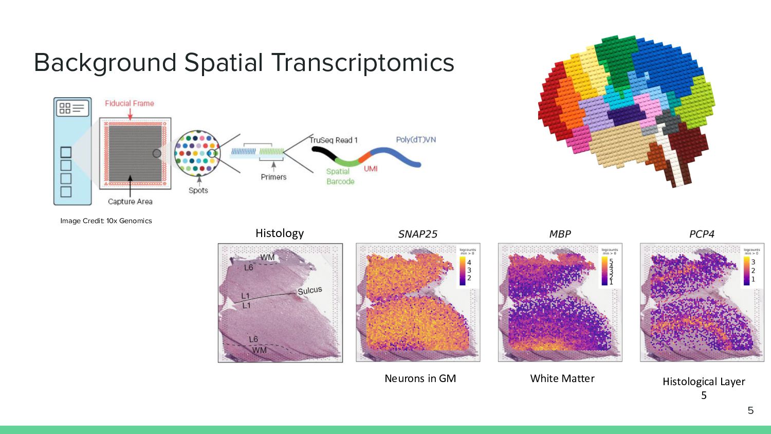
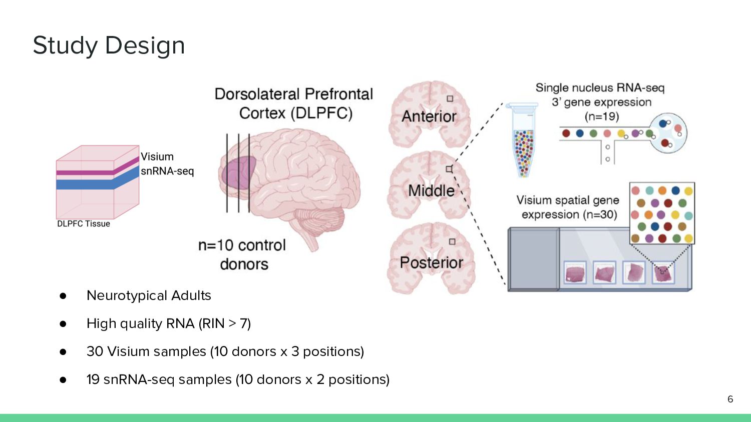
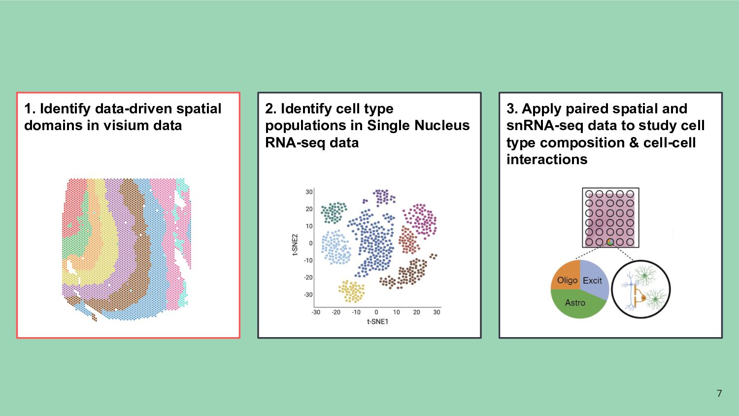
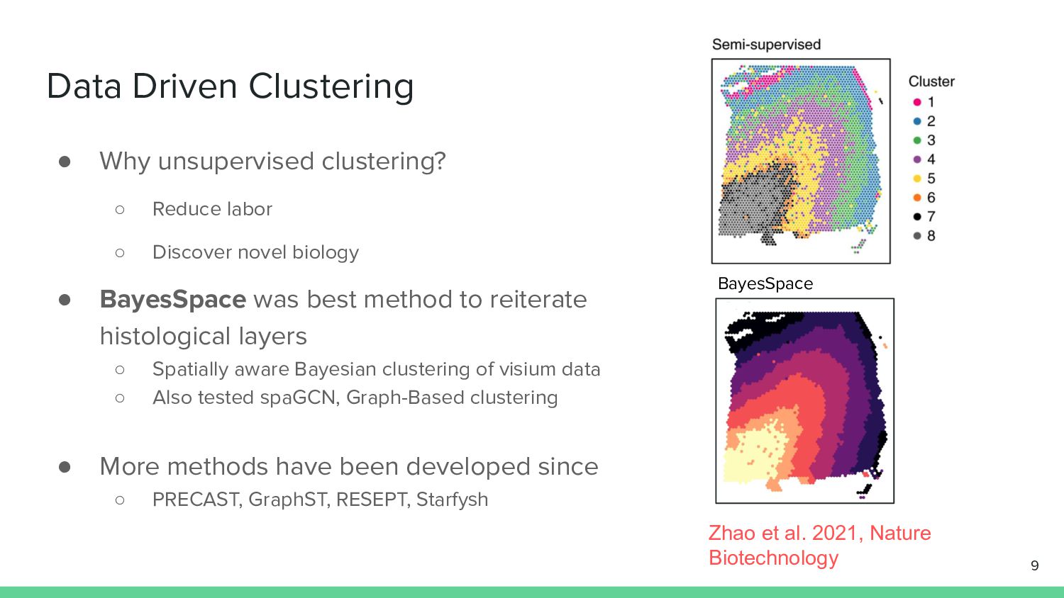
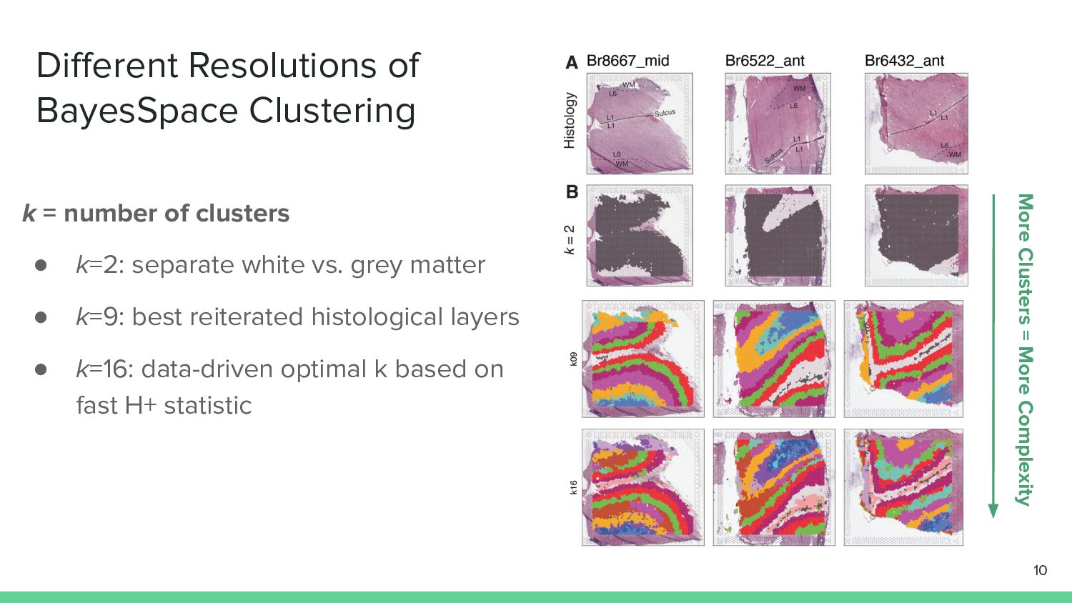
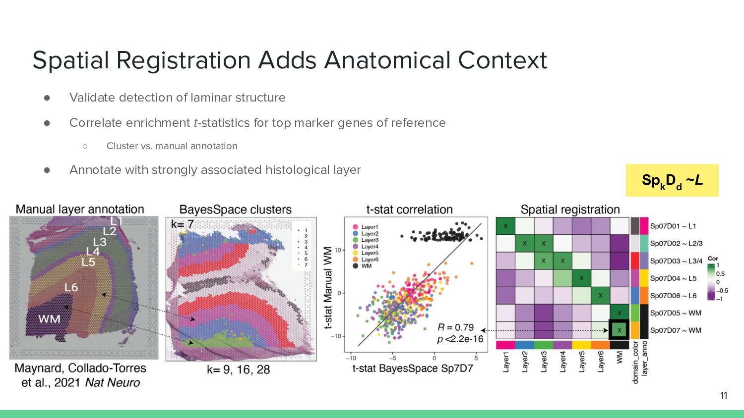
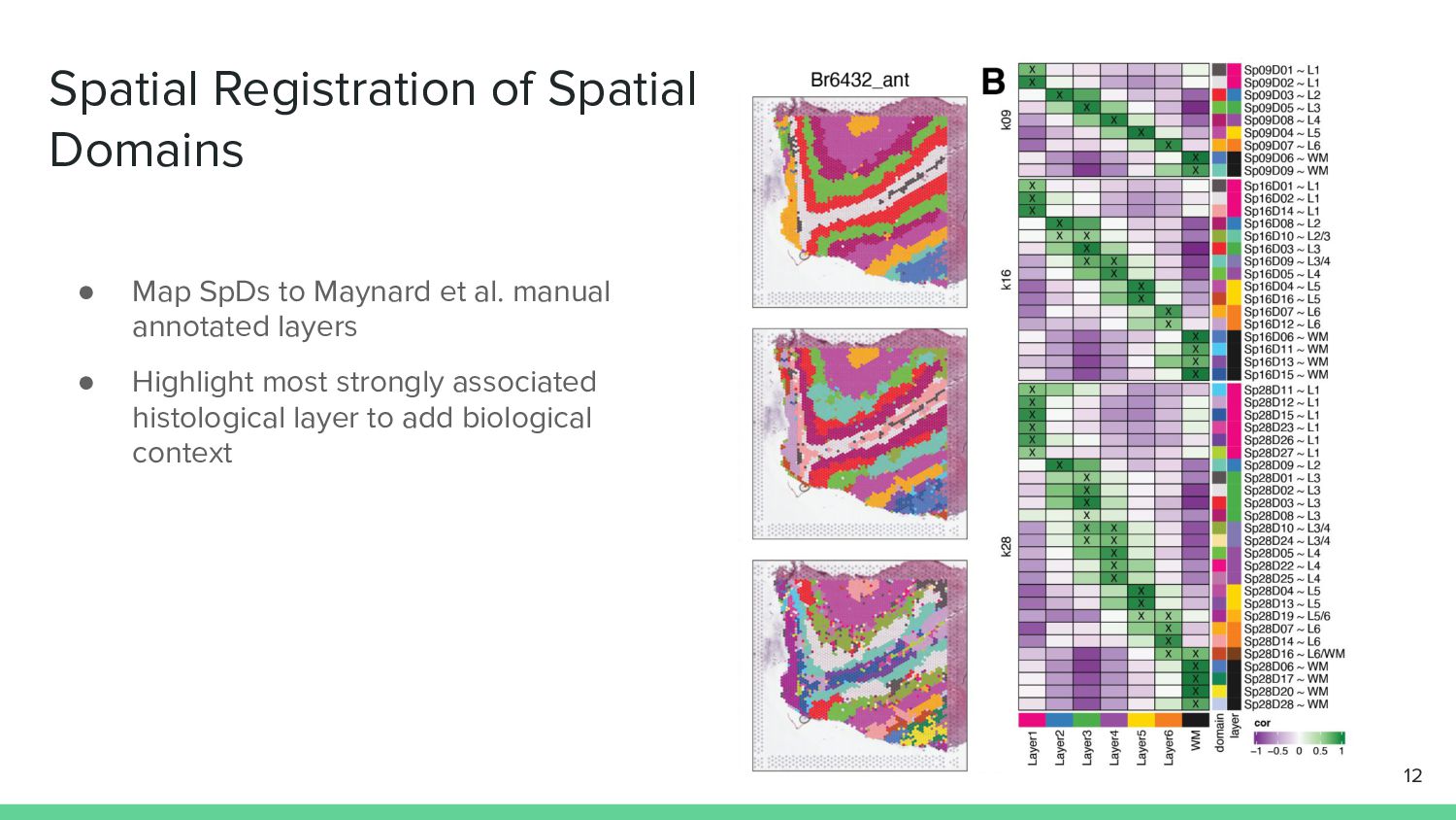
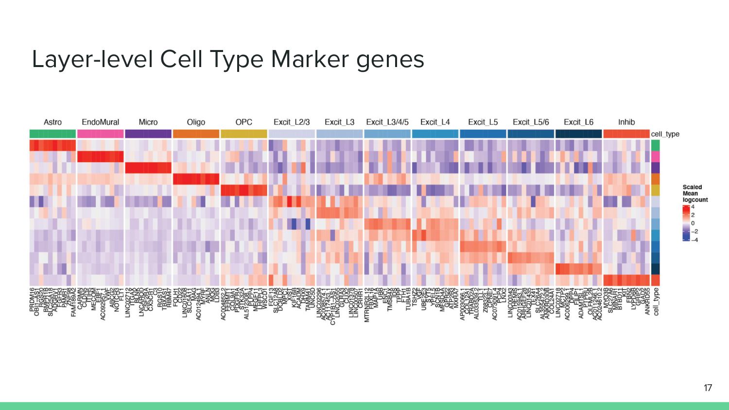
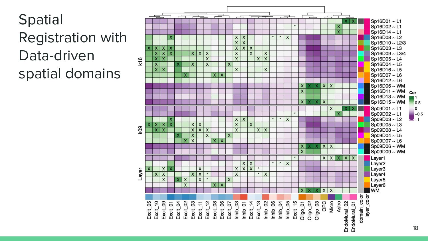
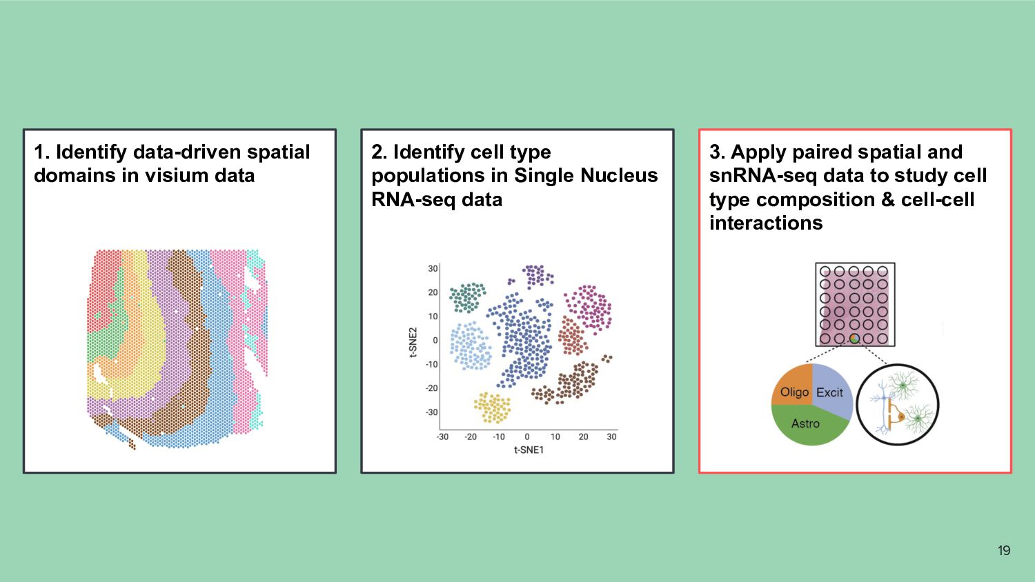
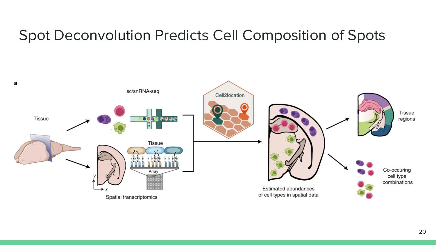
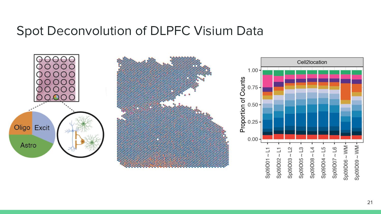
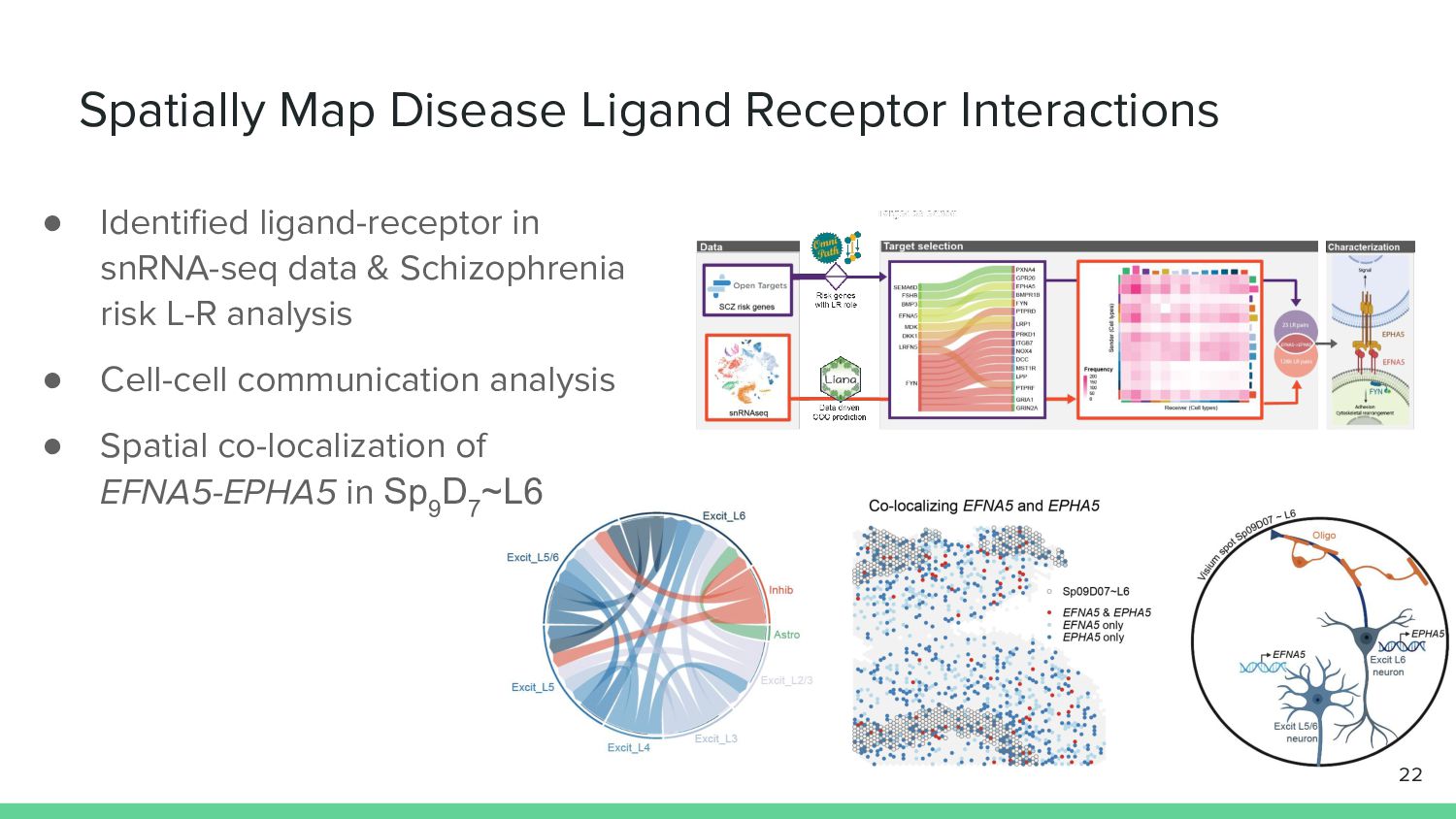
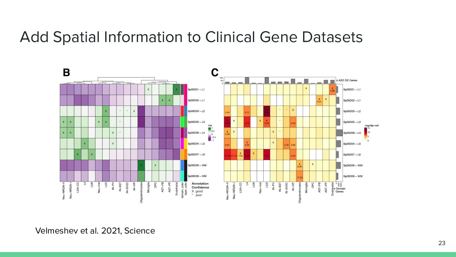
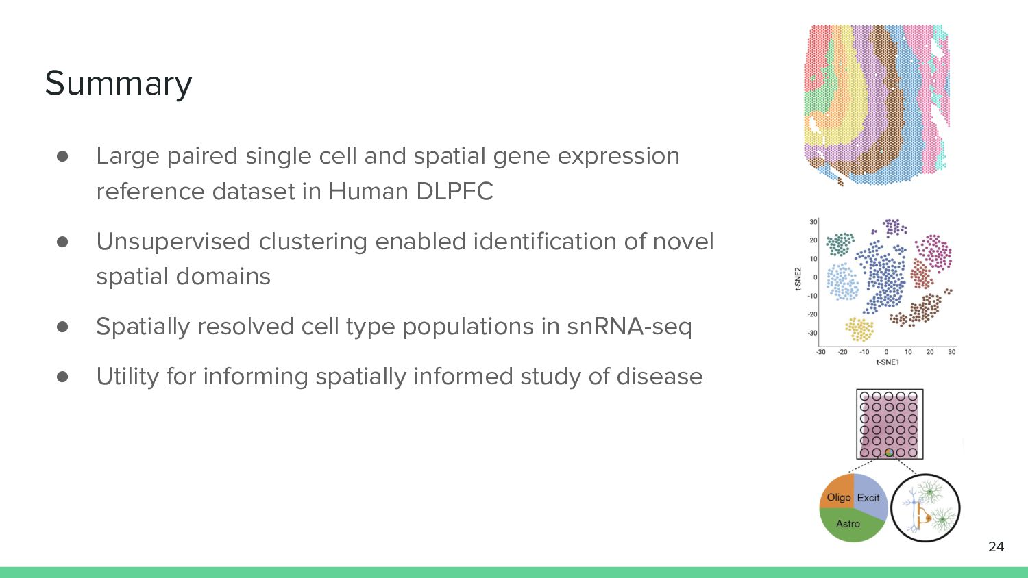
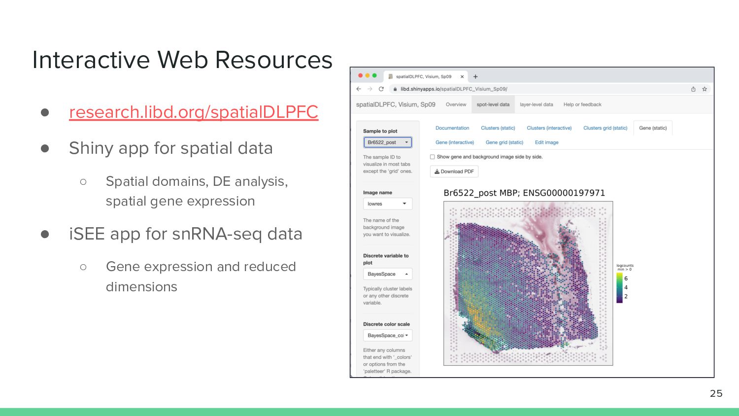
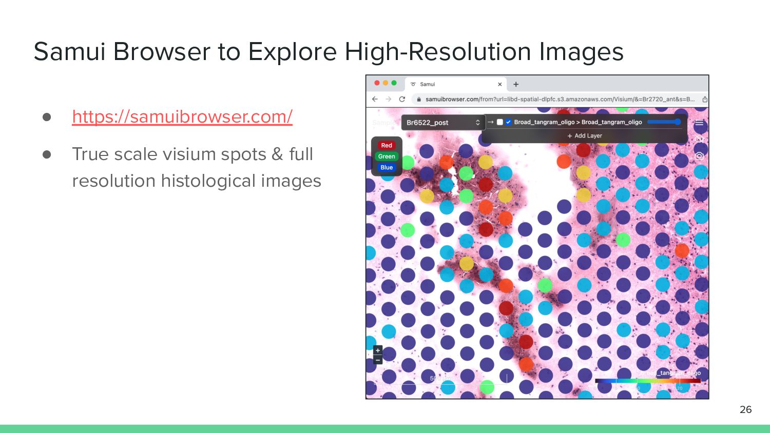
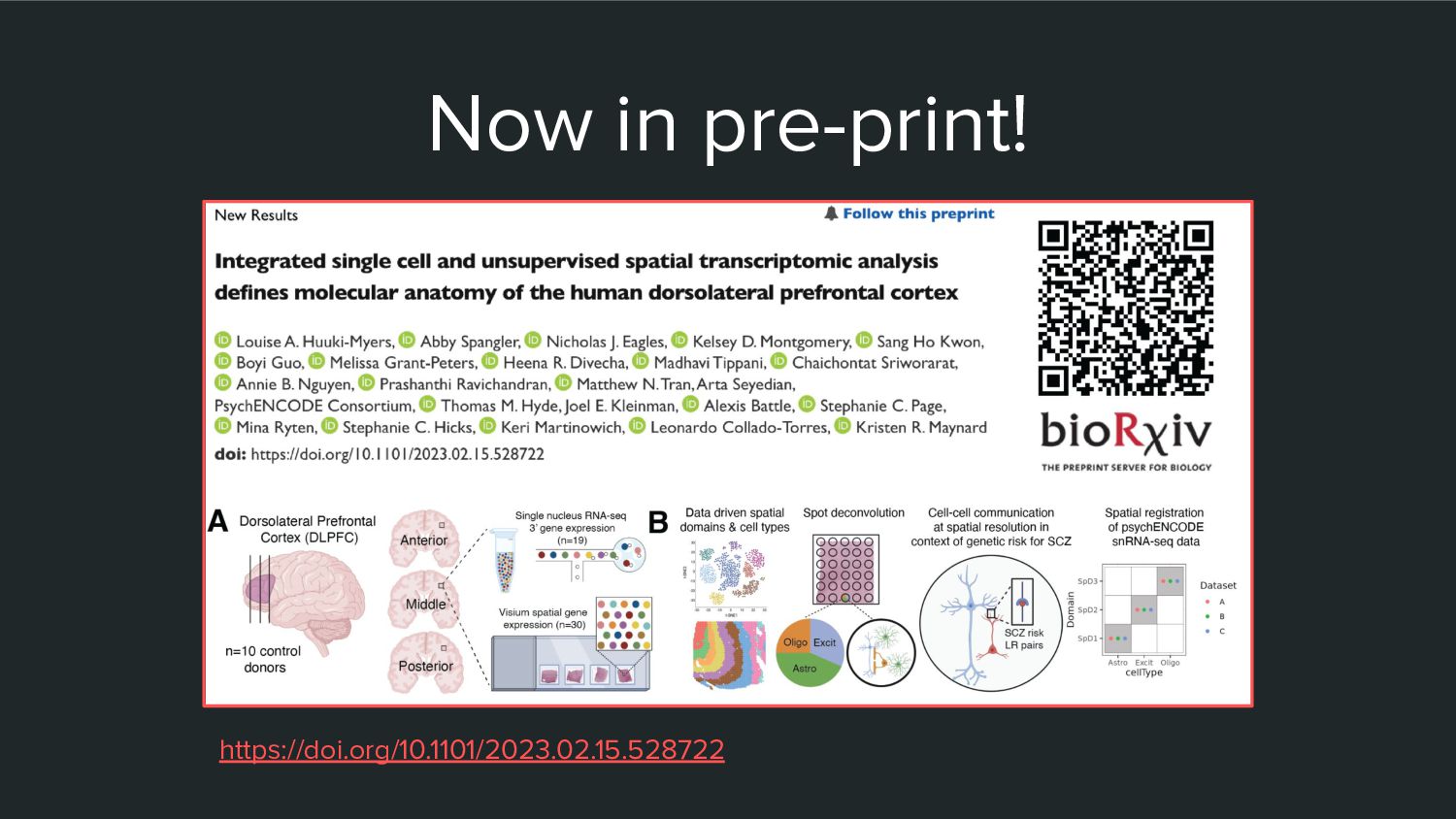
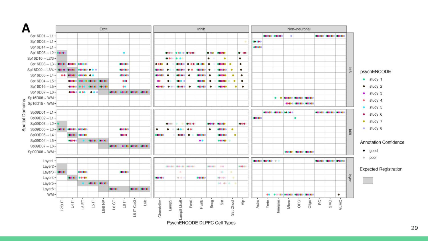
Details the "spatialDLPFC" project to build a large paired single cell and spatial gene expression reference dataset in Human DLPFC. Discusses the application of unsupervised clustering to identify of novel spatial domains, identification of spatially resolved cell type populations via snRNA-seq , and utility for this data spatially inform study of disease.

Presented at the FDA Single Cell Omics Symposium, February 23 2023.

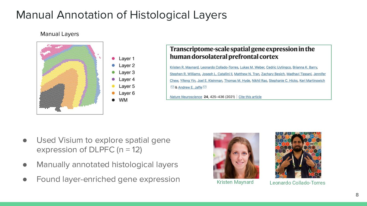
Based on work from this pre-print:
https://www.biorxiv.org/content/10.1101/2023.02.15.528722v1

Louise Huuki-Myers

February 23, 2023
Tweet

More Decks by Louise Huuki-Myers

Other Decks in Science

Transcript

  1. Single Cell and Spatial Transcriptomics in the Postmortem Brain Louise

    Huuki-Myers Staff Scientist 1 @lahuuki lahuuki.github.io Download these slides: speakerdeck.com/lahuuki
  2. Studying Gene Expression in the Human Brain Bulk RNA-seq Single

    nucleus RNA-seq Spatial Transcriptomics 3
  3. Background Single Nucleus RNA-seq 4 Image Credit: 10x Genomics Freezing

    destroys cell membrane so we are limited to nucleus
  4. 6 Study Design • Neurotypical Adults • High quality RNA

    (RIN > 7) • 30 Visium samples (10 donors x 3 positions) • 19 snRNA-seq samples (10 donors x 2 positions)
  5. 7 1. Identify data-driven spatial domains in visium data 3.

    Apply paired spatial and snRNA-seq data to study cell type composition & cell-cell interactions 2. Identify cell type populations in Single Nucleus RNA-seq data
  6. Manual Annotation of Histological Layers • Used Visium to explore

    spatial gene expression of DLPFC (n = 12) • Manually annotated histological layers • Found layer-enriched gene expression 8 Manual Layers Kristen Maynard Leonardo Collado-Torres
  7. Data Driven Clustering • Why unsupervised clustering? ◦ Reduce labor

    ◦ Discover novel biology • BayesSpace was best method to reiterate histological layers ◦ Spatially aware Bayesian clustering of visium data ◦ Also tested spaGCN, Graph-Based clustering • More methods have been developed since ◦ PRECAST, GraphST, RESEPT, Starfysh 9 Zhao et al. 2021, Nature Biotechnology BayesSpace
  8. Different Resolutions of BayesSpace Clustering k = number of clusters

    • k=2: separate white vs. grey matter • k=9: best reiterated histological layers • k=16: data-driven optimal k based on fast H+ statistic 10 More Clusters = More Complexity
  9. Spatial Registration Adds Anatomical Context • Validate detection of laminar

    structure • Correlate enrichment t-statistics for top marker genes of reference ◦ Cluster vs. manual annotation • Annotate with strongly associated histological layer 11 Sp k D d ~L
  10. Spatial Registration of Spatial Domains • Map SpDs to Maynard

    et al. manual annotated layers • Highlight most strongly associated histological layer to add biological context 12
  11. Data-driven Novel Spatial Domains in DLPFC • k=9 resolution reveals

    vascular spatial domain Sp 9 D 1 ~L1 • Enriched for vasculature genes such as CLDN5 • DEGs across spatial domains can be explored on our web resources 13
  12. 14 1. Identify data-driven spatial domains in visium data 3.

    Apply paired spatial and snRNA-seq data to study cell type composition & cell-cell interactions 2. Identify cell type populations in Single Nucleus RNA-seq data
  13. • Nucleus (not single cell) as we are working with

    frozen tissue • n = 19, from 10 donors • 56,447 high quality nuclei • 29 clusters, 7 broad cell types 15 Single Nucleus RNA-seq Endothelial/Mural
  14. Identify Layer Associated Neuron Populations 16 • Apply Spatial Registration

    with manual layers • 13 layer-level cell types ◦ Assign Excitatory Neurons histological layers ◦ Pool other cell type groups
  15. 19 1. Identify data-driven spatial domains in visium data 3.

    Apply paired spatial and snRNA-seq data to study cell type composition & cell-cell interactions 2. Identify cell type populations in Single Nucleus RNA-seq data
  16. Spatially Map Disease Ligand Receptor Interactions 22 • Identified ligand-receptor

    in snRNA-seq data & Schizophrenia risk L-R analysis • Cell-cell communication analysis • Spatial co-localization of EFNA5-EPHA5 in Sp 9 D 7 ~L6
  17. Summary • Large paired single cell and spatial gene expression

    reference dataset in Human DLPFC • Unsupervised clustering enabled identification of novel spatial domains • Spatially resolved cell type populations in snRNA-seq • Utility for informing spatially informed study of disease 24
  18. Interactive Web Resources • research.libd.org/spatialDLPFC • Shiny app for spatial

    data ◦ Spatial domains, DE analysis, spatial gene expression • iSEE app for snRNA-seq data ◦ Gene expression and reduced dimensions 25
  19. Samui Browser to Explore High-Resolution Images • https://samuibrowser.com/ • True

    scale visium spots & full resolution histological images 26
  20. 28 Acknowledgements Lieber Institute Abby Spangler Nick Eagles Kelsey D.

    Montgomery Sang Ho Kwon Heena R. Divecha Madhavi Tippani Chaichontat Sriworarat Annie B. Nguyen Matthew N. Tran Arta Seyedian Thomas M. Hyde Joel E. Kleinman Stephanie C. Page Keri Martinowich Leonardo Collado-Torres Kristen R. Maynard JHU Biostatistics Dept Boyi Guo Stephanie C. Hicks JHU Biomed Engineering Prashanthi Ravichandran Alexis Battle University College London Genetics and Genomic Medicine Melissa Grant-Peters Mina Ryten PsychENCODE Consortium Get in touch! lahuuki.github.io @lahuuki Download these slides: speakerdeck.com/lahuuki Thank you! Any Questions?
  21. 29