anatomy of the human dorsolateral prefrontal cortex Louise A. Huuki-Myers1, Abby B. Spangler1, Nicholas J. Eagles1, Kelsey D. Montgomery1, Sang Ho Kwon1,2, Heena R. Divecha1, Madhavi Tippani1, Thomas M. Hyde1, Stephanie C. Hicks3, Stephanie C. Page1, Keri Martinowich1, Leonardo Collado-Torres1,4, Kristen R. Maynard1,5 1. Lieber Institute for Brain Development, 2. Department of Neuroscience Johns Hopkins School of Medicine, 3. Department of Biostatistics Johns Hopkins Bloomberg School of Public Health, 4. Department of Psychiatry and Behavioral Sciences JHSOM, 5. Center for Computational Biology Johns Hopkins University Abstract Conclusion Acknowledgements Identification of Neuronal Subpopulation In Layers Presenter & Poster requests:
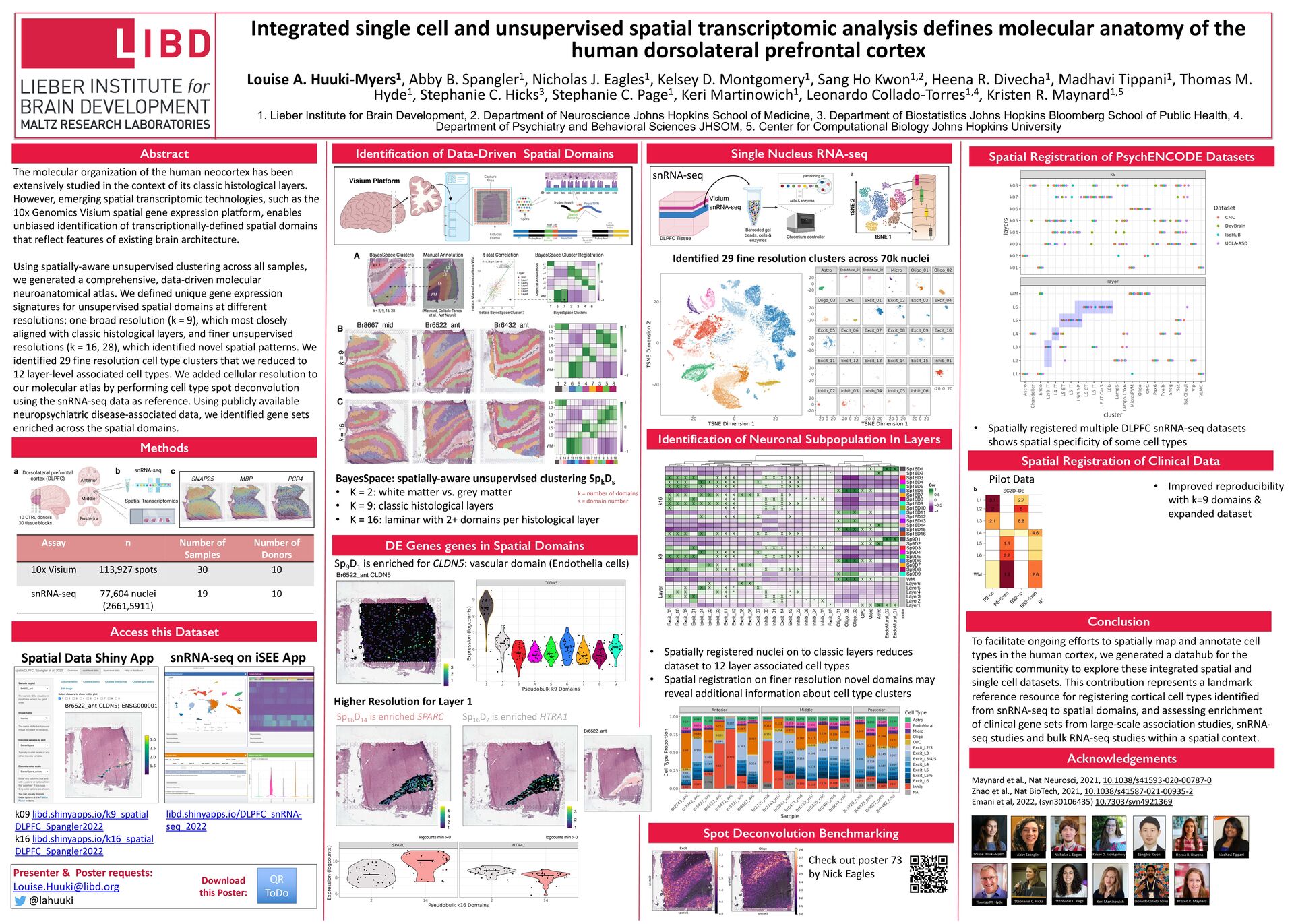
[email protected] @lahuuki Download this Poster: Methods Access this Dataset DE Genes genes in Spatial Domains Maynard et al., Nat Neurosci, 2021, 10.1038/s41593-020-00787-0 Zhao et al., Nat BioTech, 2021, 10.1038/s41587-021-00935-2 Emani et al, 2022, (syn30106435) 10.7303/syn4921369 Assay n Number of Samples Number of Donors 10x Visium 113,927 spots 30 10 snRNA-seq 77,604 nuclei (2661,5911) 19 10 snRNA-seq Identification of Data-Driven Spatial Domains BayesSpace: spatially-aware unsupervised clustering Spk Ds • K = 2: white matter vs. grey matter • K = 9: classic histological layers • K = 16: laminar with 2+ domains per histological layer Spatial Registration of PsychENCODE Datasets To facilitate ongoing efforts to spatially map and annotate cell types in the human cortex, we generated a datahub for the scientific community to explore these integrated spatial and single cell datasets. This contribution represents a landmark reference resource for registering cortical cell types identified from snRNA-seq to spatial domains, and assessing enrichment of clinical gene sets from large-scale association studies, snRNA- seq studies and bulk RNA-seq studies within a spatial context. The molecular organization of the human neocortex has been extensively studied in the context of its classic histological layers. However, emerging spatial transcriptomic technologies, such as the 10x Genomics Visium spatial gene expression platform, enables unbiased identification of transcriptionally-defined spatial domains that reflect features of existing brain architecture. Using spatially-aware unsupervised clustering across all samples, we generated a comprehensive, data-driven molecular neuroanatomical atlas. We defined unique gene expression signatures for unsupervised spatial domains at different resolutions: one broad resolution (k = 9), which most closely aligned with classic histological layers, and finer unsupervised resolutions (k = 16, 28), which identified novel spatial patterns. We identified 29 fine resolution cell type clusters that we reduced to 12 layer-level associated cell types. We added cellular resolution to our molecular atlas by performing cell type spot deconvolution using the snRNA-seq data as reference. Using publicly available neuropsychiatric disease-associated data, we identified gene sets enriched across the spatial domains. Single Nucleus RNA-seq Sp9 D1 is enriched for CLDN5: vascular domain (Endothelia cells) Higher Resolution for Layer 1 QR ToDo • Spatially registered nuclei on to classic layers reduces dataset to 12 layer associated cell types • Spatial registration on finer resolution novel domains may reveal additional information about cell type clusters • Spatially registered multiple DLPFC snRNA-seq datasets shows spatial specificity of some cell types Louise Huuki-Myers Kelsey D. Montgomery Sang Ho Kwon Stephanie C. Page Stephanie C. Hicks Kristen R. Maynard Leonardo Collado-Torres Abby Spangler Nicholas J. Eagles Keri Martinowich Heena R. Divecha Madhavi Tippani Thomas M. Hyde k09 libd.shinyapps.io/k9_spatial DLPFC_Spangler2022 k16 libd.shinyapps.io/k16_spatial DLPFC_Spangler2022 Spatial Data Shiny App snRNA-seq on iSEE App Spot Deconvolution Benchmarking Check out poster 73 by Nick Eagles Spatial Registration of Clinical Data Pilot Data • Improved reproducibility with k=9 domains & expanded dataset k = number of domains s = domain number Identified 29 fine resolution clusters across 70k nuclei Sp16 D14 is enriched SPARC Sp16 D2 is enriched HTRA1 libd.shinyapps.io/DLPFC_snRNA- seq_2022