Upgrade to Pro — share decks privately, control downloads, hide ads and more …

Brugada Syndrome (BrS)

Simon Mark
June 06, 2019
140

Brugada Syndrome (BrS)

Simon Mark

June 06, 2019
Tweet

Transcript

  1. Brugada Syndrome (BrS). B Y S I M O N

    M A R K D A L E Y ( 2 0 1 8 ) A presentation. U P D A T E D 2 0 1 9
  2. SOME HISTORY; Although ECG changes were reported in 1989, amongst

    survivors of cardiac arrest, not recognised until 1992 (by the Brugada brothers) as a distinct clinical entity. Exponential rise in the number of cases reported since then. Second Consensus Conference (2005) reported it as the second biggest killer in males <40 years (non-traumatic). Incidence higher in SE Asia than Europe and the US where it was previously known as Sudden Unexplained Nocturnal Death Syndrome (SUNDS). Mean age of sudden death is 41 years. Age at diagnosis ranging from 2 days to 84 years. More common in males due to higher penetrance of the responsible gene.
  3. WHAT IS IT? Brugada syndrome (BrS) is a genetic disorder

    characterised by abnormal ECG findings (incomplete right bundle-branch block and ST-segment elevations in the anterior precordial leads). There is an increased risk of sudden cardiac death associated with this disorder. - Dizon and Nazif (2015) BrS is an ECG abnormality with a high incidence of sudden death in patients with structurally normal hearts. - Lifeinthefastlane.com
  4. IN MORE DETAIL PLEASE; BrS is a channelopathy; a disorder

    caused by an alteration in the transmembrane ion currents that together constitute the cardiac action potential. - Dizon and Nazif (2015)
  5. Approximately 20% of cases of BrS have been shown to

    be associated with mutations in a gene that encodes for a sodium ion channel in the cell membranes of myocytes. The gene, named SCN5A is located on the short arm of the third chromosome (3p21). Over 160 mutations in this gene have been discovered to date, each having varying mechanisms and effects on function. Loss-of-function mutations in this gene lead to a loss of the action potential dome of some epicardial areas of the right ventricle. This results in transmural and epicardial dispersion of repolarisation. The transdermal dispersion underlies ST- segment elevation and the development of a vulnerable window across the ventricular wall, whereas the epicardial dispersion of repolarisation facilitates the development of phase 2 reentry,, which generates a phase 2 re-entrant extrasystole that captures the vulnerable window to precipitate VT and/or VF. EVEN MORE DETAIL; (TOO MUCH DETAIL)
  6. The majority of patients affected by BrS are not found

    to have known genetic mutations to explain the disease, as of 2015. (Sarquella-Brugada, Campuzano, Arbelo, Brugada and Brugada, 2015). Over 60 different mutations have been described so far and at least 50% are spontaneous mutations, but familial clustering and autosomal dominant inheritance has been demonstrated. GO ON; (SOMEWHAT SIMPLIFIED)
  7. It is important to understand that ECG changes may be

    transient and can be unmasked or augmented by multiple factors; Fever – very common, particularly in children Ischaemia Multiple drugs; sodium channel blockers, calcium channel blockers, alpha agonists, beta blockers, nitrates, cholinergic stimulation, cocaine, alcohol. Hypokalaemia Hypothermia Post DC Cardioversion Typical presentation is syncope or aborted sudden cardiac death (SCD) secondary to VF (less commonly VT) and symptoms typically occur at night or at rest, particularly after a large/heavy meal. WHAT ARE WE LOOKING FOR? (CLINICAL PRACTICE)
  8. Type 1 has a coved type ST elevation with at

    least 2 mm J-point elevation a gradually descending ST segment followed by a negative T-wave. Type 2 has a saddle back pattern with a least 2 mm J-point elevation and at least 1 mm ST elevation with a positive or biphasic T-wave. Type 2 pattern can occasionally be seen in healthy subjects. Type 3 has either a coved (type 1 like) or a saddle back (type 2 like) pattern with less than 2 mm J-point elevation and less than 1 mm ST elevation. Type 3 pattern is not rare in healthy subjects. An incomplete RBBB pattern can be observed, and prolongation of the PR-interval is also frequently seen. ECG PATTERNS IN BsR; (WHEN UNMASKED)
  9. Type 1 either spontaneously present or induced with the sodium-channel

    blocker challenge test is considered diagnostic. Type 2 and 3 may lead to suspicion but the drug challenge is required for diagnosis. The ECGs in the right and left panels are from the same patient before (right panel, type 3) and after (left panel, type 1) administration of 1 mg/kg of Ajmaline. ECG PATTERNS IN BsR; (WHEN UNMASKED)
  10. The sensitivity of ECG detection of BsR can be increased

    by repositioning leads v1 & v2 in the 3rd intercostal space (v1ic3 & v2ic3) [pictured right], HUNTING FOR BsR; - ECGpedia.om (2019) Higher placement of leads v1 & v2 in the 2nd intercostal space increases the sensitivity fo detecting BrS if suspected on clinical grounds. The reason for this is that the abnormal activity in BrS is thought to originate from the RV outflow tract. - Anzelevith (2008) In cases of unertainty, pharmacological provocation with intravenous administration of class 1 sodium- channel blockers such as Ajmaline or Flecainide is recommended.
  11. Resuscitated cardiac arrest and cardiac syncope are Class 1 indications

    for implantation of an ICD. All family members should be screened and those with normal or non- diagnostic ECGs should be offered ajmaline or flecainide testing. ICD implantation has a significant complication rate in BsR however, therefore this should be avoided in asymptomatic patients. Family history of sudden cardiac death (SCD) is NOT an indication for ICD implantation. Asymptomatic individuals should be advised of lifestyle measures such as avoidance of ‘Brugada drugs’*, prompt treatment of fever, avoiding excess of alcohol, particularly hot baths and big/carbohydrate rich meals at night. *So-called ‘Brugada drugs’ can be found listed at brugadadrugs.org and are not necessarily the same as QTc prolongation drugs MANAGEMENT;
  12. Event rate in those with spontaneous type 1 ECG has

    been reported as 0.24- 1.7% per year. Drug induced BrS pattern ECG patients are at minimal risk. Fragmentation of QRS on ECG (RSR/notched S/notched R), RV effective refractory period (ERP) of <200msecs, a history of syncope or AF with spontaneous type 1 pattern put the patient in a higher risk category. RISK ASSESSMENT OF ASYMPTOMATIC BrS PATIENTS; TREATMENT OF ARRHYTHMIC STORMS; Isoprenaline infusion is effective in acute situations, whilst quinidine is the only effective long-term agent.
  13. A NOTE ON MISDIAGNOSIS OF BrS; Spurious BrS type ECG

    changes can be seen in patients following cardioversion, may last for several hours and may lead to a misdiagnosis of BrS. Misdiagnosis can also occur with ECG changes of early repolarisation, athlete’s heart, RBBB, acute pericarditis, MI, prinzmetal angina, ARVC, myocarditis, Duchenne muscular dystrophy, electrolyte disturbances and hypothermia. Basically, (as always) don’t make any assumptions based on ECGs without context.
  14. Dizon, JM and Nazif, TM. (2015) Brugada Sydrome; http://emedicine.medscape.com/article/163751-overview http://lifeinthefastlane.com/ecg-library/brugada-syndrome/

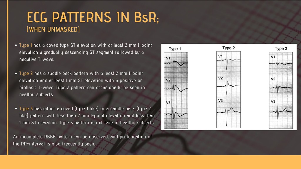
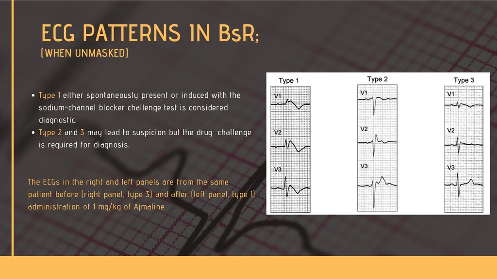
    Napolitano C, Priori SG. Brugada syndrome. Orphanet J Rare Dis. 1, 35. 2006 Sarquella-Brugada, G. Campuzano, O., Arbelo, E., Brugada, J., and Brugada, R. (2015) Brugada Syndrome: Clincal and Genetic Findings. Genetics in Medicine (2016) 18, 3-12. Postema et al. (2009) Heart Rhythm;6:1335-41 Priori, SG., Wilde, AA., Horie, M., Cho, Y., Behr, ER., Berul, C., Blom, N., Brugada, J., Chiang, C., Huikuri, H., Kannankeril, P., Krahn, A., Leenhardt, A., Moss, A., Schwartz, PJ., Shimizu, W., Tomaselli, G. and Tracy, T. (2013) HRS/EHRA/APHRS Expert Consensus Statement on the Diagnosis and Management of Patients with Inherited Primary Arrhythmia Syndromes. Available online at http://dx.doi.org/10.1016/j.hrthm.2013.05.014 Vohra, J. and Rajagopalan, S. (2015) Update on the Diagnosis and Management of Brugada Syndrome. Heart, Lung and Circulation. Vol 24, Iss 12 p1141-1148. https://en.ecgpedia.org/index.php?title=Brugada_Syndrome&mobileaction=toggle_view_mobile https://www.ncbi.nlm.nih.gov/pubmed/18715534 https://www.ncbi.nlm.nih.gov/pubmed/27186380 REFERENCES