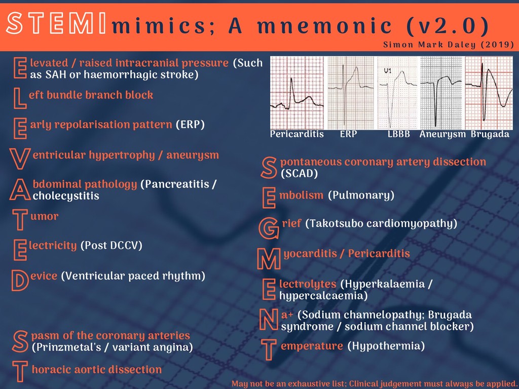
levated / raised intracranial pressure (Such as SAH or haemorrhagic stroke) S E G M E N T eft bundle branch block entricular hypertrophy / aneurysm pontaneous coronary artery dissection (SCAD) umor evice (Ventricular paced rhythm) emperature (Hypothermia) pasm of the coronary arteries (Prinzmetal's / variant angina) mbolism (Pulmonary) rief (Takotsubo cardiomyopathy) yocarditis / Pericarditis lectrolytes (Hyperkalaemia / hypercalcaemia) a+ (Sodium channelopathy; Brugada syndrome / sodium channel blocker) horacic aortic dissection S T E M I S i m o n M a r k D a l e y ( 2 0 1 9 ) m i m i c s ; A m n e m o n i c ( v 2 . 0 ) arly repolarisation pattern (ERP) bdominal pathology (Pancreatitis / cholecystitis lectricity (Post DCCV) Pericarditis ERP LBBB Aneurysm Brugada May not be an exhaustive list; Clinical judgement must always be applied.
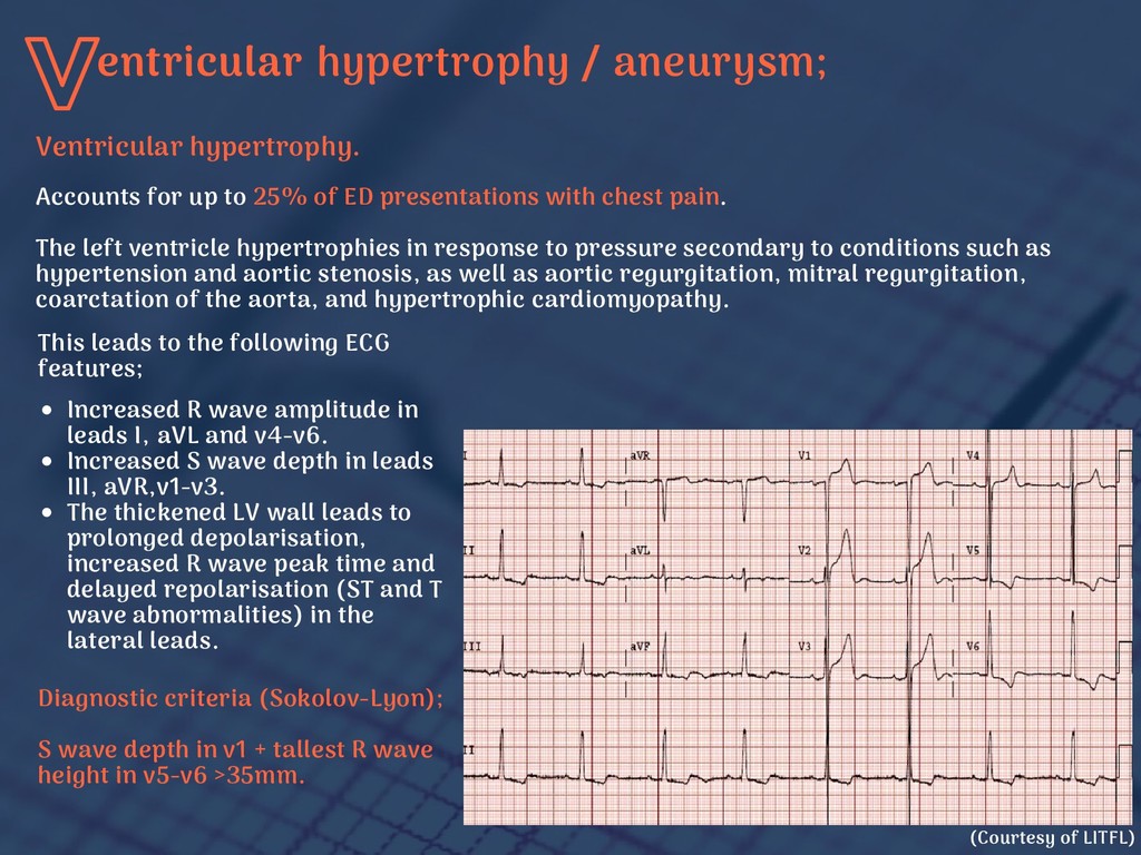
ED presentations with chest pain. The left ventricle hypertrophies in response to pressure secondary to conditions such as hypertension and aortic stenosis, as well as aortic regurgitation, mitral regurgitation, coarctation of the aorta, and hypertrophic cardiomyopathy. Increased R wave amplitude in leads I, aVL and v4-v6. Increased S wave depth in leads III, aVR,v1-v3. The thickened LV wall leads to prolonged depolarisation, increased R wave peak time and delayed repolarisation (ST and T wave abnormalities) in the lateral leads. Diagnostic criteria (Sokolov-Lyon); S wave depth in v1 + tallest R wave height in v5-v6 >35mm. (Courtesy of LITFL) This leads to the following ECG features; Ventricular hypertrophy.
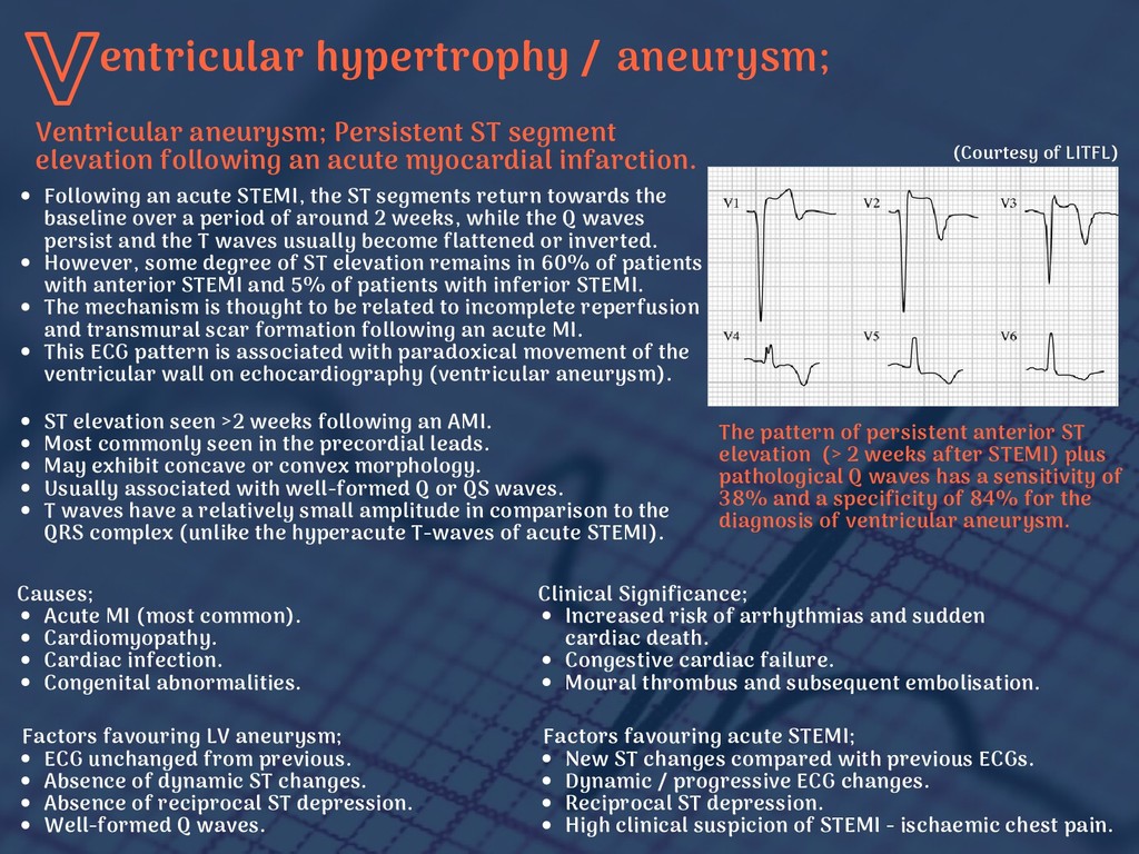
an AMI. Most commonly seen in the precordial leads. May exhibit concave or convex morphology. Usually associated with well-formed Q or QS waves. T waves have a relatively small amplitude in comparison to the QRS complex (unlike the hyperacute T-waves of acute STEMI). (Courtesy of LITFL) The pattern of persistent anterior ST elevation (> 2 weeks after STEMI) plus pathological Q waves has a sensitivity of 38% and a specificity of 84% for the diagnosis of ventricular aneurysm. Following an acute STEMI, the ST segments return towards the baseline over a period of around 2 weeks, while the Q waves persist and the T waves usually become flattened or inverted. However, some degree of ST elevation remains in 60% of patients with anterior STEMI and 5% of patients with inferior STEMI. The mechanism is thought to be related to incomplete reperfusion and transmural scar formation following an acute MI. This ECG pattern is associated with paradoxical movement of the ventricular wall on echocardiography (ventricular aneurysm). Ventricular aneurysm; Persistent ST segment elevation following an acute myocardial infarction. Clinical Significance; Increased risk of arrhythmias and sudden cardiac death. Congestive cardiac failure. Moural thrombus and subsequent embolisation. Causes; Acute MI (most common). Cardiomyopathy. Cardiac infection. Congenital abnormalities. Factors favouring LV aneurysm; ECG unchanged from previous. Absence of dynamic ST changes. Absence of reciprocal ST depression. Well-formed Q waves. Factors favouring acute STEMI; New ST changes compared with previous ECGs. Dynamic / progressive ECG changes. Reciprocal ST depression. High clinical suspicion of STEMI - ischaemic chest pain.
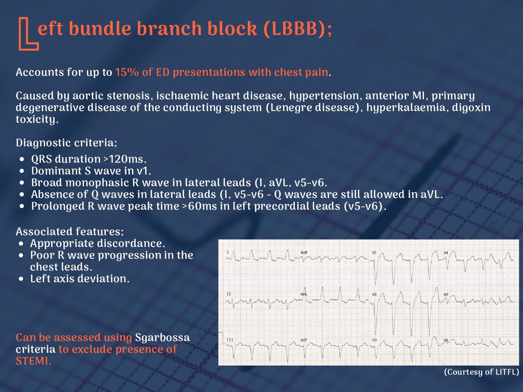
of ED presentations with chest pain. Caused by aortic stenosis, ischaemic heart disease, hypertension, anterior MI, primary degenerative disease of the conducting system (Lenegre disease), hyperkalaemia, digoxin toxicity. Diagnostic criteria; QRS duration >120ms. Dominant S wave in v1. Broad monophasic R wave in lateral leads (I, aVL, v5-v6. Absence of Q waves in lateral leads (I, v5-v6 - Q waves are still allowed in aVL. Prolonged R wave peak time >60ms in left precordial leads (v5-v6). Associated features; Appropriate discordance. Poor R wave progression in the chest leads. Left axis deviation. Can be assessed using Sgarbossa criteria to exclude presence of STEMI. (Courtesy of LITFL)
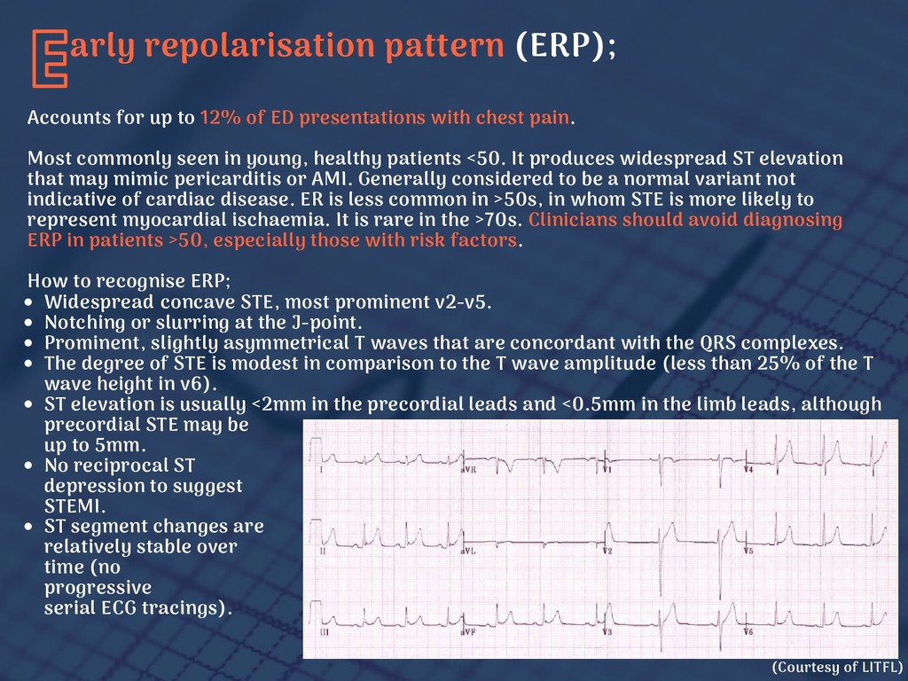
ED presentations with chest pain. Most commonly seen in young, healthy patients <50. It produces widespread ST elevation that may mimic pericarditis or AMI. Generally considered to be a normal variant not indicative of cardiac disease. ER is less common in >50s, in whom STE is more likely to represent myocardial ischaemia. It is rare in the >70s. Clinicians should avoid diagnosing ERP in patients >50, especially those with risk factors. How to recognise ERP; Widespread concave STE, most prominent v2-v5. Notching or slurring at the J-point. Prominent, slightly asymmetrical T waves that are concordant with the QRS complexes. The degree of STE is modest in comparison to the T wave amplitude (less than 25% of the T wave height in v6). ST elevation is usually <2mm in the precordial leads and <0.5mm in the limb leads, although precordial STE may be up to 5mm. No reciprocal ST depression to suggest STEMI. ST segment changes are relatively stable over time (no progressive on serial ECG tracings). (Courtesy of LITFL)
to changes in autonomic tone: diminishing with increased sympathetic tone / exercise / tachycardia or increasing when the HR slows. The ST elevation may gradually disappear over time as the patient ages: up to 30% of patients with ERP will have resolution of ST elevation on ECGs taken several years later. Early repolarisation no longer a "benign" finding devoid of clinical significance. Studies have demonstrated a 2 to 3-fold increased risk of death vs those without. Absolute risk remains exceedingly low in otherwise healthy individuals. Incidental finding of ERP should not be interpreted as a high-risk marker for arrhythmic death due to the relatively low odds of SCD based on ERP alone. In the context of ERP observed in the inferior/lateral territory in a patient presenting with aborted cardiac arrest, VF or polymorphic VT, ER syndrome can be diagnosed. This lies on the spectrum with Brugada syndrome and is an indication for ICD implantation. A consensus paper in 2015 concluded that in the absence of syncope, or strong family history of SCD, the finding of ERP does not merit further investigation. arly repolarisation pattern (ERP);
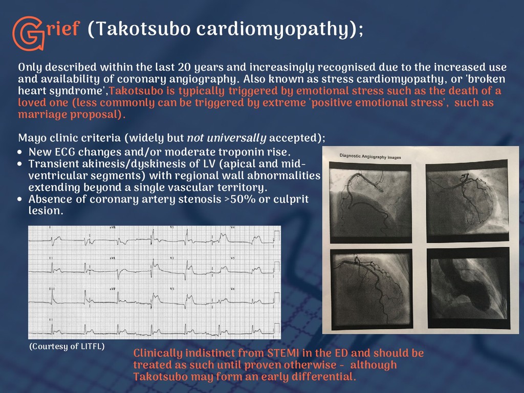
and increasingly recognised due to the increased use and availability of coronary angiography. Also known as stress cardiomyopathy, or 'broken heart syndrome',Takotsubo is typically triggered by emotional stress such as the death of a loved one (less commonly can be triggered by extreme 'positive emotional stress', such as marriage proposal). Mayo clinic criteria (widely but not universally accepted); New ECG changes and/or moderate troponin rise. Transient akinesis/dyskinesis of LV (apical and mid- ventricular segments) with regional wall abnormalities extending beyond a single vascular territory. Absence of coronary artery stenosis >50% or culprit lesion. Clinically indistinct from STEMI in the ED and should be treated as such until proven otherwise - although Takotsubo may form an early differential. (Courtesy of LITFL)
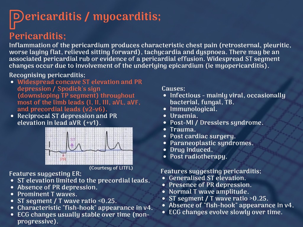
pain (retrosternal, pleuritic, worse laying flat, relieved sitting forward), tachycardia and dyspnoea. There may be an associated pericardial rub or evidence of a pericardial effusion. Widespread ST segment changes occur due to involvement of the underlying epicardium (ie myopericarditis). Recognising pericarditis; Widespread concave ST elevation and PR depression / Spodick's sign (downsloping TP segment) throughout most of the limb leads (I, II, III, aVL, aVF, and precordial leads (v2-v6). Reciprocal ST depression and PR elevation in lead aVR (+v1). Causes; Infectious - mainly viral, occasionally bacterial, fungal, TB. Immunological. Uraemia. Post-MI / Dresslers syndrome. Trauma. Post cardiac surgery. Paraneoplastic syndromes. Drug induced. Post radiotherapy. Features suggesting ER; ST elevation limited to the precordial leads. Absence of PR depression. Prominent T waves. ST segment / T wave ratio <0.25. Characteristic "fish-hook" appearance in v4. ECG changes usually stable over time (non- progressive). Features suggesting pericarditis; Generalised ST elevation. Presence of PR depression. Normal T wave amplitude. ST segment / T wave ratio >0.25. Absence of "fish-hook" appearance in v4. ECG changes evolve slowly over time. Pericarditis; (Courtesy of LITFL)
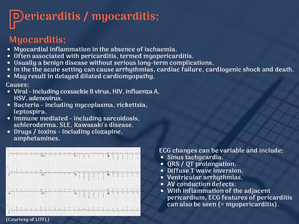
Often associated with pericarditis, termed myopericarditis. Usually a benign disease without serious long-term complications. In the the acute setting can cause arrhythmias, cardiac failure, cardiogenic shock and death. May result in delayed dilated cardiomyopathy. Causes; Viral - including coxsackie B virus, HIV, influenza A, HSV, adenovirus. Bacteria - including mycoplasma, rickettsia, leptospira. Immune mediated - including sarcoidosis, schleroderma, SLE, Kawasaki's disease. Drugs / toxins - including clozapine, amphetamines. ECG changes can be variable and include; Sinus tachycardia. QRS / QT prolongation. Diffuse T wave inversion. Ventricular arrhythmias. AV conduction defects. With inflammation of the adjacent pericardium, ECG features of pericarditis can also be seen (= myopericarditis). Myocarditis; (Courtesy of LITFL)
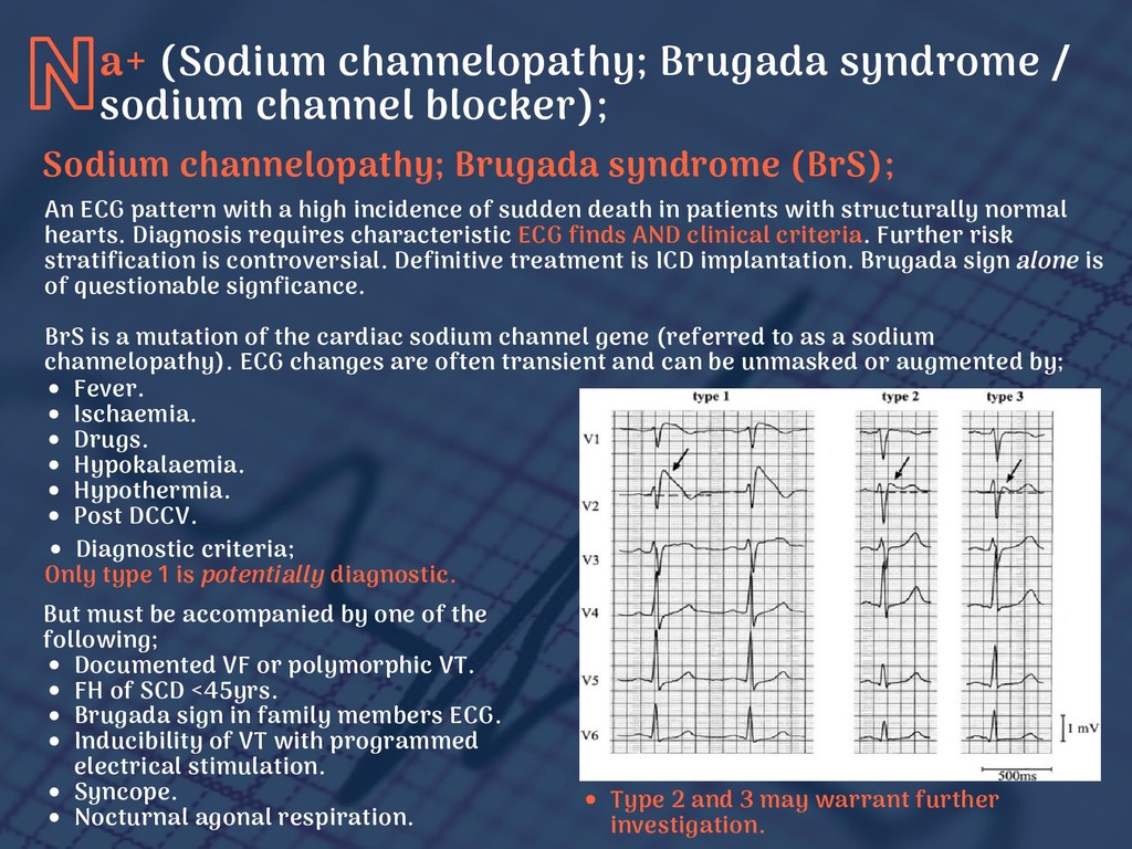
death in patients with structurally normal hearts. Diagnosis requires characteristic ECG finds AND clinical criteria. Further risk stratification is controversial. Definitive treatment is ICD implantation. Brugada sign alone is of questionable signficance. BrS is a mutation of the cardiac sodium channel gene (referred to as a sodium channelopathy). ECG changes are often transient and can be unmasked or augmented by; Fever. Ischaemia. Drugs. Hypokalaemia. Hypothermia. Post DCCV. Diagnostic criteria; Only type 1 is potentially diagnostic. But must be accompanied by one of the following; Documented VF or polymorphic VT. FH of SCD <45yrs. Brugada sign in family members ECG. Inducibility of VT with programmed electrical stimulation. Syncope. Nocturnal agonal respiration. Type 2 and 3 may warrant further investigation. a+ (Sodium channelopathy; Brugada syndrome / sodium channel blocker); Sodium channelopathy; Brugada syndrome (BrS);
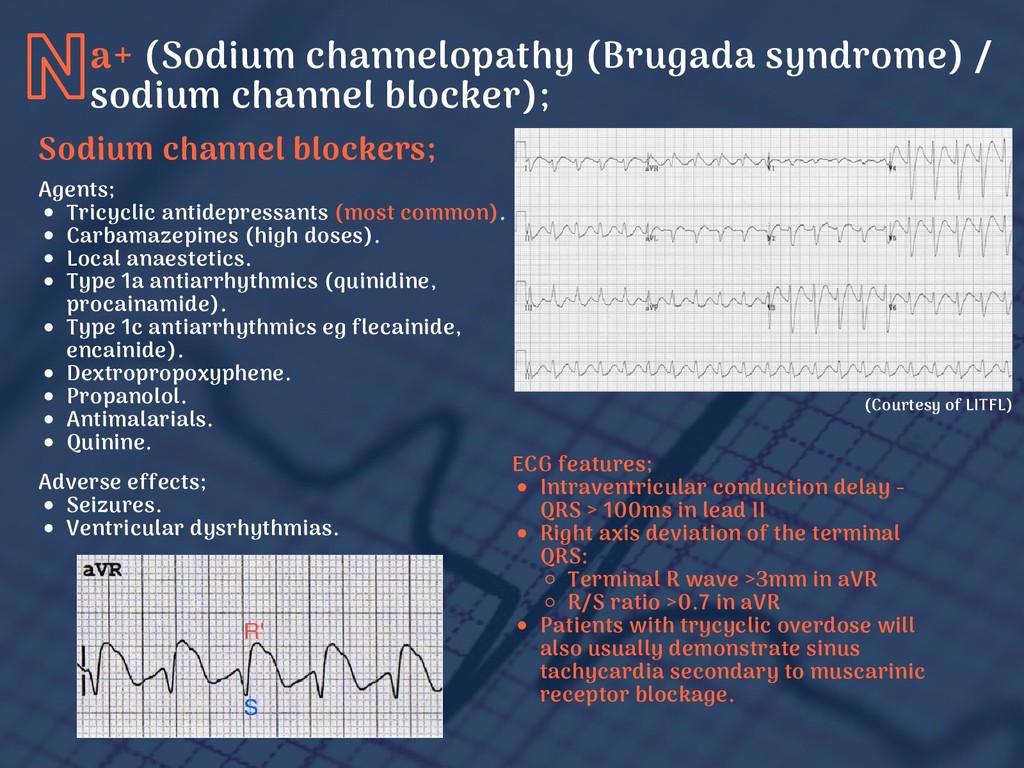
anaestetics. Type 1a antiarrhythmics (quinidine, procainamide). Type 1c antiarrhythmics eg flecainide, encainide). Dextropropoxyphene. Propanolol. Antimalarials. Quinine. a+ (Sodium channelopathy (Brugada syndrome) / sodium channel blocker); Sodium channel blockers; ECG features; Intraventricular conduction delay - QRS > 100ms in lead II Right axis deviation of the terminal QRS: Terminal R wave >3mm in aVR R/S ratio >0.7 in aVR Patients with trycyclic overdose will also usually demonstrate sinus tachycardia secondary to muscarinic receptor blockage. Adverse effects; Seizures. Ventricular dysrhythmias. (Courtesy of LITFL)
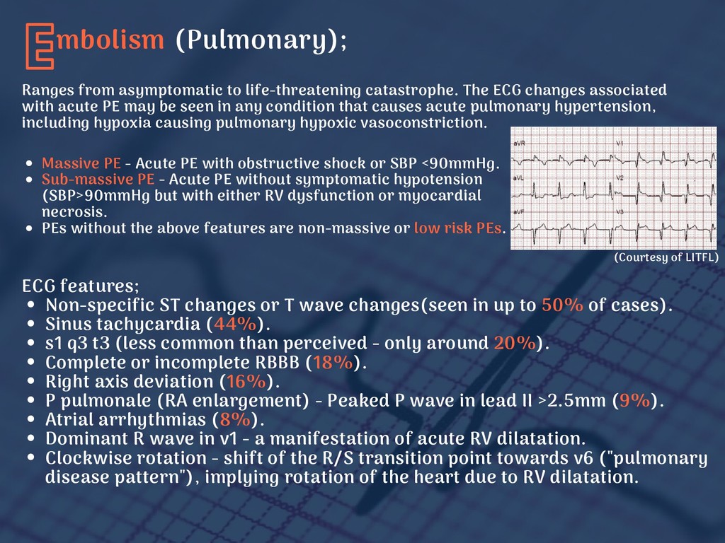
changes associated with acute PE may be seen in any condition that causes acute pulmonary hypertension, including hypoxia causing pulmonary hypoxic vasoconstriction. Massive PE - Acute PE with obstructive shock or SBP <90mmHg. Sub-massive PE - Acute PE without symptomatic hypotension (SBP>90mmHg but with either RV dysfunction or myocardial necrosis. PEs without the above features are non-massive or low risk PEs. ECG features; Non-specific ST changes or T wave changes(seen in up to 50% of cases). Sinus tachycardia (44%). s1 q3 t3 (less common than perceived - only around 20%). Complete or incomplete RBBB (18%). Right axis deviation (16%). P pulmonale (RA enlargement) - Peaked P wave in lead II >2.5mm (9%). Atrial arrhythmias (8%). Dominant R wave in v1 - a manifestation of acute RV dilatation. Clockwise rotation - shift of the R/S transition point towards v6 ("pulmonary disease pattern"), implying rotation of the heart due to RV dilatation. (Courtesy of LITFL)
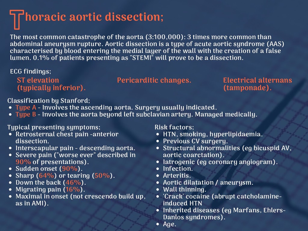
(3:100,000): 3 times more common than abdominal aneurysm rupture. Aortic dissection is a type of acute aortic syndrome (AAS) characterised by blood entering the medial layer of the wall with the creation of a false lumen. 0.1% of patients presenting as "STEMI" will prove to be a dissection. ECG findings; Classification by Stanford; Type A - Involves the ascending aorta. Surgery usually indicated. Type B - Involves the aorta beyond left subclavian artery. Managed medically. Typical presenting symptoms; Retrosternal chest pain -anterior dissection. Interscapular pain - descending aorta. Severe pain ("worse ever" described in 90% of presentations). Sudden onset (90%). Sharp (64%) or tearing (50%). Down the back (46%). Migrating pain (16%). Maximal in onset (not crescendo build up, as in AMI). Risk factors; HTN, smoking, hyperlipidaemia. Previous CV surgery. Structural abnormalities (eg bicuspid AV, aortic coarctation). Iatrogenic (eg coronary angiogram). Infection. Arteritis. Aortic dilatation / aneurysm. Wall thinning. 'Crack' cocaine (abrupt catcholamine- induced HTN Inherited diseases (eg Marfans, Ehlers- Danlos syndromes). Age. ST elevation (typically inferior). Electrical alternans (tamponade). Pericarditic changes.
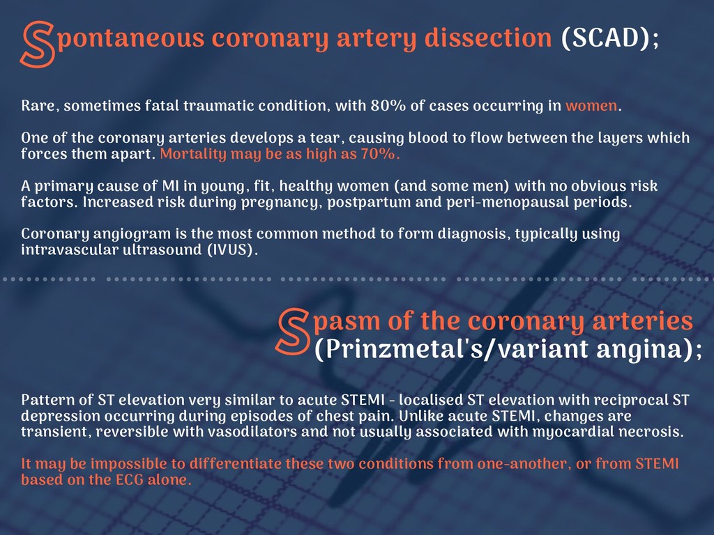
with 80% of cases occurring in women. One of the coronary arteries develops a tear, causing blood to flow between the layers which forces them apart. Mortality may be as high as 70%. A primary cause of MI in young, fit, healthy women (and some men) with no obvious risk factors. Increased risk during pregnancy, postpartum and peri-menopausal periods. Coronary angiogram is the most common method to form diagnosis, typically using intravascular ultrasound (IVUS). pasm of the coronary arteries (Prinzmetal's/variant angina); S Pattern of ST elevation very similar to acute STEMI - localised ST elevation with reciprocal ST depression occurring during episodes of chest pain. Unlike acute STEMI, changes are transient, reversible with vasodilators and not usually associated with myocardial necrosis. It may be impossible to differentiate these two conditions from one-another, or from STEMI based on the ECG alone.
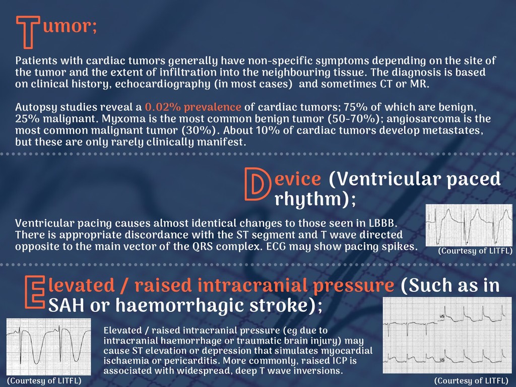
identical changes to those seen in LBBB. There is appropriate discordance with the ST segment and T wave directed opposite to the main vector of the QRS complex. ECG may show pacing spikes. levated / raised intracranial pressure (Such as in SAH or haemorrhagic stroke); E Elevated / raised intracranial pressure (eg due to intracranial haemorrhage or traumatic brain injury) may cause ST elevation or depression that simulates myocardial ischaemia or pericarditis. More commonly, raised ICP is associated with widespread, deep T wave inversions. Patients with cardiac tumors generally have non-specific symptoms depending on the site of the tumor and the extent of infiltration into the neighbouring tissue. The diagnosis is based on clinical history, echocardiography (in most cases) and sometimes CT or MR. Autopsy studies reveal a 0.02% prevalence of cardiac tumors; 75% of which are benign, 25% malignant. Myxoma is the most common benign tumor (50-70%); angiosarcoma is the most common malignant tumor (30%). About 10% of cardiac tumors develop metastates, but these are only rarely clinically manifest. (Courtesy of LITFL) (Courtesy of LITFL) (Courtesy of LITFL)
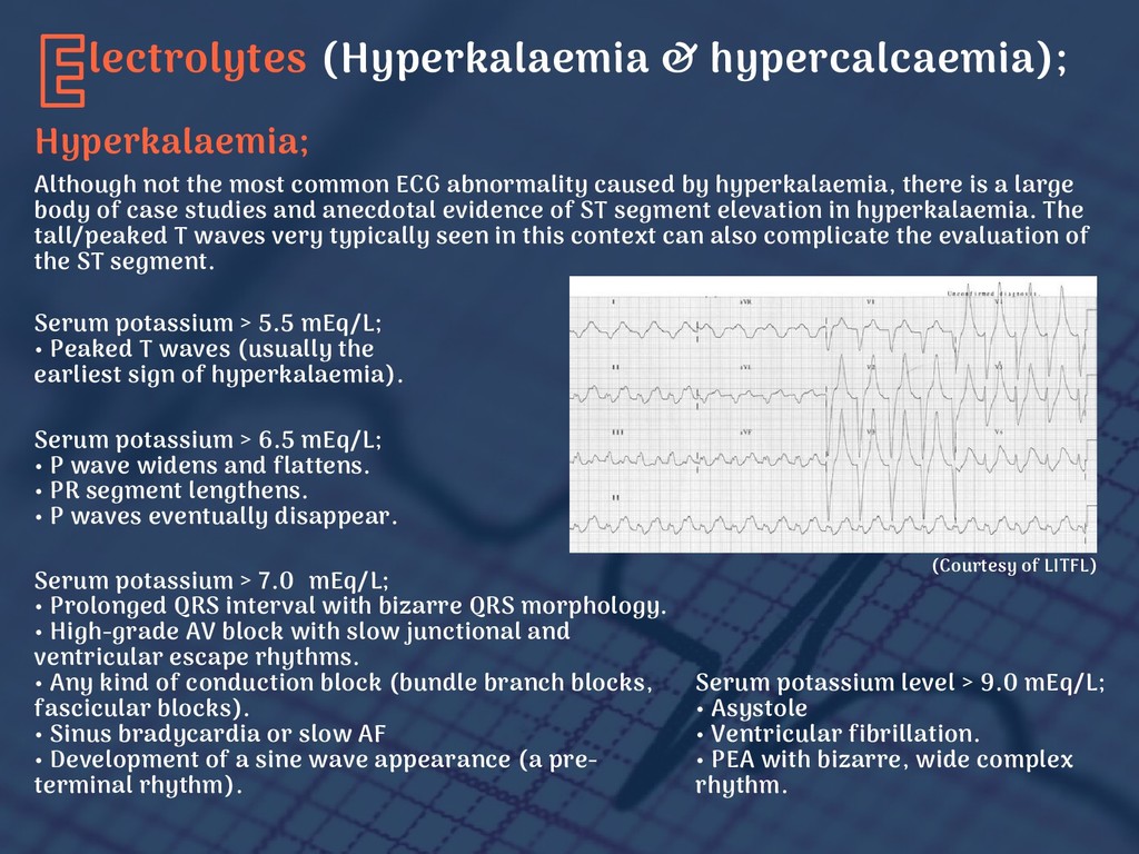
abnormality caused by hyperkalaemia, there is a large body of case studies and anecdotal evidence of ST segment elevation in hyperkalaemia. The tall/peaked T waves very typically seen in this context can also complicate the evaluation of the ST segment. Serum potassium > 7.0 mEq/L; • Prolonged QRS interval with bizarre QRS morphology. • High-grade AV block with slow junctional and ventricular escape rhythms. • Any kind of conduction block (bundle branch blocks, fascicular blocks). • Sinus bradycardia or slow AF • Development of a sine wave appearance (a pre- terminal rhythm). Serum potassium > 5.5 mEq/L; • Peaked T waves (usually the earliest sign of hyperkalaemia). Serum potassium > 6.5 mEq/L; • P wave widens and flattens. • PR segment lengthens. • P waves eventually disappear. Serum potassium level > 9.0 mEq/L; • Asystole • Ventricular fibrillation. • PEA with bizarre, wide complex rhythm. (Courtesy of LITFL) Hyperkalaemia;
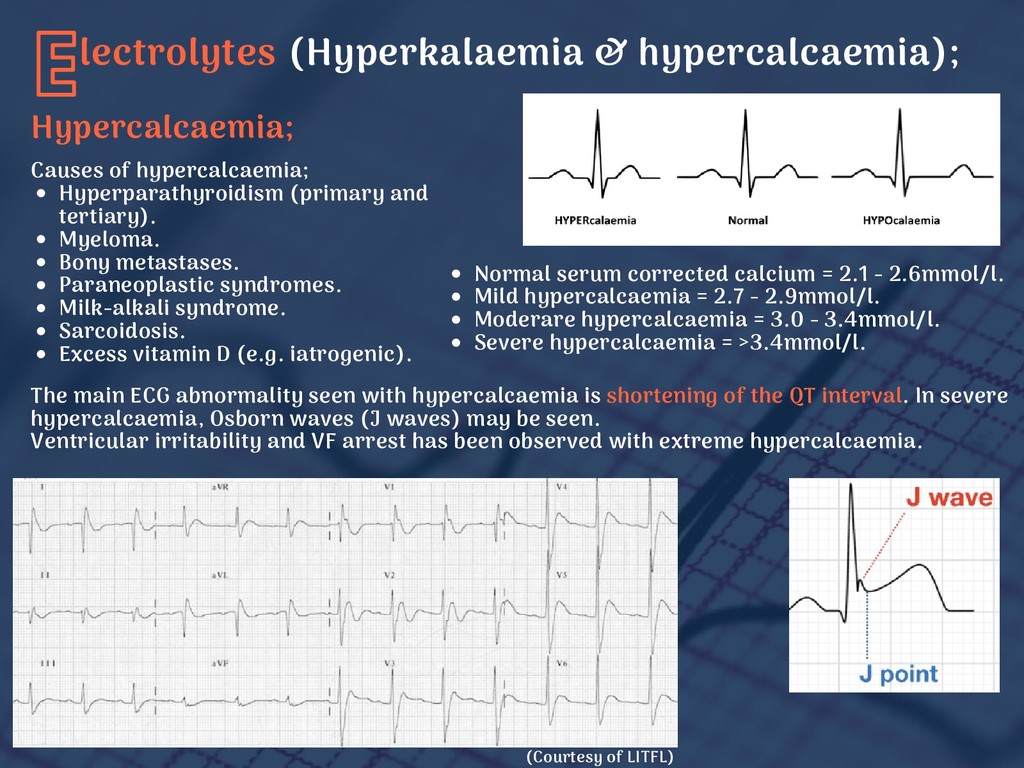
hypercalcaemia; Hyperparathyroidism (primary and tertiary). Myeloma. Bony metastases. Paraneoplastic syndromes. Milk-alkali syndrome. Sarcoidosis. Excess vitamin D (e.g. iatrogenic). Normal serum corrected calcium = 2.1 - 2.6mmol/l. Mild hypercalcaemia = 2.7 - 2.9mmol/l. Moderare hypercalcaemia = 3.0 - 3.4mmol/l. Severe hypercalcaemia = >3.4mmol/l. The main ECG abnormality seen with hypercalcaemia is shortening of the QT interval. In severe hypercalcaemia, Osborn waves (J waves) may be seen. Ventricular irritability and VF arrest has been observed with extreme hypercalcaemia.
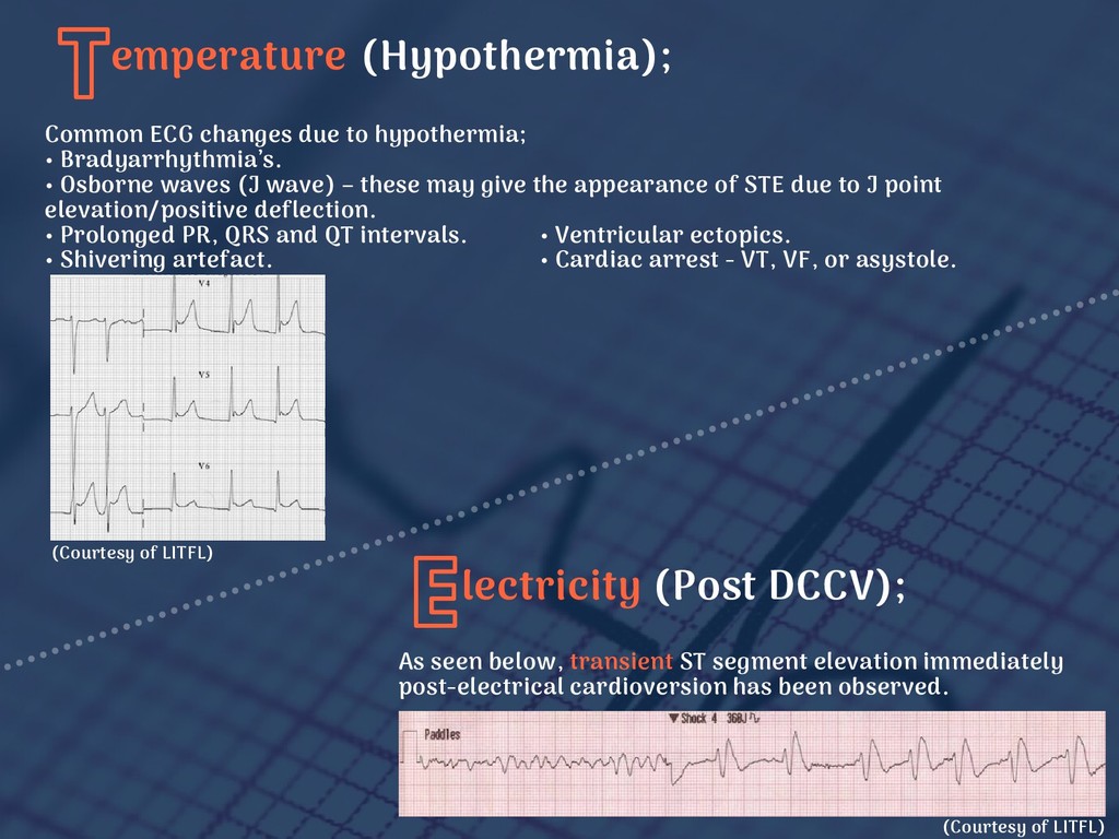
asystole. Common ECG changes due to hypothermia; • Bradyarrhythmia’s. • Osborne waves (J wave) – these may give the appearance of STE due to J point elevation/positive deflection. • Prolonged PR, QRS and QT intervals. • Shivering artefact. emperature (Hypothermia); T (Courtesy of LITFL) lectricity (Post DCCV); E (Courtesy of LITFL) As seen below, transient ST segment elevation immediately post-electrical cardioversion has been observed.
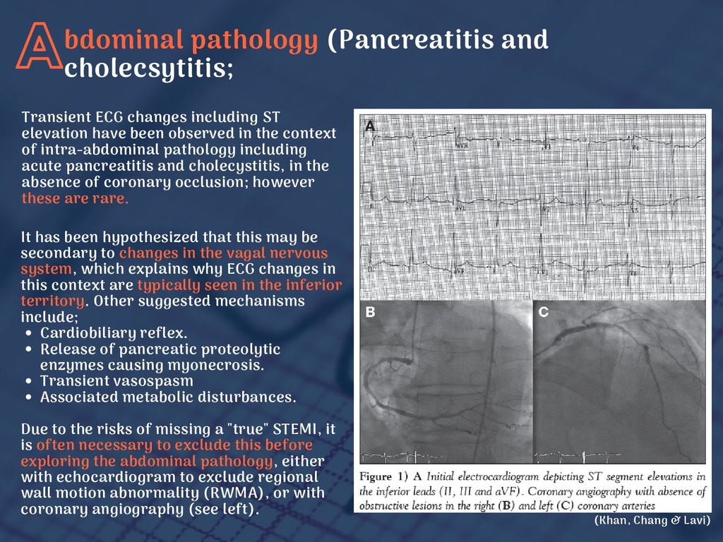
this may be secondary to changes in the vagal nervous system, which explains why ECG changes in this context are typically seen in the inferior territory. Other suggested mechanisms include; Cardiobiliary reflex. Release of pancreatic proteolytic enzymes causing myonecrosis. Transient vasospasm Associated metabolic disturbances. Due to the risks of missing a "true" STEMI, it is often necessary to exclude this before exploring the abdominal pathology, either with echocardiogram to exclude regional wall motion abnormality (RWMA), or with coronary angiography (see left). Transient ECG changes including ST elevation have been observed in the context of intra-abdominal pathology including acute pancreatitis and cholecystitis, in the absence of coronary occlusion; however these are rare. (Khan, Chang & Lavi)