Share
Embed
Raccoon Roundworm
The following presentation is an introduction to raccoon roundworm (Baylisascaris procyonis), a parasite common in North America infecting raccoons, pets, and, rarely, humans.
Raccoon Roundworm © 2022 by U. Nomi vs. Parasites, licensed under CC BY-NC-SA 4.0
U. Nomi vs. Parasites
March 03, 2026
Transcript
31 slides
-
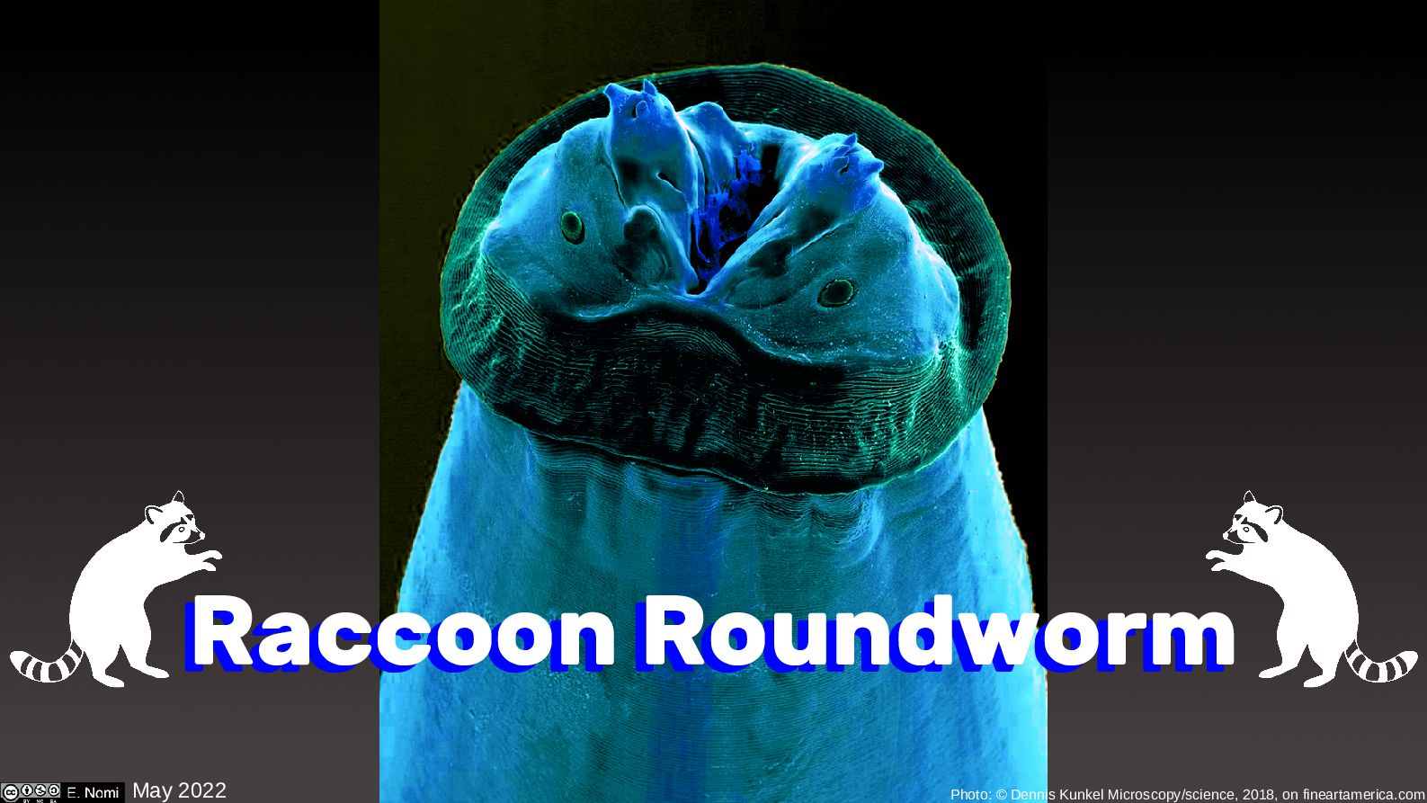
May 2022 Raccoon Roundworm Raccoon Roundworm Photo: © Dennis Kunkel
Microscopy/science, 2018, on fineartamerica.com -
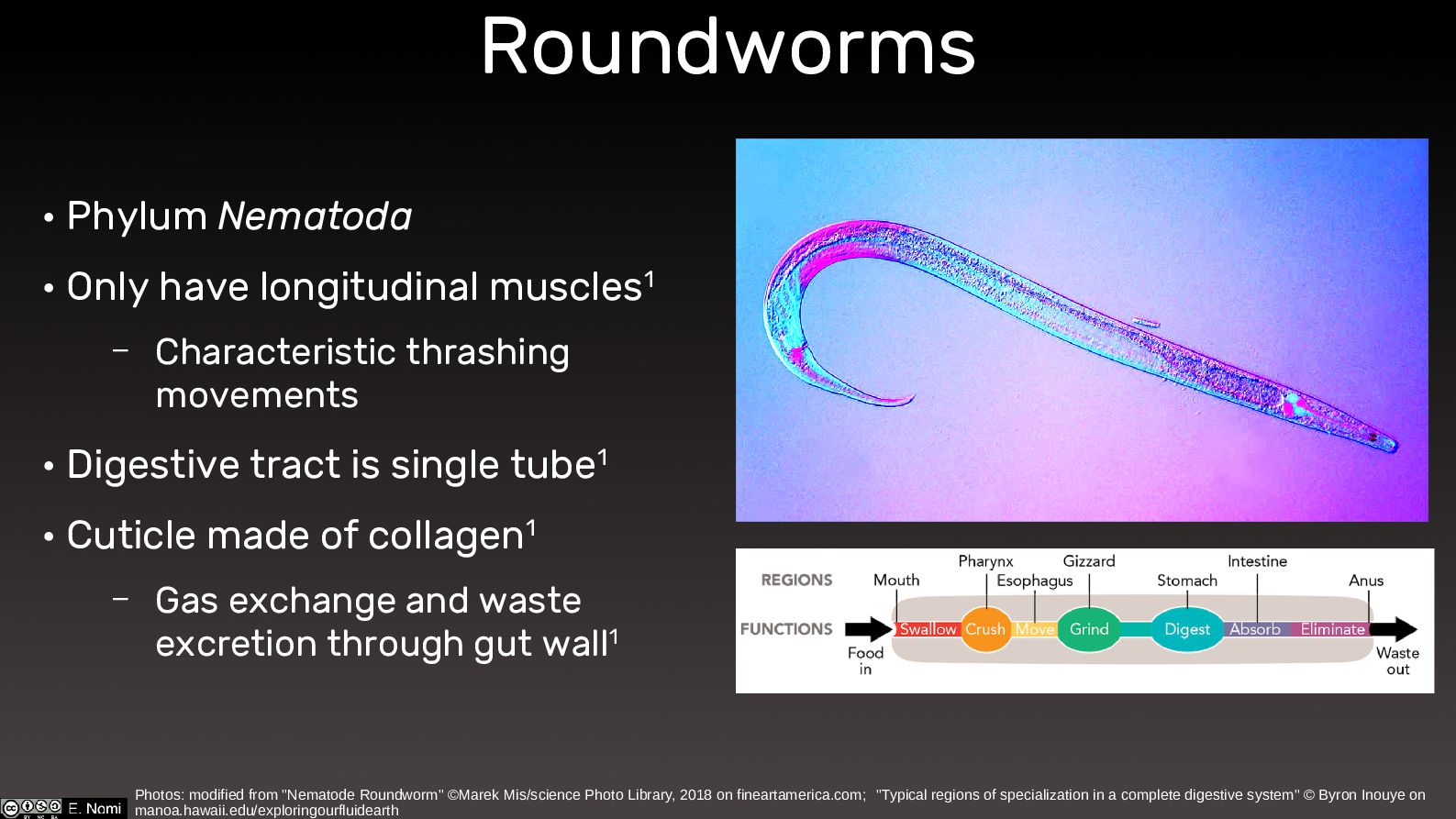
Roundworms • Phylum Nematoda • Only have longitudinal muscles1 –
Characteristic thrashing movements • Digestive tract is single tube1 • Cuticle made of collagen1 – Gas exchange and waste excretion through gut wall1 Photos: modified from "Nematode Roundworm" ©Marek Mis/science Photo Library, 2018 on fineartamerica.com; "Typical regions of specialization in a complete digestive system" © Byron Inouye on manoa.hawaii.edu/exploringourfluidearth -
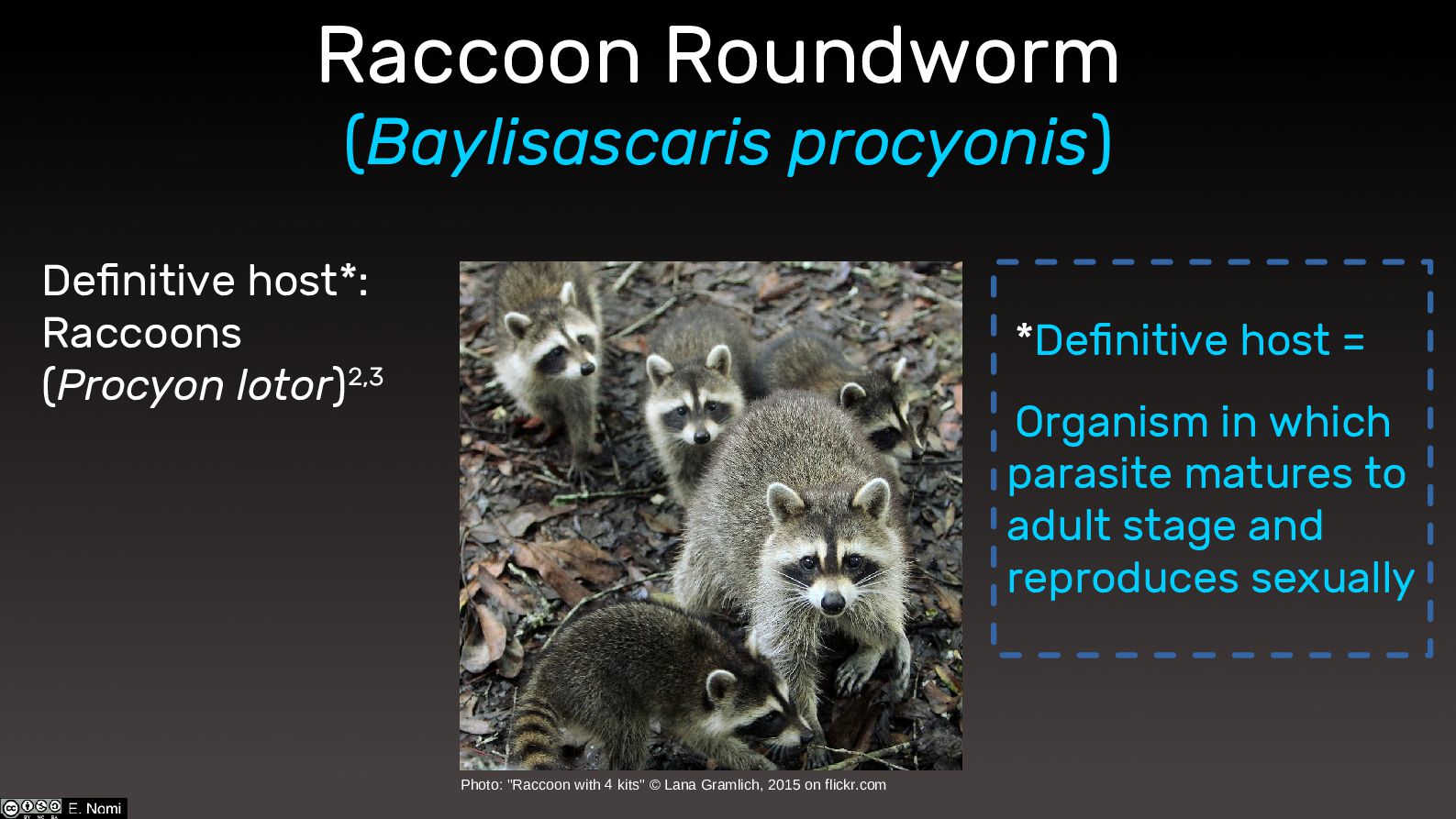
Definitive host*: Raccoons (Procyon lotor)2,3 Photo: "Raccoon with 4 kits"
© Lana Gramlich, 2015 on flickr.com *Definitive host = Organism in which parasite matures to adult stage and reproduces sexually Raccoon Roundworm (Baylisascaris procyonis) -
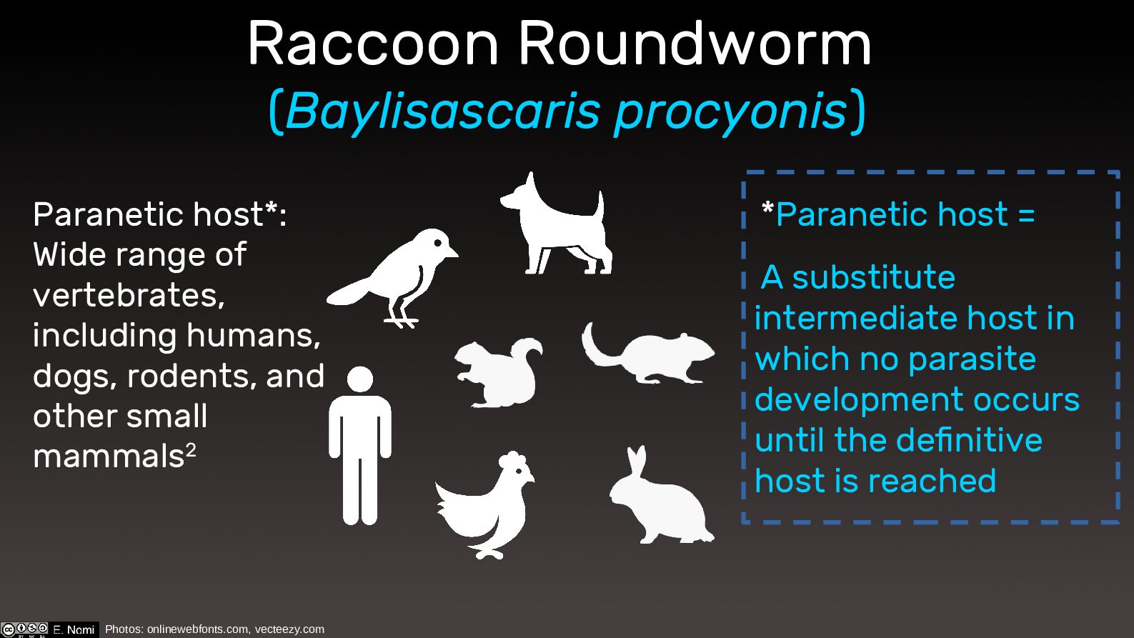
Photos: onlinewebfonts.com, vecteezy.com Paranetic host*: Wide range of vertebrates, including
humans, dogs, rodents, and other small mammals2 *Paranetic host = A substitute intermediate host in which no parasite development occurs until the definitive host is reached Raccoon Roundworm (Baylisascaris procyonis) -
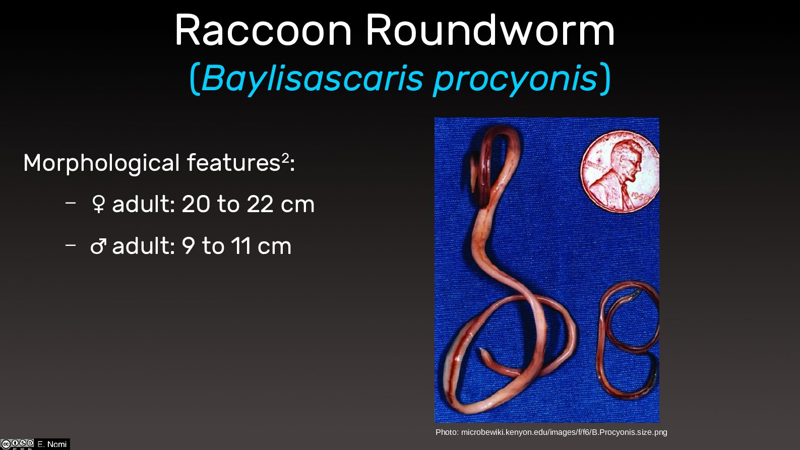
Raccoon Roundworm (Baylisascaris procyonis) Morphological features2: – ♀ adult: 20
to 22 cm – ♂ adult: 9 to 11 cm Photo: microbewiki.kenyon.edu/images/f/f6/B.Procyonis.size.png -
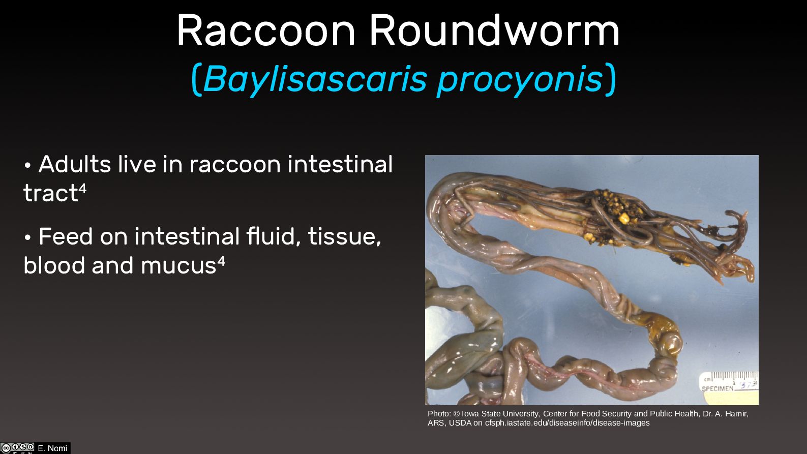
• Adults live in raccoon intestinal tract4 • Feed on
intestinal fluid, tissue, blood and mucus4 Raccoon Roundworm (Baylisascaris procyonis) Photo: © Iowa State University, Center for Food Security and Public Health, Dr. A. Hamir, ARS, USDA on cfsph.iastate.edu/diseaseinfo/disease-images -
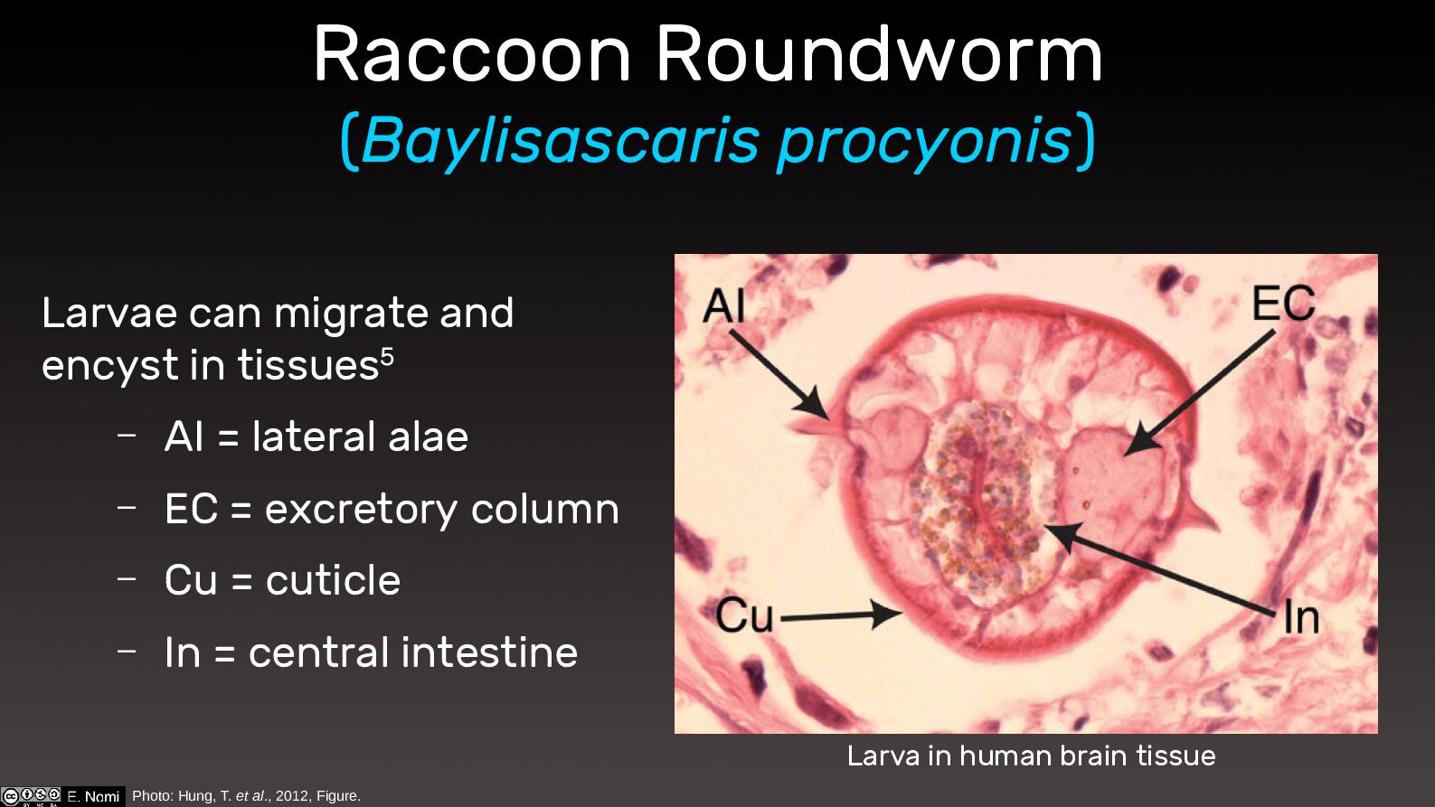
Raccoon Roundworm (Baylisascaris procyonis) Larvae can migrate and encyst in
tissues5 – AI = lateral alae – EC = excretory column – Cu = cuticle – In = central intestine Photo: Hung, T. et al., 2012, Figure. Larva in human brain tissue -
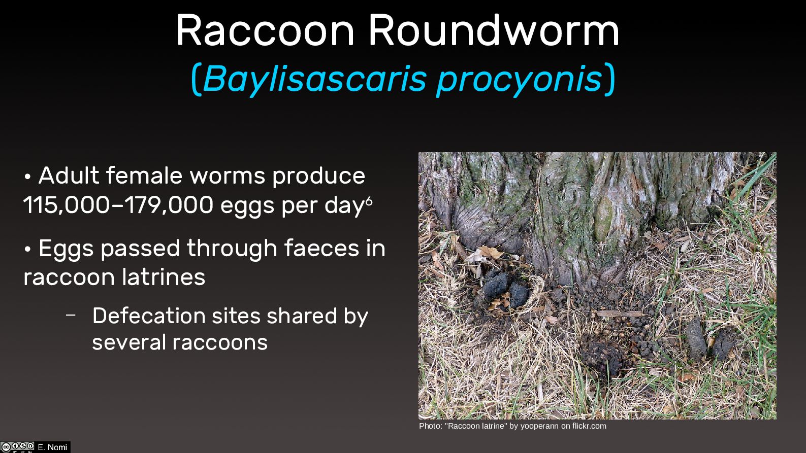
Raccoon Roundworm (Baylisascaris procyonis) • Adult female worms produce 115,000–179,000
eggs per day6 • Eggs passed through faeces in raccoon latrines – Defecation sites shared by several raccoons Photo: "Raccoon latrine" by yooperann on flickr.com -
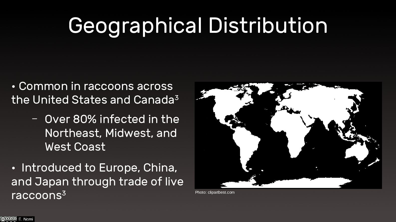
Geographical Distribution • Common in raccoons across the United States
and Canada3 – Over 80% infected in the Northeast, Midwest, and West Coast • Introduced to Europe, China, and Japan through trade of live raccoons3 Photo: clipartbest.com -
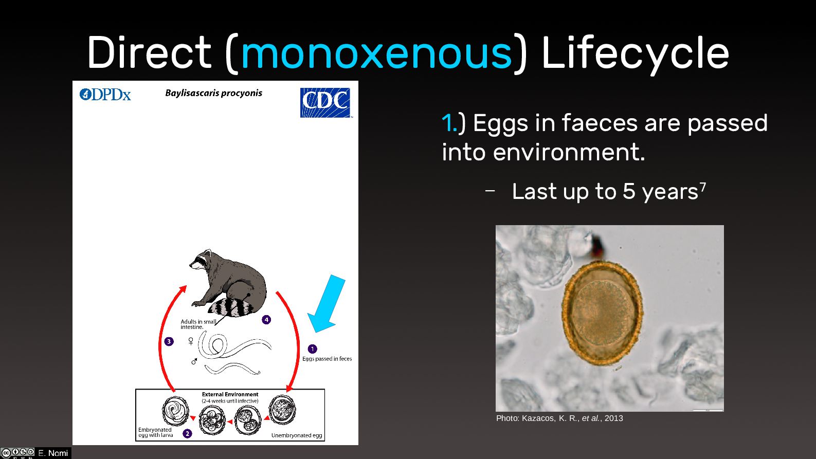
Direct (monoxenous) Lifecycle 1.) Eggs in faeces are passed into
environment. – Last up to 5 years7 Photo: Kazacos, K. R., et al., 2013 -
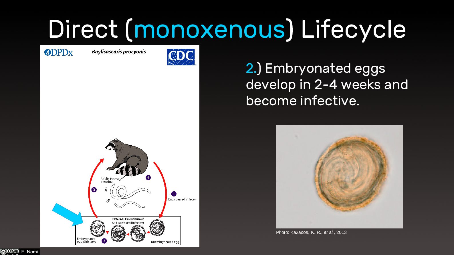
Direct (monoxenous) Lifecycle 2.) Embryonated eggs develop in 2-4 weeks
and become infective. Photo: Kazacos, K. R., et al., 2013 -
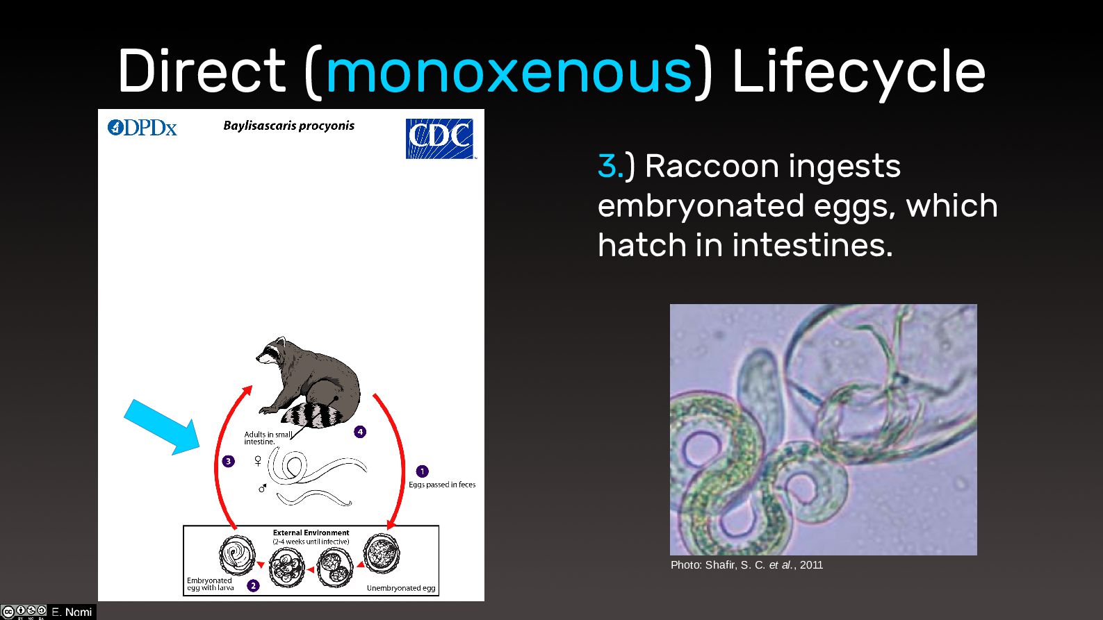
Direct (monoxenous) Lifecycle 3.) Raccoon ingests embryonated eggs, which hatch
in intestines. Photo: Shafir, S. C. et al., 2011 -
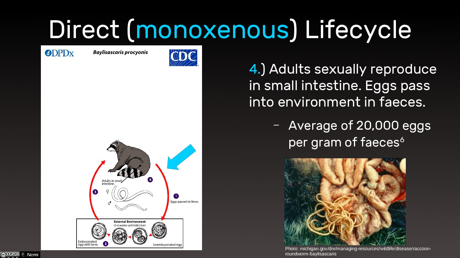
Direct (monoxenous) Lifecycle 4.) Adults sexually reproduce in small intestine.
Eggs pass into environment in faeces. – Average of 20,000 eggs per gram of faeces6 Photo: michigan.gov/dnr/managing-resources/wildlife/disease/raccoon- roundworm-baylisascaris -
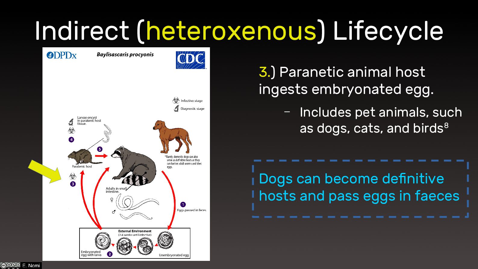
Indirect (heteroxenous) Lifecycle 3.) Paranetic animal host ingests embryonated egg.
– Includes pet animals, such as dogs, cats, and birds8 Dogs can become definitive hosts and pass eggs in faeces -
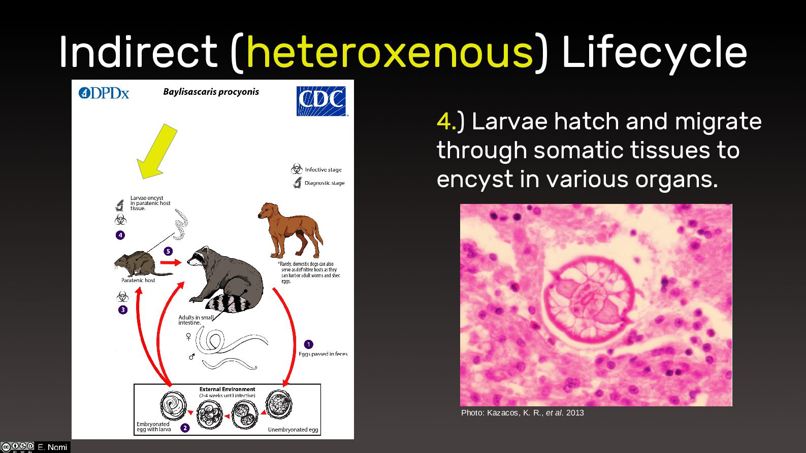
Indirect (heteroxenous) Lifecycle 4.) Larvae hatch and migrate through somatic
tissues to encyst in various organs. Photo: Kazacos, K. R., et al. 2013 -
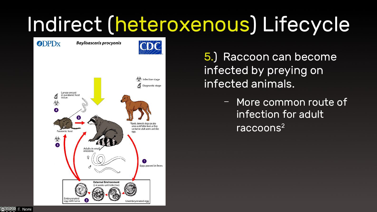
Indirect (heteroxenous) Lifecycle 5.) Raccoon can become infected by preying
on infected animals. – More common route of infection for adult raccoons2 -
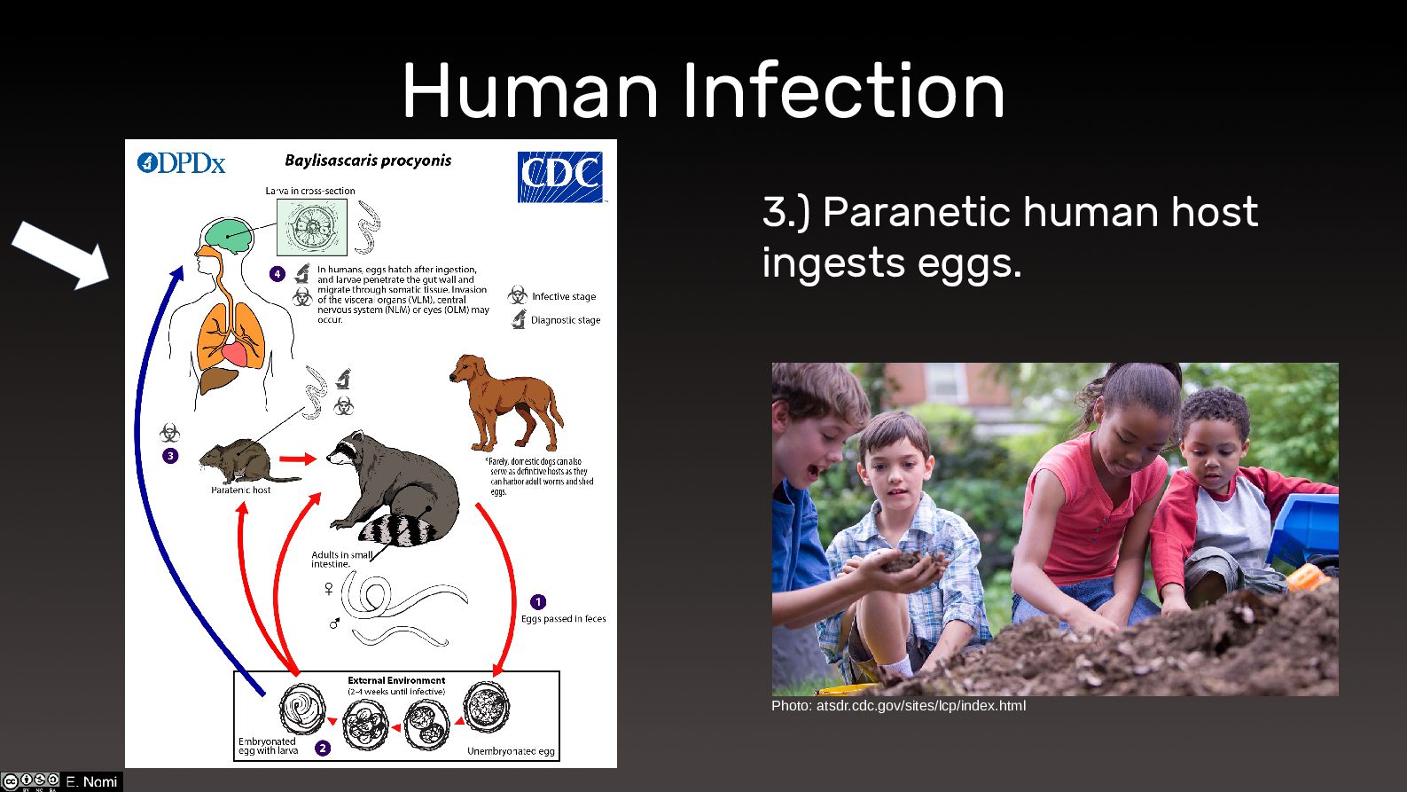
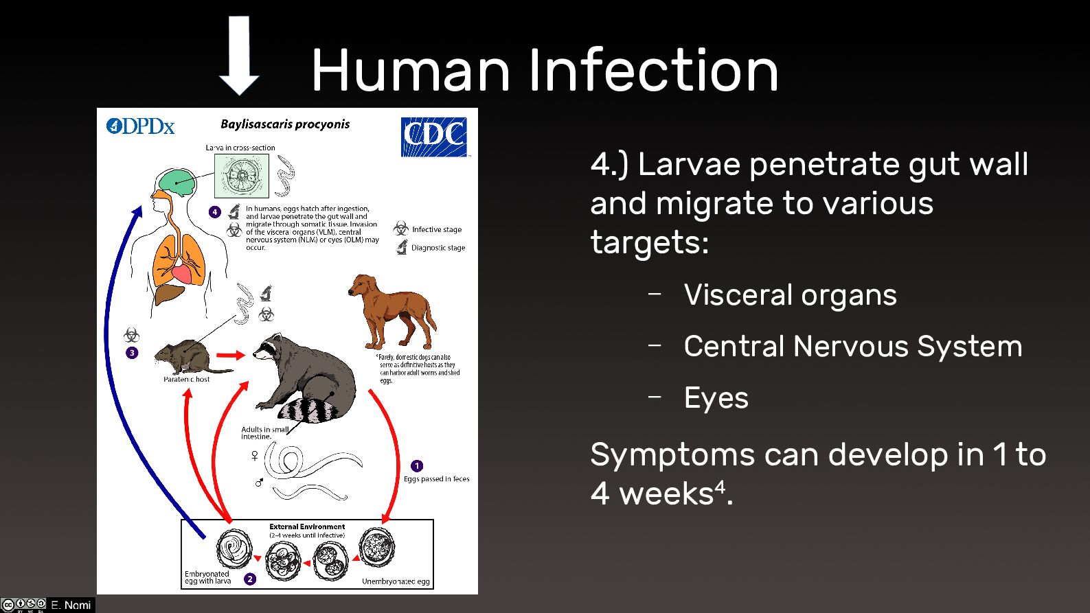
Human Infection 4.) Larvae penetrate gut wall and migrate to
various targets: – Visceral organs – Central Nervous System – Eyes Symptoms can develop in 1 to 4 weeks4. -
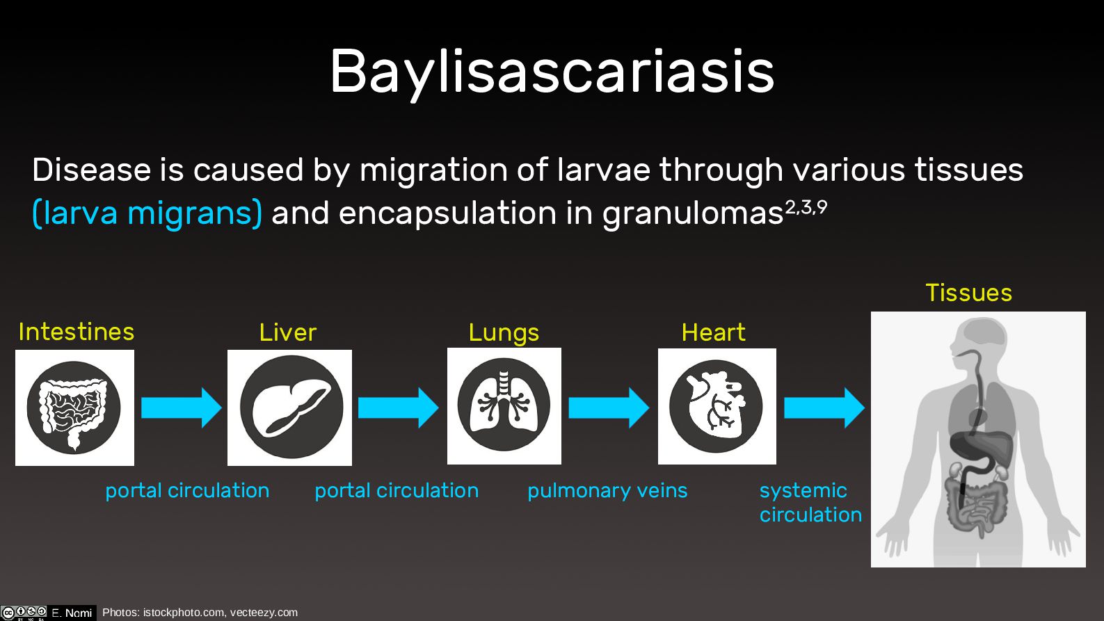
Baylisascariasis Disease is caused by migration of larvae through various
tissues (larva migrans) and encapsulation in granulomas2,3,9 Intestines Liver Lungs Heart Tissues portal circulation portal circulation pulmonary veins systemic circulation Photos: istockphoto.com, vecteezy.com -
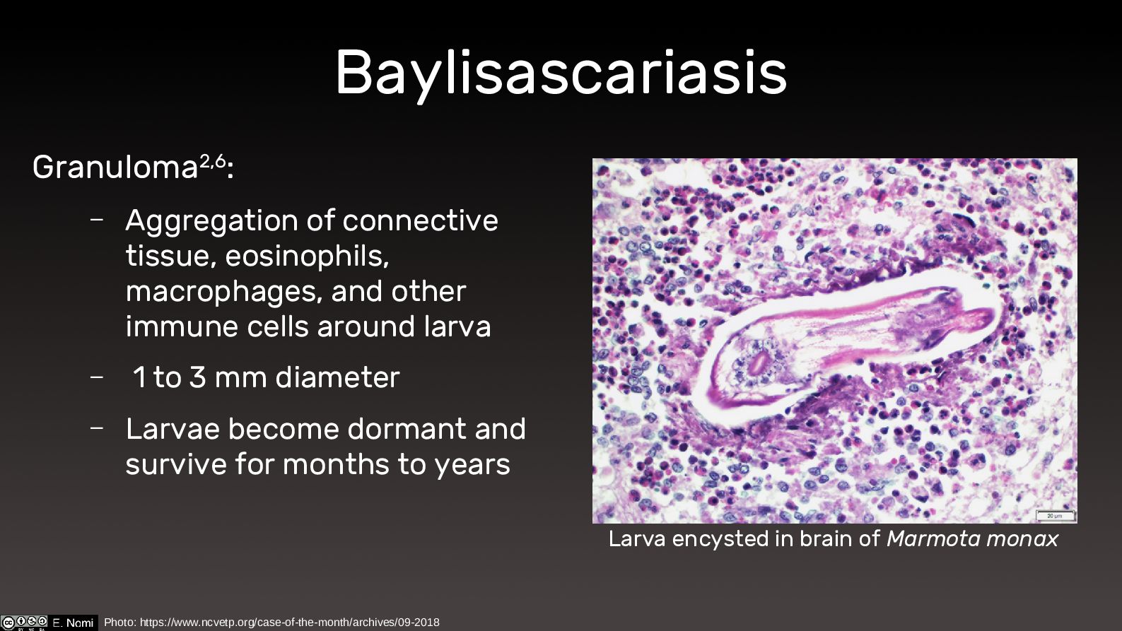
Baylisascariasis Granuloma2,6: – Aggregation of connective tissue, eosinophils, macrophages, and
other immune cells around larva – 1 to 3 mm diameter – Larvae become dormant and survive for months to years Larva encysted in brain of Marmota monax Photo: https://www.ncvetp.org/case-of-the-month/archives/09-2018 -
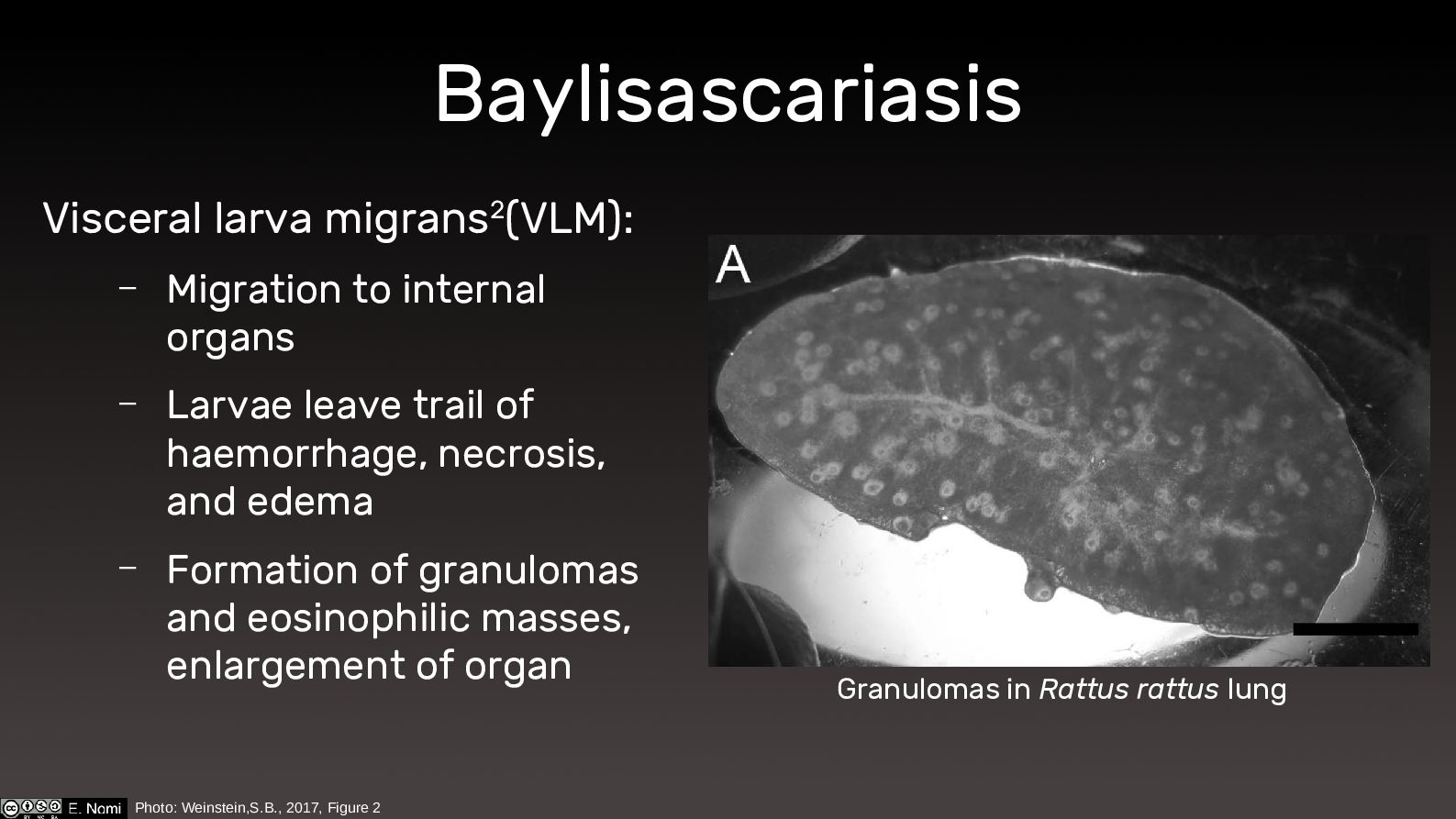
Baylisascariasis Visceral larva migrans2(VLM): – Migration to internal organs –
Larvae leave trail of haemorrhage, necrosis, and edema – Formation of granulomas and eosinophilic masses, enlargement of organ Photo: Weinstein,S.B., 2017, Figure 2 Granulomas in Rattus rattus lung -
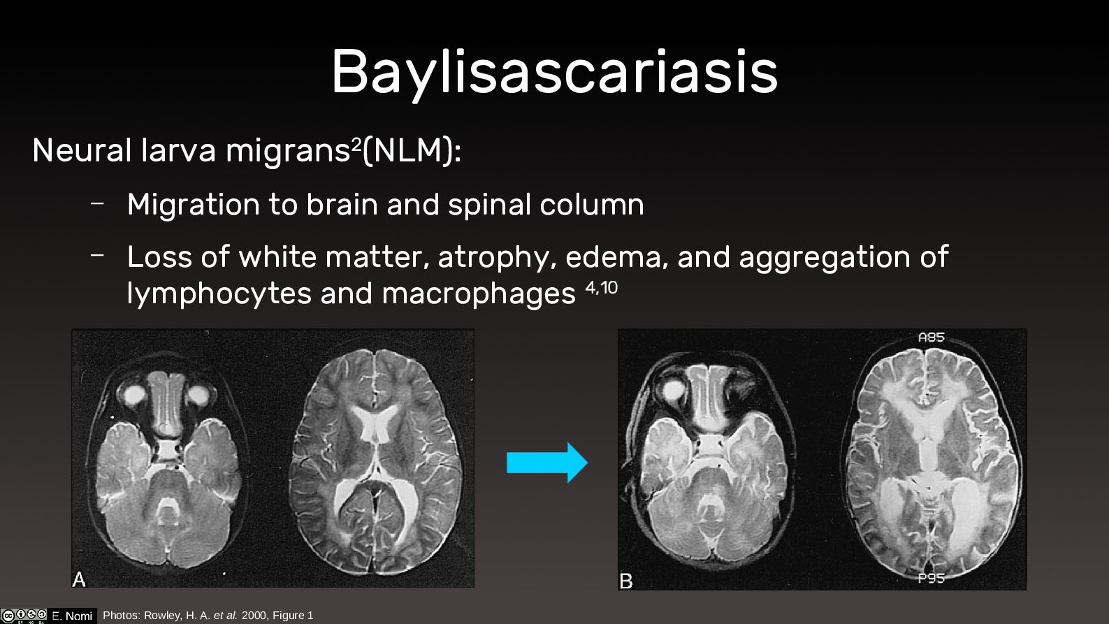
Baylisascariasis Neural larva migrans2(NLM): – Migration to brain and spinal
column – Loss of white matter, atrophy, edema, and aggregation of lymphocytes and macrophages 4,10 Photos: Rowley, H. A. et al. 2000, Figure 1 -
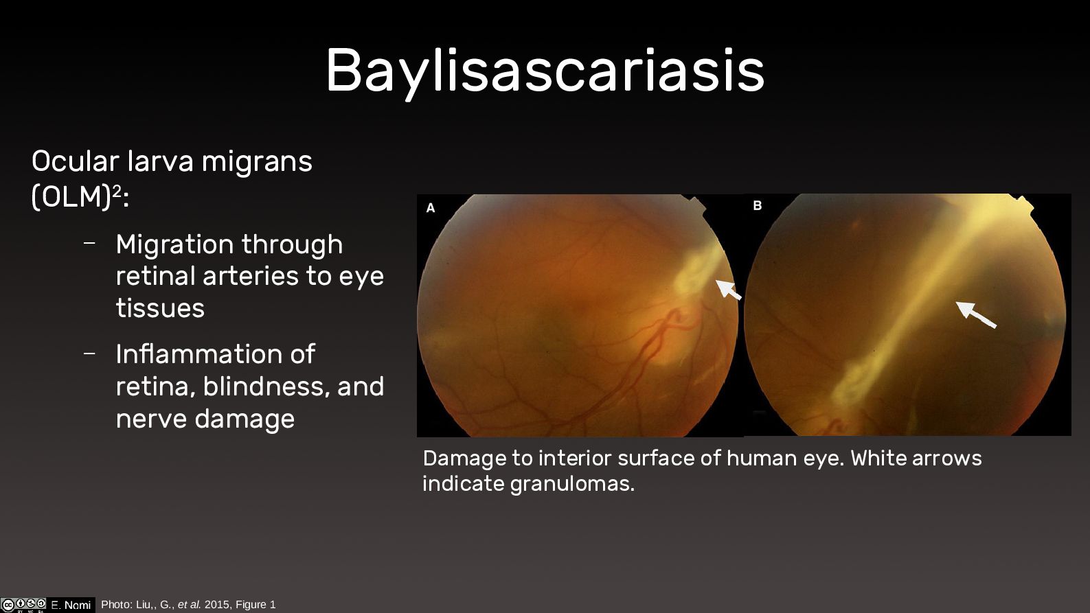
Baylisascariasis Ocular larva migrans (OLM)2: – Migration through retinal arteries
to eye tissues – Inflammation of retina, blindness, and nerve damage Photo: Liu,, G., et al. 2015, Figure 1 Damage to interior surface of human eye. White arrows indicate granulomas. -
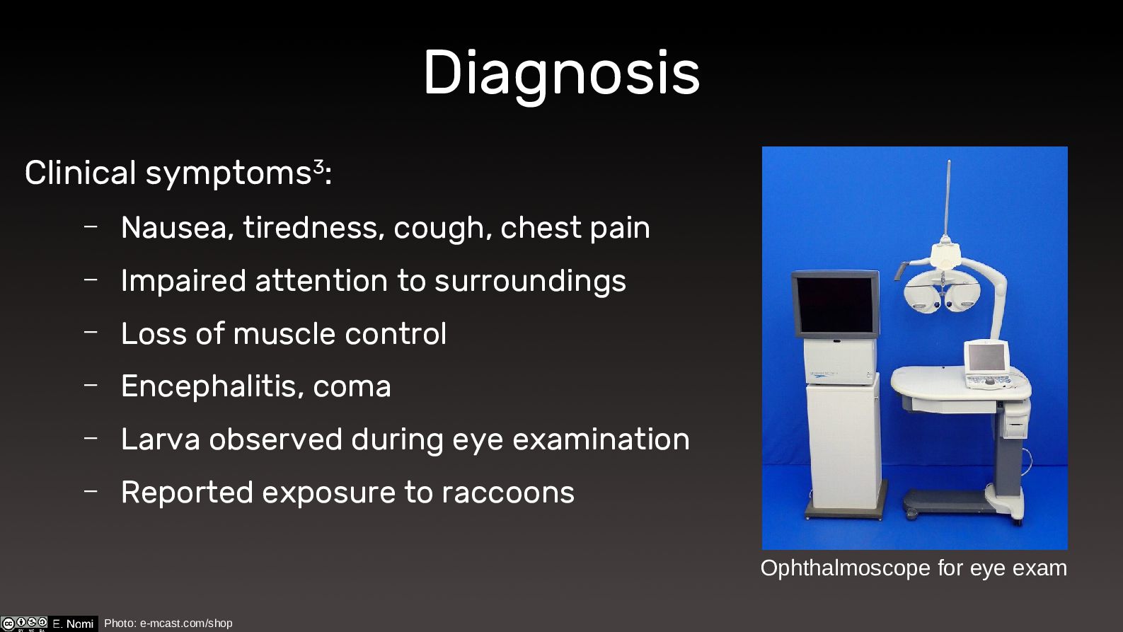
Diagnosis Clinical symptoms3: – Nausea, tiredness, cough, chest pain –
Impaired attention to surroundings – Loss of muscle control – Encephalitis, coma – Larva observed during eye examination – Reported exposure to raccoons Ophthalmoscope for eye exam Photo: e-mcast.com/shop -
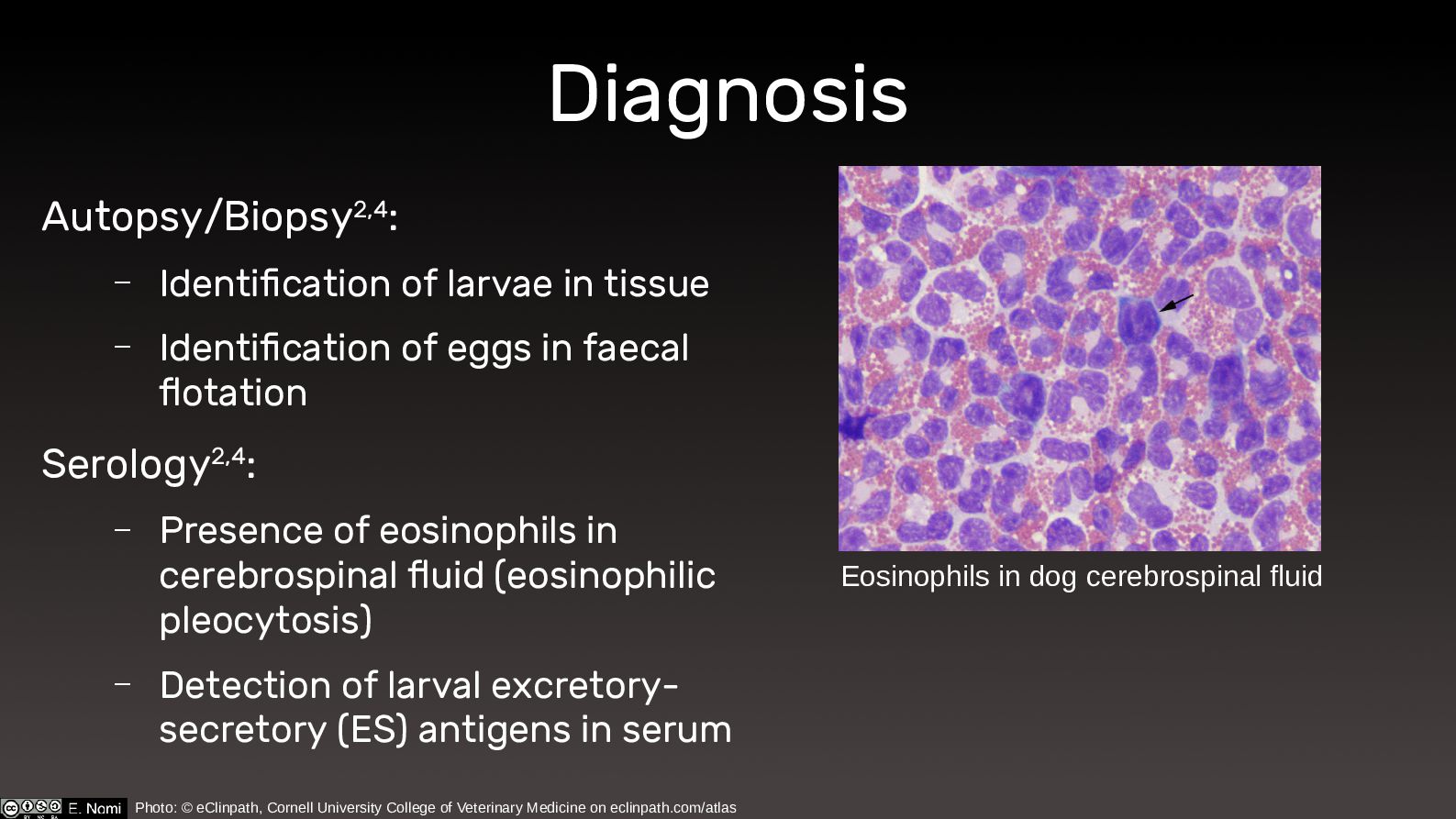
Diagnosis Autopsy/Biopsy2,4: – Identification of larvae in tissue – Identification
of eggs in faecal flotation Serology2,4: – Presence of eosinophils in cerebrospinal fluid (eosinophilic pleocytosis) – Detection of larval excretory- secretory (ES) antigens in serum Eosinophils in dog cerebrospinal fluid Photo: © eClinpath, Cornell University College of Veterinary Medicine on eclinpath.com/atlas -
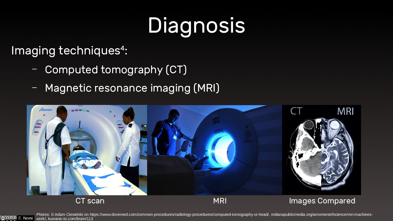
Diagnosis Imaging techniques4: – Computed tomography (CT) – Magnetic resonance
imaging (MRI) Photos: © Adam Ciesielski on https://www.dovemed.com/common-procedures/radiology-procedures/computed-tomography-ct-head/, indianapublicmedia.org/amomentofscience/mri-machines- work/, kuwana-sc.com/brain/113 CT scan MRI Images Compared -
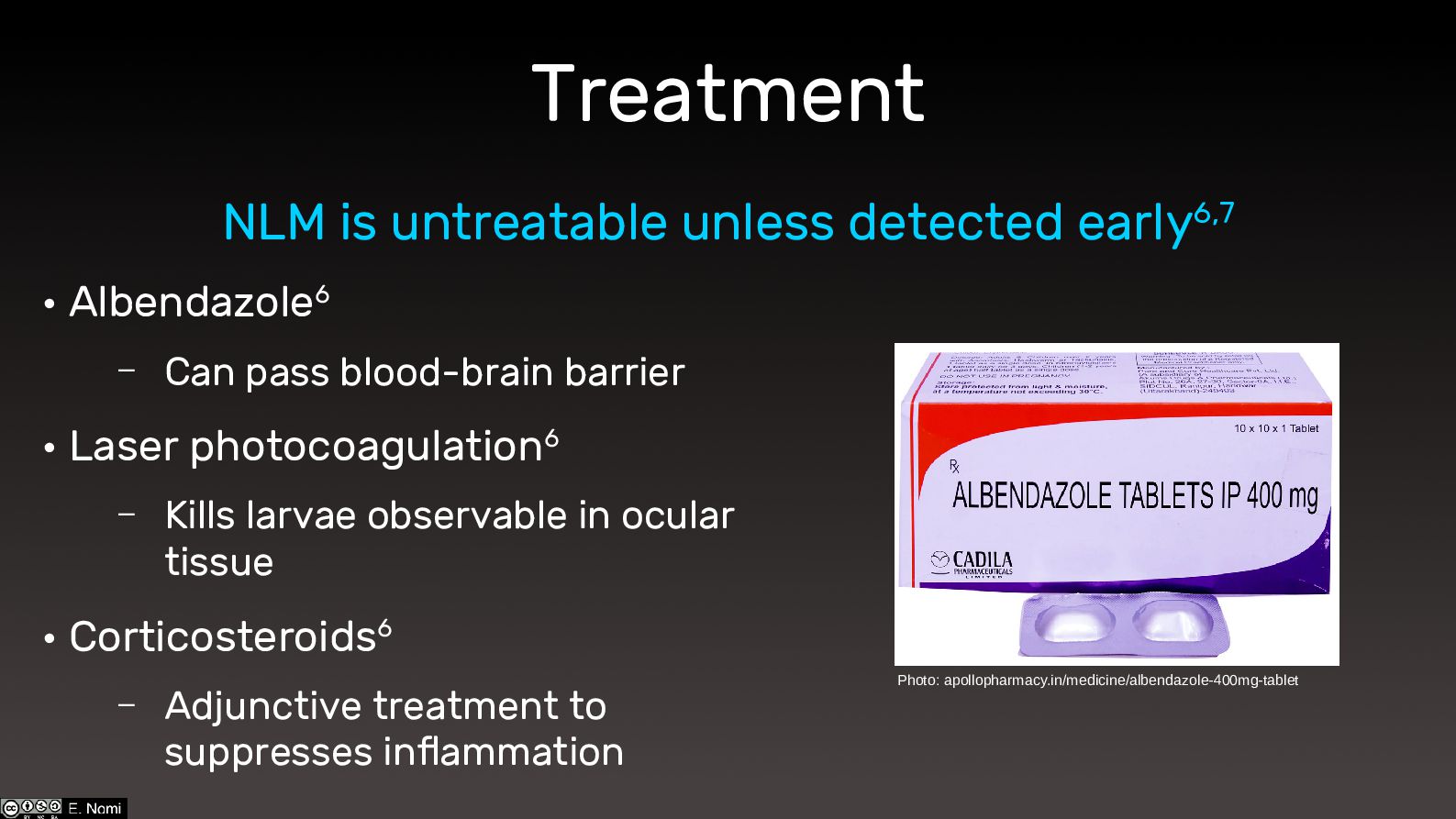
Treatment • Albendazole6 – Can pass blood-brain barrier • Laser
photocoagulation6 – Kills larvae observable in ocular tissue • Corticosteroids6 – Adjunctive treatment to suppresses inflammation NLM is untreatable unless detected early6,7 Photo: apollopharmacy.in/medicine/albendazole-400mg-tablet -
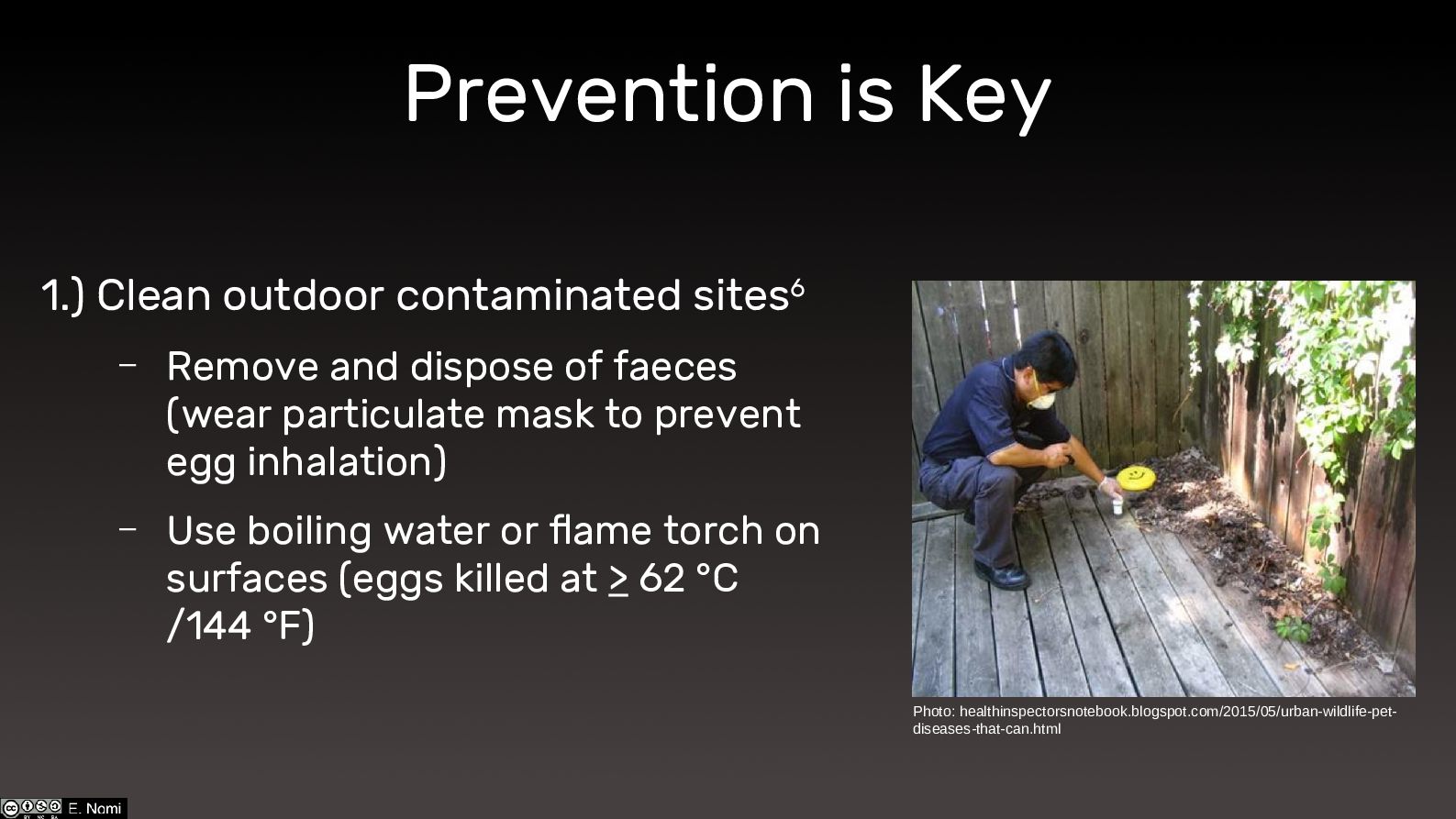
Prevention is Key 1.) Clean outdoor contaminated sites6 – Remove
and dispose of faeces (wear particulate mask to prevent egg inhalation) – Use boiling water or flame torch on surfaces (eggs killed at > 62 °C /144 °F) Photo: healthinspectorsnotebook.blogspot.com/2015/05/urban-wildlife-pet- diseases-that-can.html -
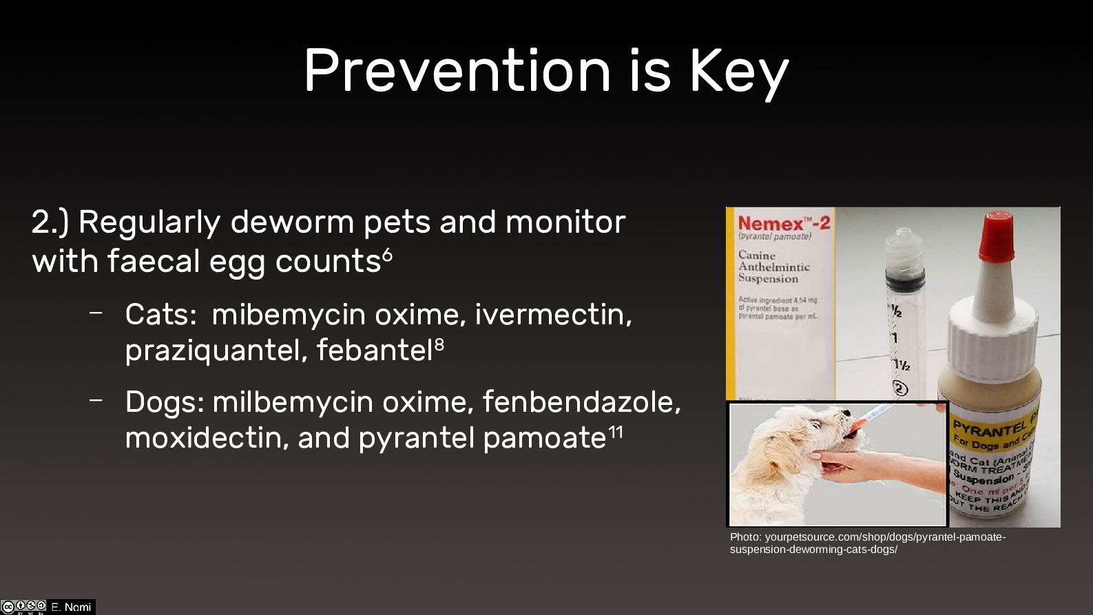
Prevention is Key 2.) Regularly deworm pets and monitor with
faecal egg counts6 – Cats: mibemycin oxime, ivermectin, praziquantel, febantel8 – Dogs: milbemycin oxime, fenbendazole, moxidectin, and pyrantel pamoate11 Photo: yourpetsource.com/shop/dogs/pyrantel-pamoate- suspension-deworming-cats-dogs/ -
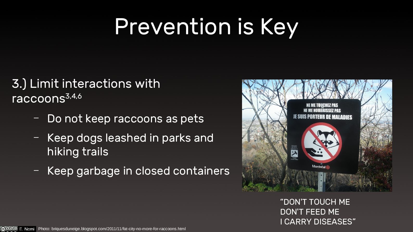
Prevention is Key 3.) Limit interactions with raccoons3,4,6 – Do
not keep raccoons as pets – Keep dogs leashed in parks and hiking trails – Keep garbage in closed containers Photo: briquesduneige.blogspot.com/2011/11/fat-city-no-more-for-raccoons.html “DON'T TOUCH ME DON'T FEED ME I CARRY DISEASES” -
References 1.) University of Hawai‘i. (2022) Worms: Phyla Platyhelmintes, Nematoda,
and Annelida. Retrieved May 10, 2022 from https://manoa.hawaii.edu/exploringourfluidearth/biological/invertebrates/worms-phyla-platyhelmintes-nematoda-and-annelida. 2.) Graeff-Teixeira, C., Morassutti, A. L., & Kazacos, K. R. (2016). Update on Baylisascariasis, a Highly Pathogenic Zoonotic Infection. Clinical microbiology reviews, 29(2), 375–399. https://doi.org/10.1128/CMR.00044-15. 3.) The Centers for Disease Control and Prevention. (2018, April 11). Parasites - Baylisascaris infection. Retrieved May 9, 2022 from https://www.cdc.gov/parasites/baylisascaris/index.html. 4.) Kazacos, K.R., 2016, Baylisascaris Larva Migrans: U.S. Geological Survey Circular 1412, 122 p., 3 appendixes, http://dx.doi.org/10.3133/cir1412. 5.) Hung, T., Neafie, R. C., & Mackenzie, I. R. (2012). Baylisascaris procyonis infection in elderly person, British Columbia, Canada. Emerging infectious diseases, 18(2), 341–342. https://doi.org/10.3201/eid1802.111046. 6.) Kazacos, K. R., Jelicks, L. A., & Tanowitz, H. B. (2013). Baylisascaris larva migrans. Handbook of clinical neurology, 114, 251–262. 7.) Sapp, S., Handali, S., Weinstein, S. B., & Yabsley, M. J. (2018). Detection and Evaluation of Antibody Response to a Baylisascaris-Specific Antigen in Rodent Hosts with the Use of Western Blotting and Elisa. The Journal of parasitology, 104(6), 651–659. https://doi.org/10.1645/18-48. 8.) PetMD Editorial. (2009, March 06). Baylisascariasis in Cats. Retrieved May 26, 2022 from https://www.petmd.com/cat/conditions/infectious-parasitic/c_ct_baylisascariasis. 9.) Gavin, P. J., Kazacos, K. R., & Shulman, S. T. (2005). Baylisascariasis. Clinical microbiology reviews, 18(4), 703–718. https://doi.org/10.1128/CMR.18.4.703-718.2005. 10.) Rowley, H. A., Uht, R. M., Kazacos, K. R., Sakanari, J., Wheaton, W. V., Barkovich, A. J., & Bollen, A. W. (2000). Radiologic-pathologic findings in raccoon roundworm (Baylisascaris procyonis) encephalitis. AJNR. American journal of neuroradiology, 21(2), 415–420. 11.) CAPC Vet. (2015, July 01). Baylisascaris procyonis for Dog. Retrieved May 26, 2022 from https://capcvet.org/guidelines/baylisascaris-procyonis.