Upgrade to Pro — share decks privately, control downloads, hide ads and more …

Arm & Superficial muscles of forearm

Arm & Superficial muscles of forearm

This presentation illustrates the Anatomy of Arm & Superficial muscles of forearm

Dr.Sherif Fahmy

July 31, 2019
Tweet

More Decks by Dr.Sherif Fahmy

Other Decks in Science

Transcript

  1. Humerus Medial intermuscular septum Lateral intermuscular septum Anterior (Flexor )

    compartment Posterior (extensor) compartment Brachial fascia Deep fascia of the arm
  2. Dr. Sherif Fahmy Coraco-brachialis muscle Origin: tip of coracoid process

    Insertion: Middle of medial border of shaft of humerus
  3. Action of Coracobrachialis Muscle 1- Assist in adduction of arm

    at shoulder joint. 2- Assist in flexion of arm at shoulder joint. Nerve supply: - Musculo-cutaneous nerve (C5,6,7)
  4. Dr. Sherif Fahmy Short head of biceps Coracoid process Long

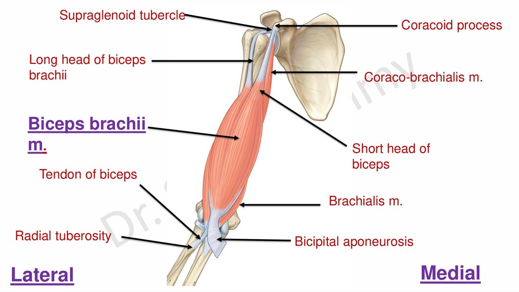
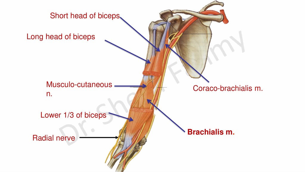
    head of biceps brachii Coraco-brachialis m. Brachialis m. Tendon of biceps Bicipital aponeurosis Biceps brachii m. Supraglenoid tubercle Lateral Medial Radial tuberosity
  5. Capsule of shoulder joint Tendon of long head of biceps

    Short head of biceps Tendon of long head of biceps Transverse humeral ligament Intracapsular extrasynovial course of long head of biceps m.
  6. Action of biceps brachii muscle • It flexes the elbow.

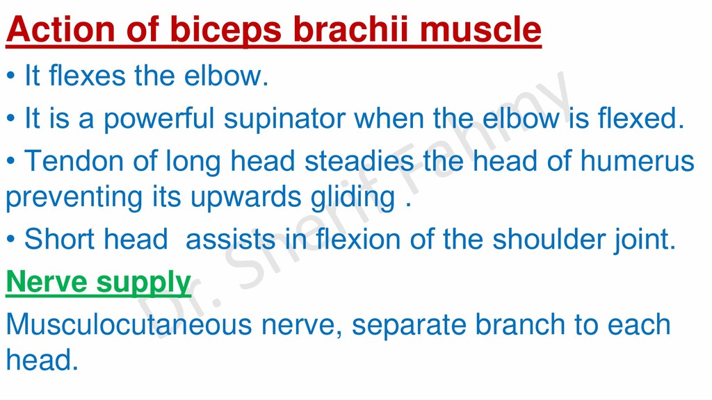
    • It is a powerful supinator when the elbow is flexed. • Tendon of long head steadies the head of humerus preventing its upwards gliding . • Short head assists in flexion of the shoulder joint. Nerve supply Musculocutaneous nerve, separate branch to each head.
  7. Long head of biceps Short head of biceps Lower 1/3

    of biceps Musculo-cutaneous n. Brachialis m. Coraco-brachialis m. Radial nerve
  8. Action of brachialis muscle: - Main flexor of elbow. Nerve

    supply: - Musculo-cutaneous nerve. - Radial nerve to its lateral part.
  9. Dr. Sherif Fahmy Infraglenoid tubercle Upper lip of spiral groove

    Lower lip of spiral groove Upper surface of olecranon process
  10. Origin of long head of triceps Origin of lateral head

    of triceps Origin of medial head of triceps Insertion of triceps Spiral groove
  11. Nerve Supply Radial nerve through the following branches: -To long

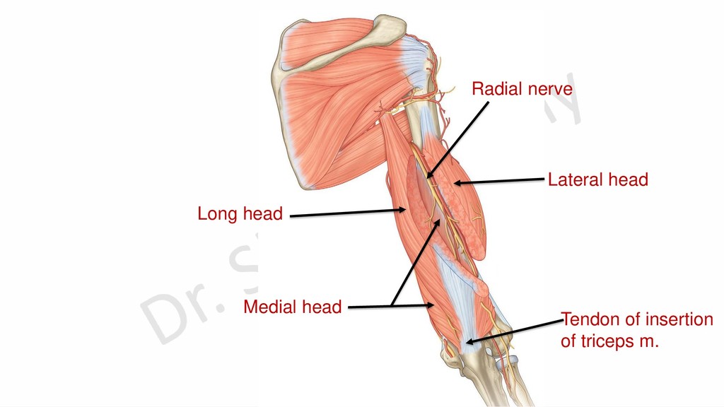
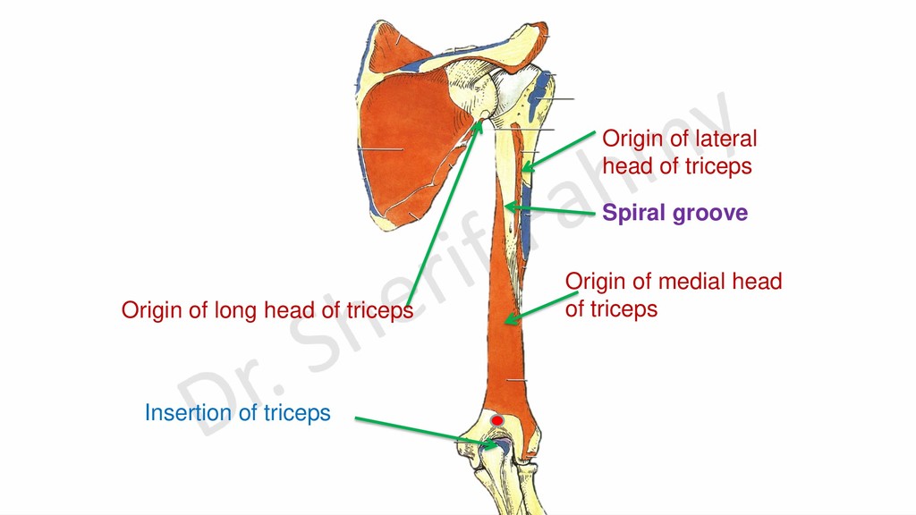
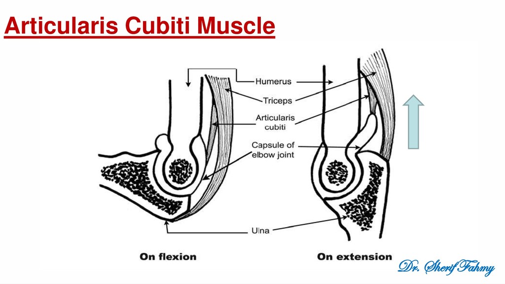
    and medial heads from branches of radial nerve before spiral groove (In axilla). -To lateral and medial heads from branches of radial nerve in the spiral groove. N.B.: Articularis cubiti is supplied by a branch from nerve to medial head of triceps.
  12. Action of Triceps -Triceps is the main extensor of elbow.

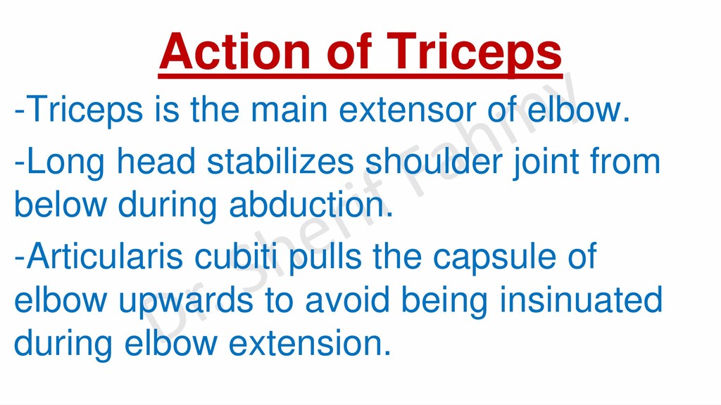
    -Long head stabilizes shoulder joint from below during abduction. -Articularis cubiti pulls the capsule of elbow upwards to avoid being insinuated during elbow extension.
  13. Branches Muscular: 1- Coracobrachialis m. 2- Biceps brachii m 3-

    Medial part of brachialis m. Cutaneous: - Skin of lateral surface of forearm till the proximal part of thenar eminence.
  14. Arrangement of Muscles of the front of Forearm Superficial Group

    A- Superficial layer: 1) Pronator teres. 2) Flexor carpi radialis. 3) Palmaris longus. 4) Flexor carpi ulnaris. B- Deep layer: 1) Flexor digitorum superficialis. Deep Group A- Superficial layer: 1) Flexor pollicis longus. 2) Flexor digitorum profundus. B- Deep layer: 1) Pronator quadratus
  15. Common rules of superficial flexors ➢Muscles are named after their

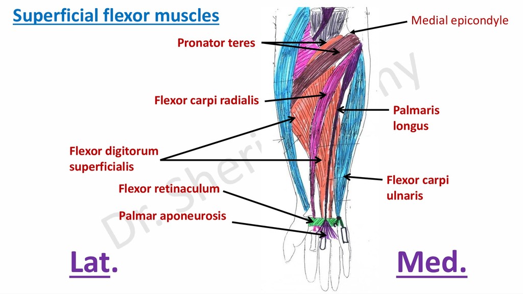
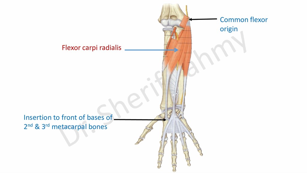
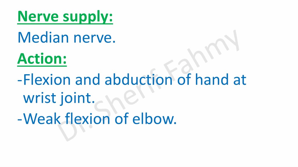
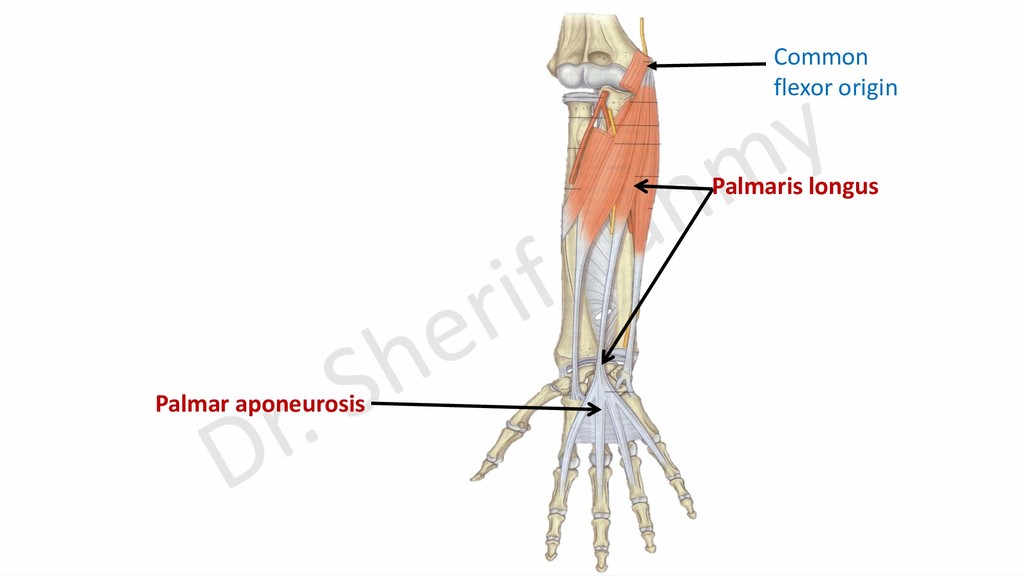
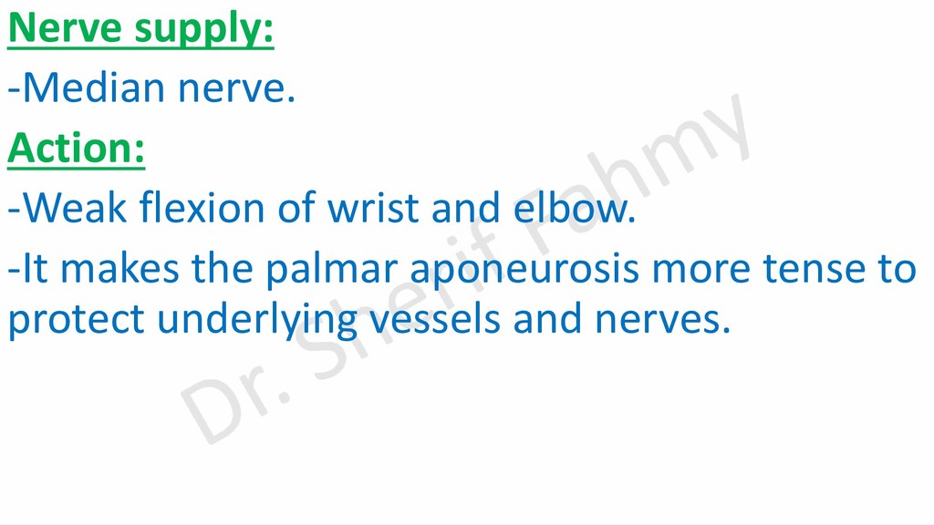
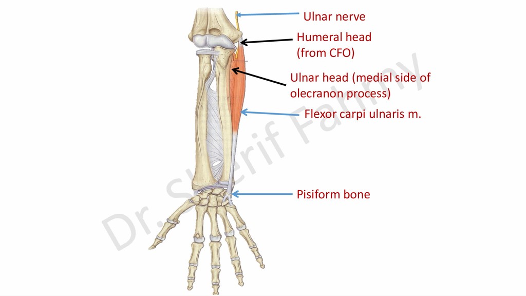
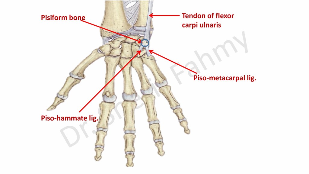
    action. ➢All muscles arises partially or totally from common flexor origin (front of medial epicondyle). ➢All muscles have 2 heads except flexor carpi radialis and palmaris longus. ➢Each 2 headed muscle has a nerve that passes between 2 heads and supplies the muscle. Nerve supply: ➢Median nerve except flexor carpi ulnaris is supplied by ulnar nerve. Common action: ➢All are week flexors of elbow. ➢All are flexors of wrist except pronator teres.
  16. Pronator teres Flexor carpi radialis Palmaris longus Flexor carpi ulnaris

    Flexor digitorum superficialis Flexor retinaculum Palmar aponeurosis Superficial flexor muscles Med. Lat. Medial epicondyle
  17. Relations of 8 flexor tendons to flexor retinaculum: ➢Tendons of

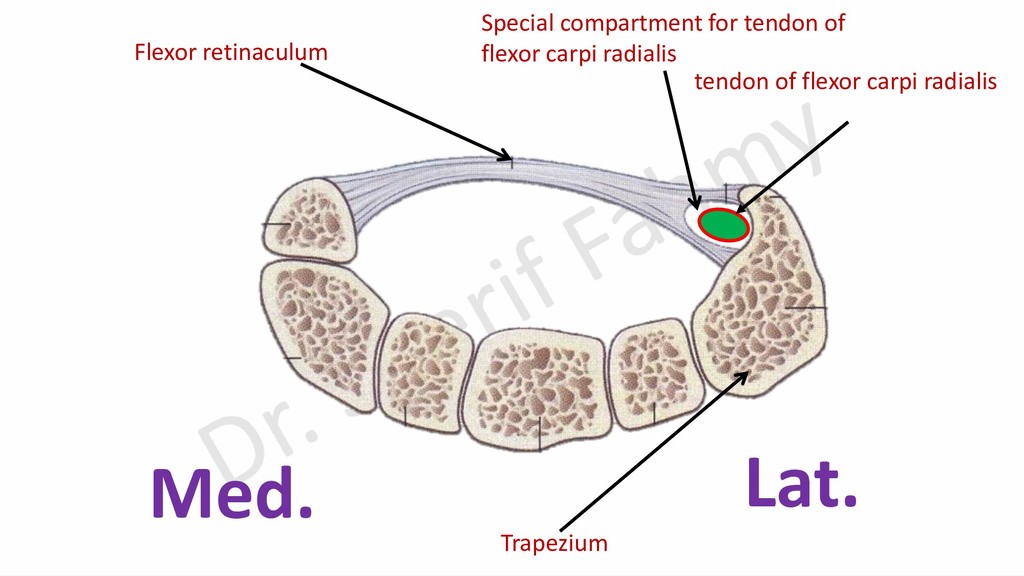
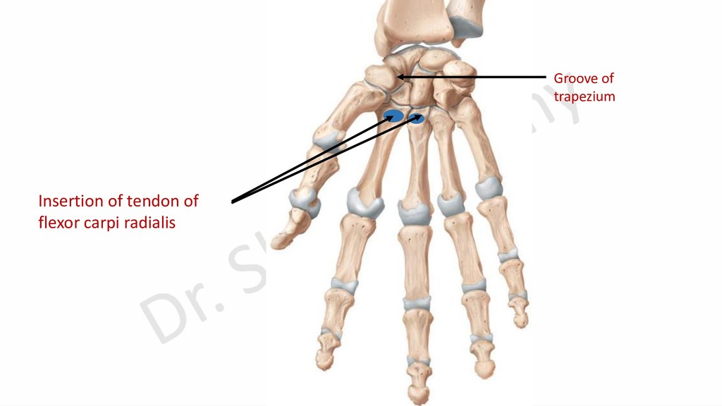
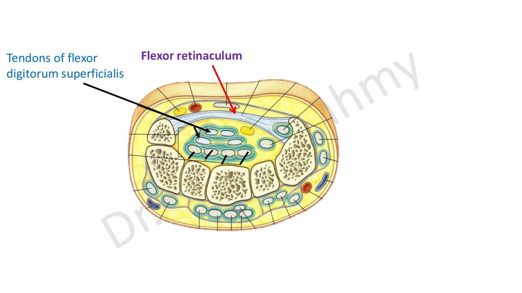
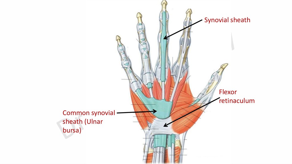
    flexor digitorum (superficialis & profundus) and flexor pollicis longus pass deep to it. ➢Tendon of palmaris longus passes superficial to it. ➢Tendon of flexor carpi radialis passes through a special compartment in the lateral attachment of the retinaculum. ➢Tendon of flexor carpi ulnaris on its medial margin. ➢Tendons of 2 pronator muscles have no relations.
  18. Superficial (humeral) head Deep (ulnar) head Insertion of pronator teres

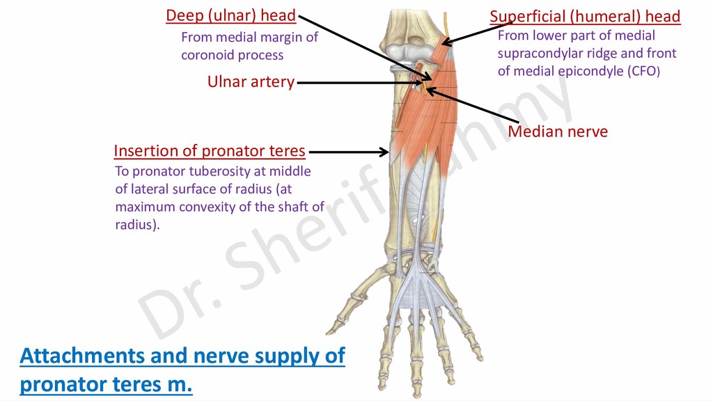
    Median nerve Attachments and nerve supply of pronator teres m. From lower part of medial supracondylar ridge and front of medial epicondyle (CFO) From medial margin of coronoid process To pronator tuberosity at middle of lateral surface of radius (at maximum convexity of the shaft of radius). Ulnar artery
  19. Trapezium Flexor retinaculum Special compartment for tendon of flexor carpi

    radialis Med. Lat. tendon of flexor carpi radialis
  20. Nerve supply: -Median nerve. Action: -Weak flexion of wrist and

    elbow. -It makes the palmar aponeurosis more tense to protect underlying vessels and nerves.
  21. Humeral head (from CFO) Ulnar head (medial side of olecranon

    process) Flexor carpi ulnaris m. Pisiform bone Ulnar nerve
  22. Radial head of flexor digitorum superficialis Humero-ulnar head of flexor

    digitorum superficialis Origin of flexor digitorum superficialis Anterior oblique line
  23. Humero-ulnar head Radial head Tendinous arch Flexor retinaculum Insertion of

    flexor digitorum superficialis Flexor digitorum superficialis Attachments of flexor digitorum superficialis
  24. Nerve supply: Median nerve. Action: -Flexion of medial 4 fingers

    at proximal inter- phalangeal joints and metacarpo-phalangeal joints. -Flexion of wrist. -Weak flexion of elbow.