Upgrade to Pro — share decks privately, control downloads, hide ads and more …

Mammary gland, palm spaces, Arteries of the hand

Mammary gland, palm spaces, Arteries of the hand

This presentation describes Mammary gland, palm spaces, Arteries of the hand

Dr.Sherif Fahmy

August 01, 2019
Tweet

More Decks by Dr.Sherif Fahmy

Other Decks in Science

Transcript

  1. Mammary Gland (Breast) Modified sweat gland that is responsible for

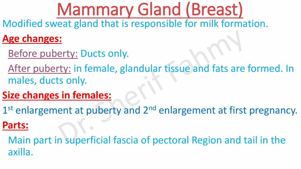
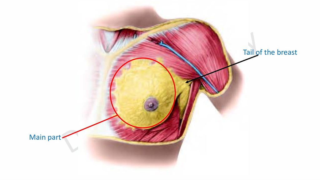
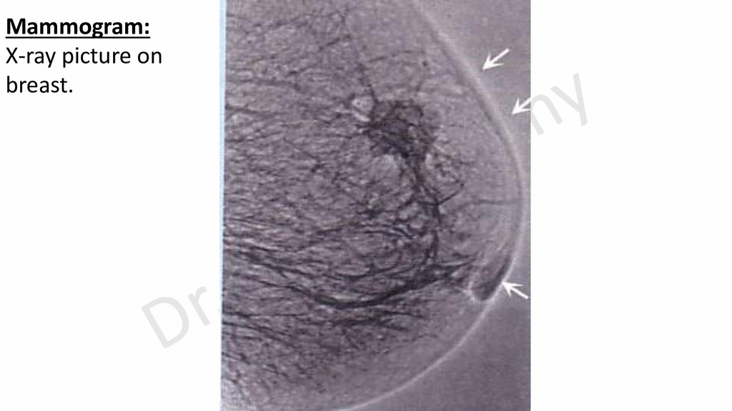
    milk formation. Age changes: Before puberty: Ducts only. After puberty: in female, glandular tissue and fats are formed. In males, ducts only. Size changes in females: 1st enlargement at puberty and 2nd enlargement at first pregnancy. Parts: Main part in superficial fascia of pectoral Region and tail in the axilla.
  2. External Features • Shape: It is hemispherical. It has base

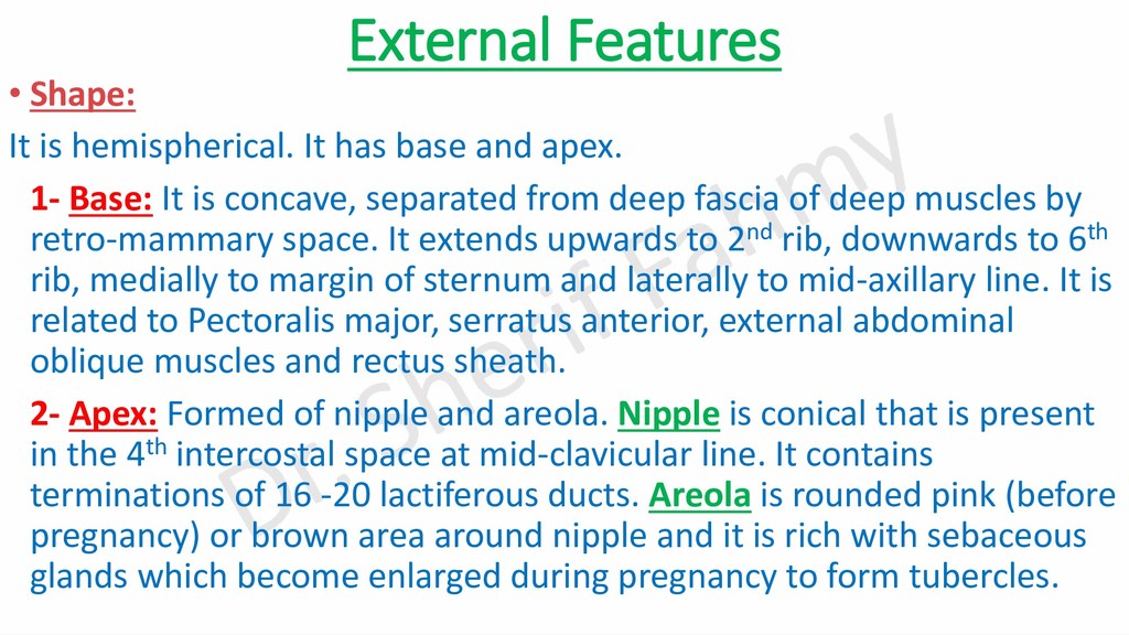
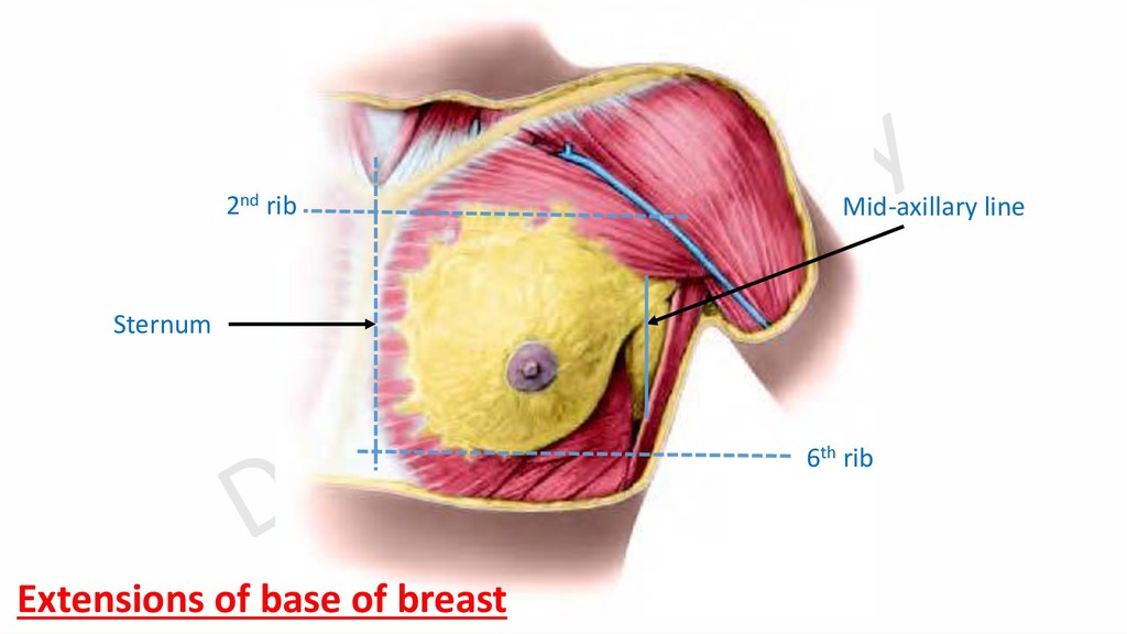
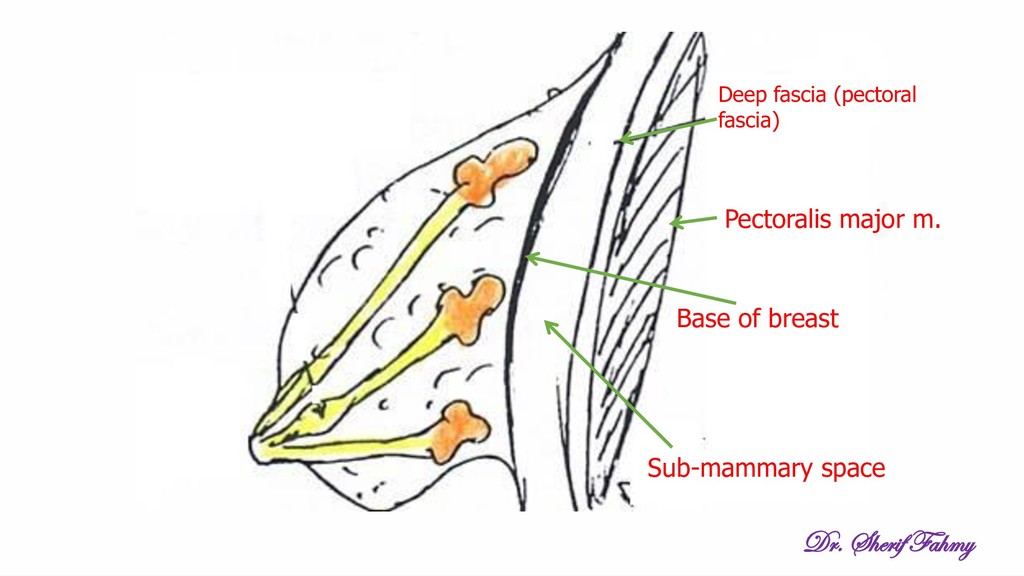
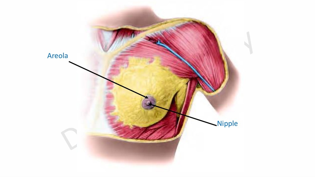
    and apex. 1- Base: It is concave, separated from deep fascia of deep muscles by retro-mammary space. It extends upwards to 2nd rib, downwards to 6th rib, medially to margin of sternum and laterally to mid-axillary line. It is related to Pectoralis major, serratus anterior, external abdominal oblique muscles and rectus sheath. 2- Apex: Formed of nipple and areola. Nipple is conical that is present in the 4th intercostal space at mid-clavicular line. It contains terminations of 16 -20 lactiferous ducts. Areola is rounded pink (before pregnancy) or brown area around nipple and it is rich with sebaceous glands which become enlarged during pregnancy to form tubercles.
  3. Internal Structure •Breast is composed of 16 – 20 radially

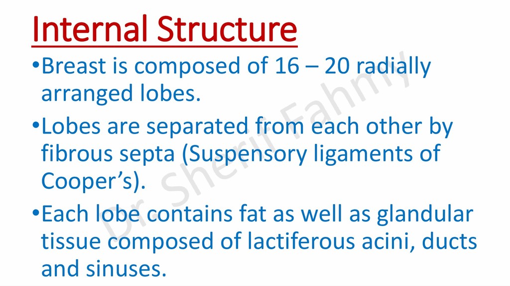
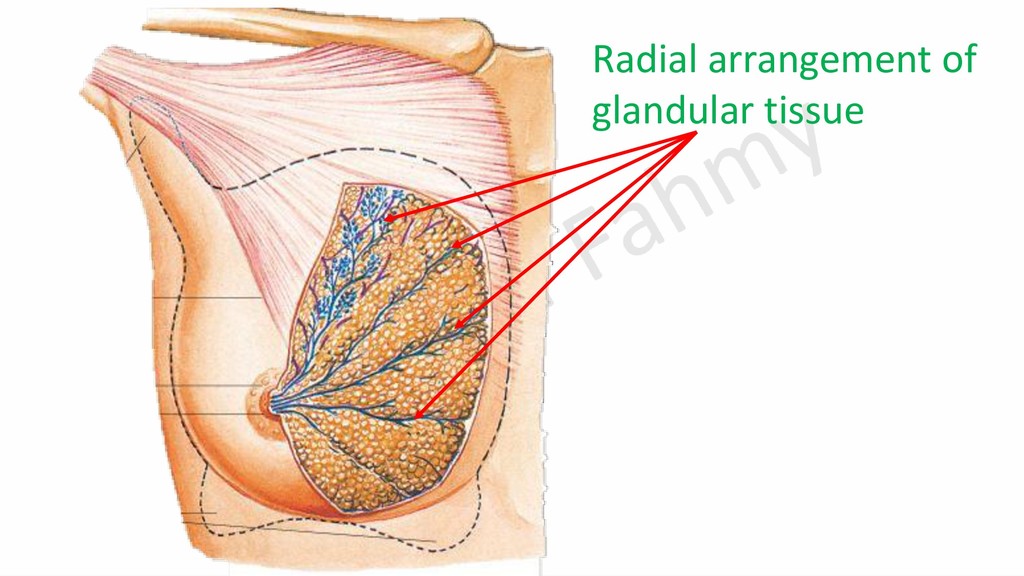
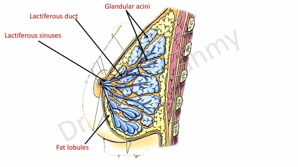
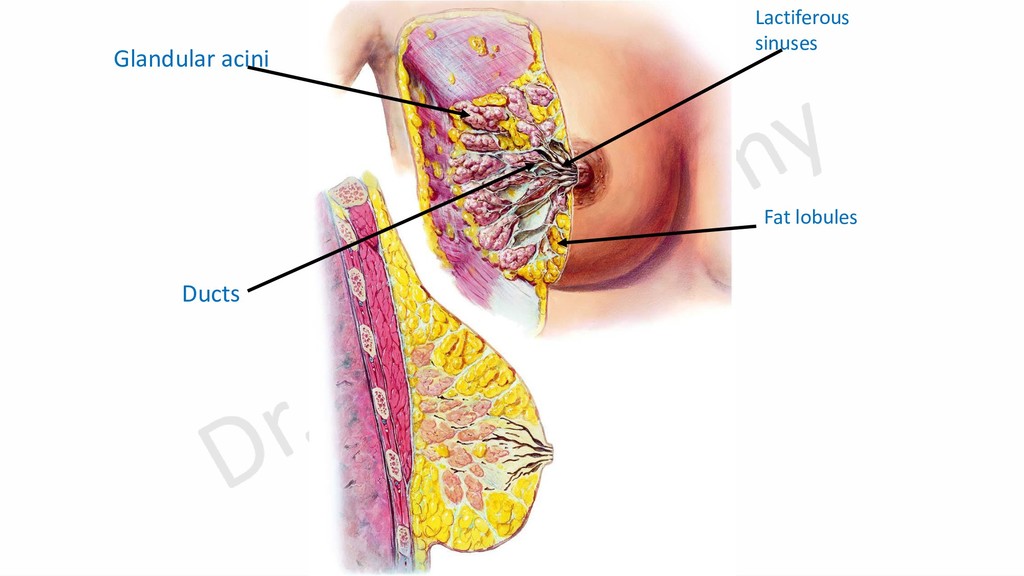
    arranged lobes. •Lobes are separated from each other by fibrous septa (Suspensory ligaments of Cooper’s). •Each lobe contains fat as well as glandular tissue composed of lactiferous acini, ducts and sinuses.
  4. A- Arterial Supply 1- Perforating branches of internal thoracic artery.

    2- Lateral thoracic artery (from axillary artery). 3- Lateral cutaneous branches of posterior intercostal arteries. 4- Pectoral branch of acromio-thoracic artery.
  5. Pectoral branch from acromiothoracic artery Lateral thoracic astery Mammary branches

    from lateral cutaneous branches of posterior intercostal arteries Perforating branches of internal thoracic artery
  6. Venous drainage Lateral thoracic vein to axillary vein Perforating veins

    to internal thoracic vein Mammary veins to posterior intercostal veins to azygos or heniazygos veins
  7. Lymphatic Drainage Lymphatic plexuses: 1- Subareolar plexus: deep to areola

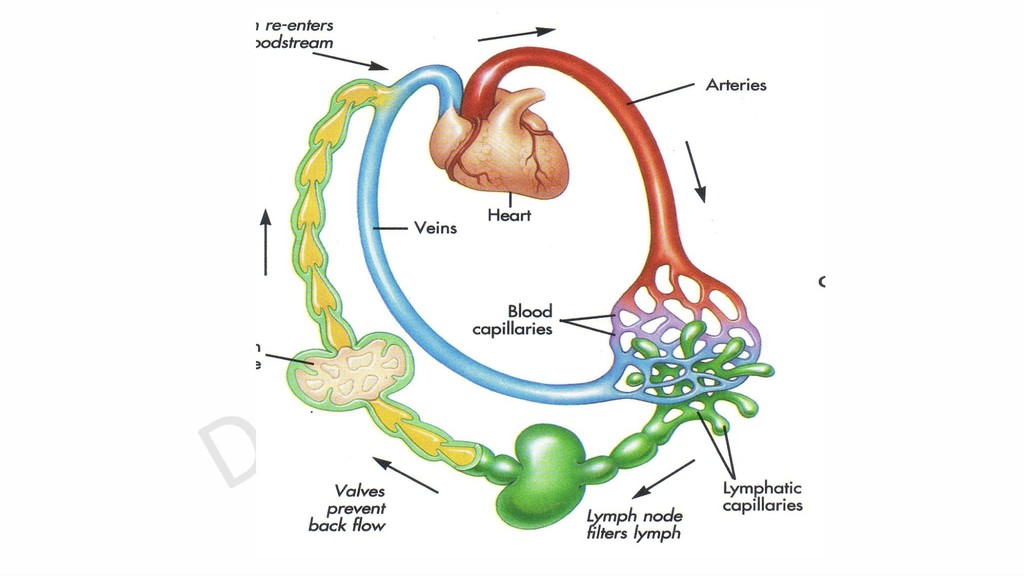
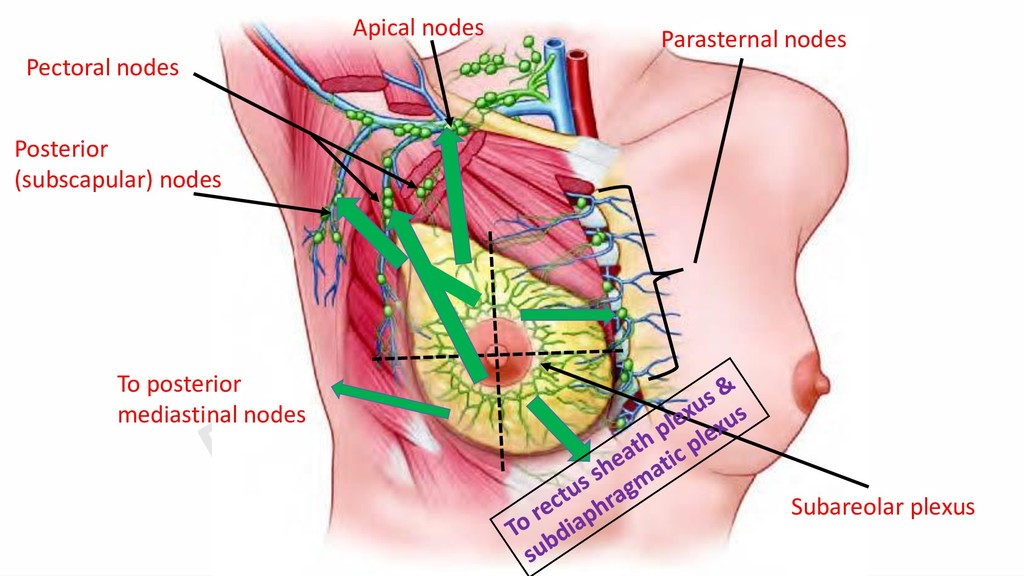
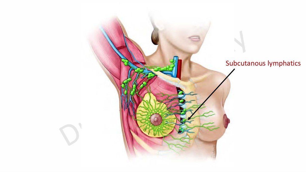
    and nipple drains superficial part of the breast. 2- Pectoral plexus: on pectoral fascia drains deep part of the breast. Both plexuses communicate together by interlobular lymphatics and sends efferent vessels to lymph nodes as follows:
  8. Palmar Compartments 1- lateral compartment (Thenar). 2- Medial compartment (Hypothenar).

    3- Intermediate compartment: A- Mid-palmar space. B- Thenar space.
  9. Lateral compartment Medial Compartment Adductor pollicis m. A line that

    divides middle compartment into mid-palmar (Medial) & thenar and adductor (Lateral)
  10. Lat. Med. Palmar aponeurosis Medial intermuscular septum Lateral intermuscular septum

    Intermediate intermuscular septum adductor pollicis muscle Thenar space Mid-palmar space
  11. Lat. Med. Superficial palmar arch & Branches of median and

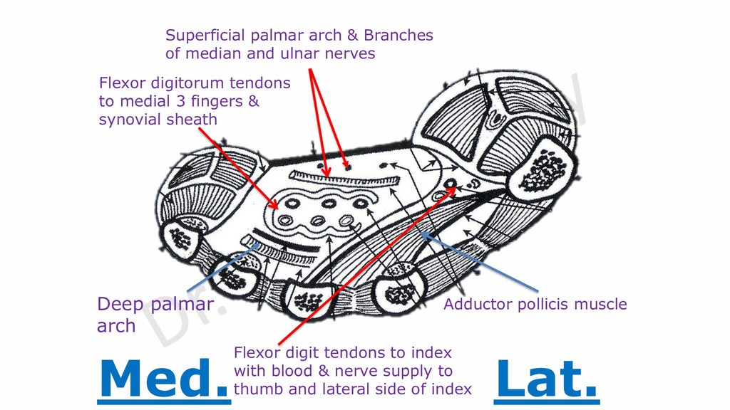
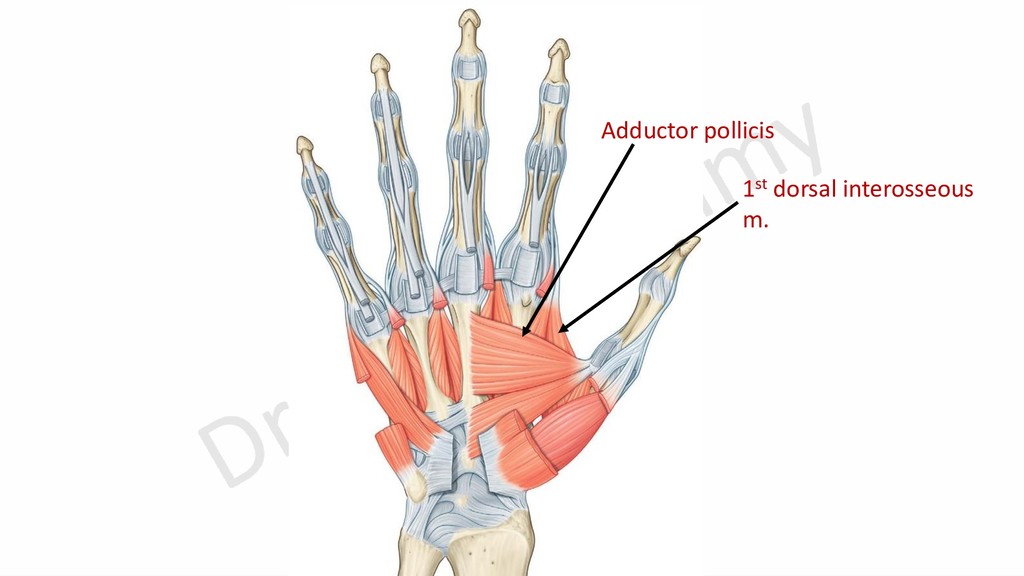
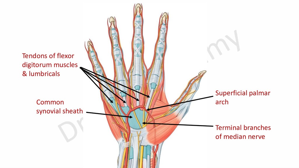
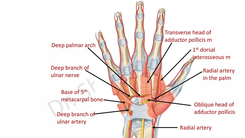
    ulnar nerves Flexor digitorum tendons to medial 3 fingers & synovial sheath Adductor pollicis muscle Flexor digit tendons to index with blood & nerve supply to thumb and lateral side of index Deep palmar arch
  12. Pisiform bone Hook of hamate Lower end of radius Base

    of 5th metacarpal bone 1st intermetacarpal space
  13. Palmar aponeurosis Adductor pollicis m. Palmaris brevis m. Abductor pollicis

    brevis m. Abductor digiti minimi m. Flexor digiti minimi brevis m. Flexor pollicis brevis m.
  14. Arterial Arches in the Hand 1- Anterior carpal arch: gives

    articular blood supply to wrist joint. 2- Superficial palmar arch: gives 3 common palmar digital arteries. 3- Posterior carpal arch: gives 2nd, 3rd and 4th dorsal metacarpal arteries. 4- Deep palmar arch: gives 3 palmar metacarpal arteries.
  15. Ulnar artery Flexor retinaculum Deep br. of ulnar artery Thenar

    eminence Superficial palmar arch Superficial palmar branch from radial artery Pisiform bone Superficial branch of ulnar artery Hook of hamate Ulnar nerve
  16. Lateral to pisiform bone Medial to hook of hamate A

    curved line at level of distal margin of outstretched thumb
  17. Relations of Superficial Palmar Arch Superficial: Skin, superficial fascia and

    palmar aponeurosis. Deep: Terminal branches of median nerve, flexor digitorum tendons, common synovial sheath and lumbricals.
  18. Superficial palmar arch Terminal branches of median nerve Tendons of

    flexor digitorum muscles & lumbricals Common synovial sheath
  19. Branches of Superficial Palmar Arch 1- Palmar digital branch of

    little finger. 2- Three common palmar digital branches.
  20. Palmar metacarpal branch Communication between Branches of Deep Palmar Arch

    and branches of superficial palmar arch Deep palmar arch Common palmar digital branches
  21. Radial artery in the forearm Radial artery in anatomical snuff

    box Radial artery passes between 2 heads of 1st dorsal interosseous muscle Abductor pollicis longus tendon Extensor pollicis brevis tendon Extensor pollicis longus tendon
  22. Transverse head of adductor pollicis m Oblique head of adductor

    pollicis Deep palmar arch Base of 5th metacarpal bone Deep branch of ulnar artery Radial artery in the palm Radial artery 1st dorsal interosseous m Deep branch of ulnar nerve
  23. Curved line 1/2 inch proximal to superficial palmar arch A

    curved line at level of distal margin of outstretched thumb
  24. Relations of Deep Palmar Arch Superficial: -Flexor digitorum tendons and

    lumbricals. Deep: Metacarpal bones (just distal to their bases) and interossei muscles. Concavity: Deep branch of ulnar nerve.
  25. Branches of Radial Artery in the Hand Dorsum of hand:

    1- Dorsal (posterior) carpal branch. 2- Dorsal digital branch. 3- First dorsal metacarpal branch. In the 1st intermetacarpal space: 1- Radialis indices. 2- Princips pollicis. In the palm (Deep palmar arch): 1- Three palmar metacarpal branches. 2- Proximal perforating arteries. 3- Recurrent branch to anterior carpal arch.
  26. 2- Dorsal digital branch 3-1st dorsal metacarpal br. 1-Posterior carpal

    branch Radial artery Branches of Radial Artery on the Dorsum Dorsal (posterior) branch of ulnar artery Dorsal (posterior) carpal arch 2nd, 3rd and 4th dorsal metacarpal arteries
  27. Perforating arteries: A- Proximal perforating arteries: Between dorsal metacarpal arteries

    and deep palmar arch. B- Distal perforating arteries: Between dorsal metacarpal arteries and palmar metacarpal arteries.
  28. 2- Priceps pollicis artery 1- Radialis indices Branches of Radial

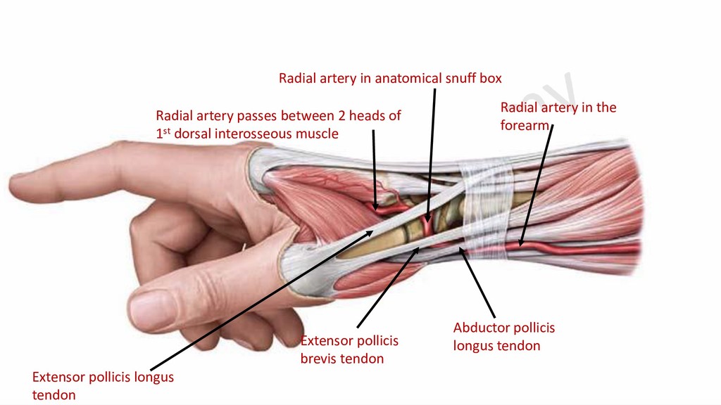
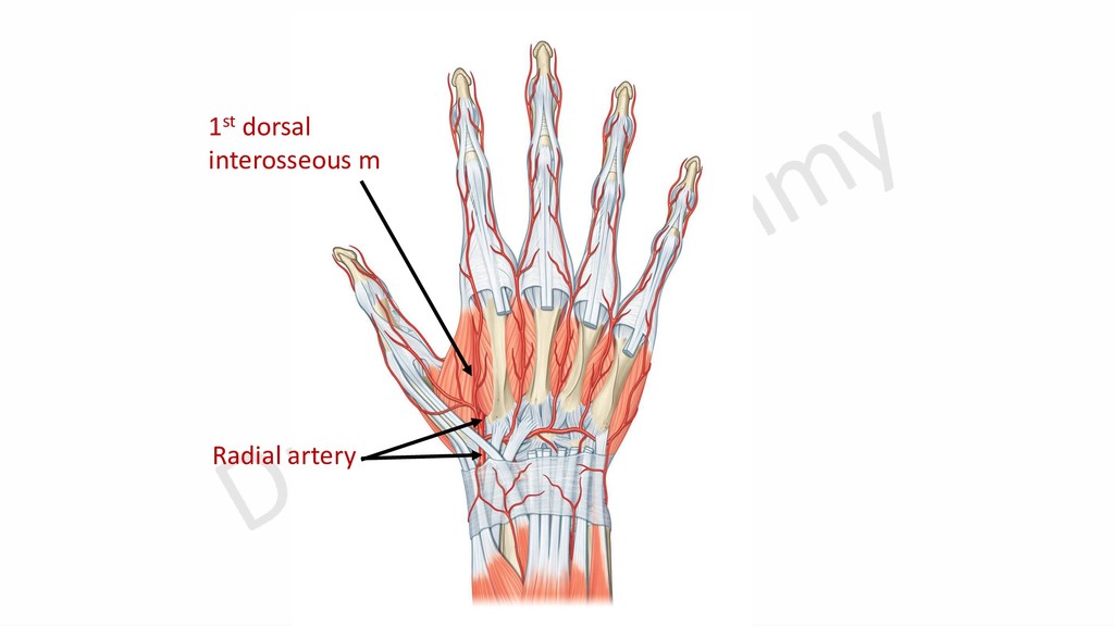
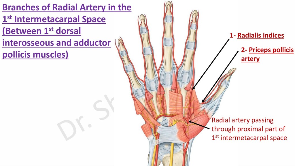
    Artery in the 1st Intermetacarpal Space (Between 1st dorsal interosseous and adductor pollicis muscles) Radial artery passing through proximal part of 1st intermetacarpal space
  29. Arterial Arches in the Hand Superficial to flexor digitorum tendons.

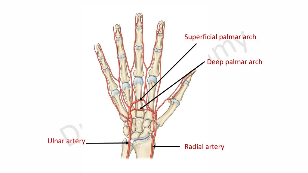
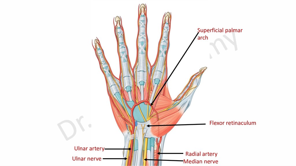
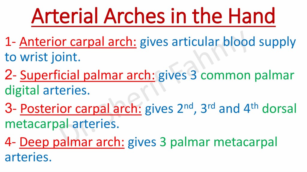
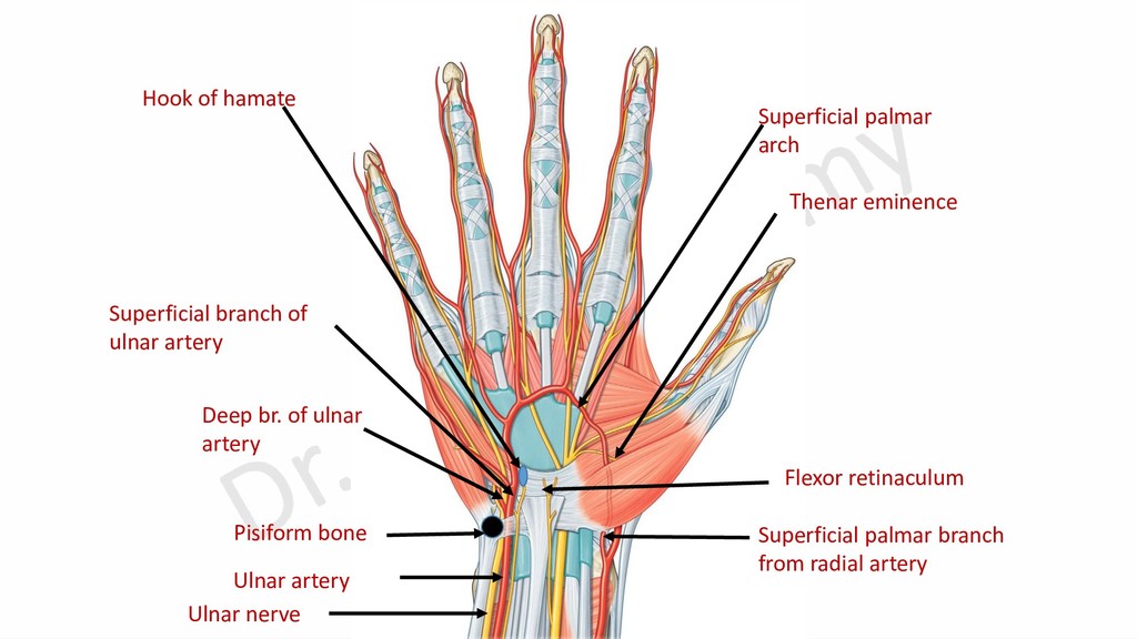
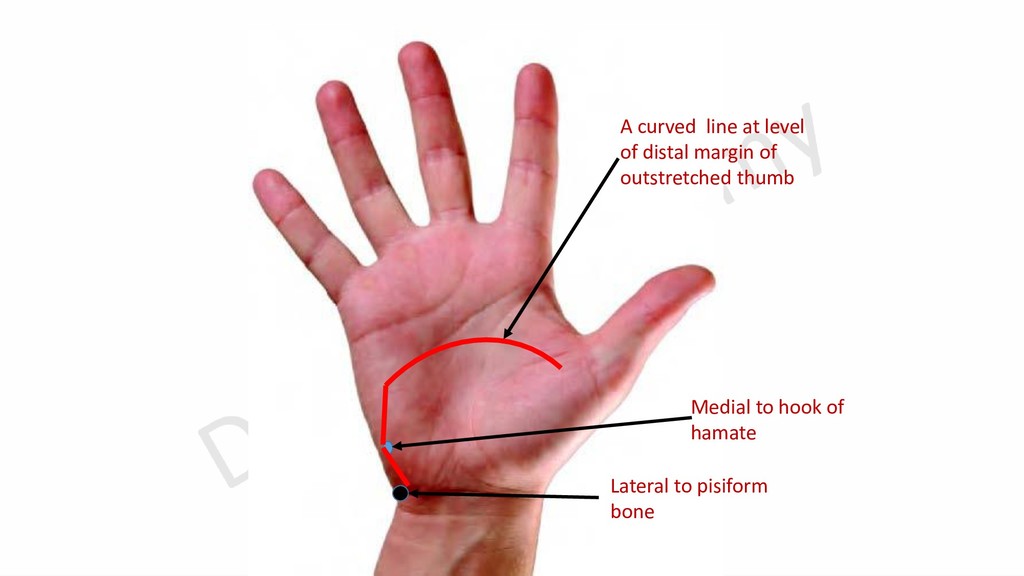
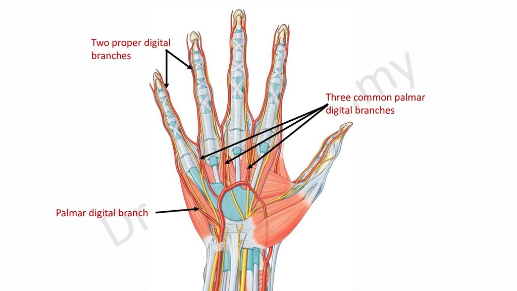
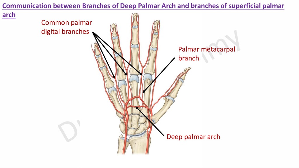
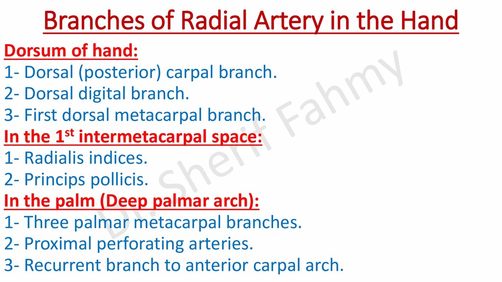
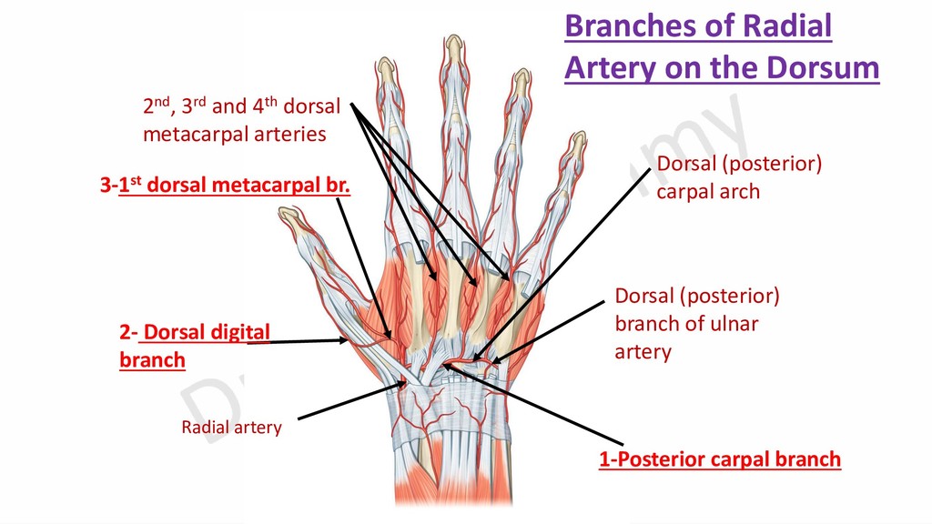
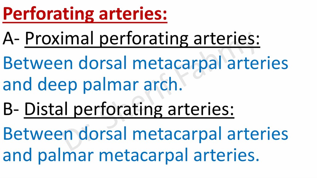
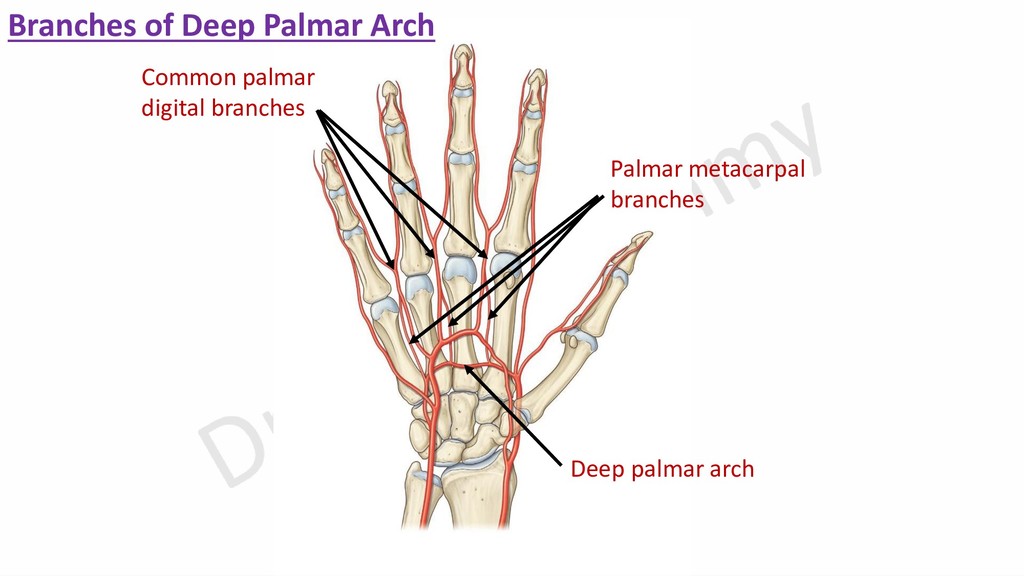
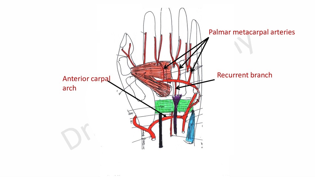
    At the level of middle of shaft of metacarpal bones. Deep to flexor digitorum tendons. At level just distal to bases of metacarpal bones. Anterior to carpal bones, deep to flexor tendons. Posterior to carpal bones, deep to extensor tendons. -Mainly superficial branch of ulnar artery. -Superficial palmar branch of radial a. -Mainly radial artery. -Deep branch of ulnar artery. -Anterior carpal branches of radial& ulnar arteries. -Descending branch of anterior interosseous artery. -Recurrent branch from deep palmar arch. It is a cruciate anastomosis. -Posterior carpal branches of radial& ulnar arteries. -Anterior & posterior interosseous artery. -3 common palmar digital arteries. -Palmar digital artery. -3 palmar metacarpal arteries. -Proximal perforating arteries. -Recurrent branch. -Articular branches to wrist joint. -2nd, 3rd and 4th dorsal metacarpal arteries. Site Formation Branches Superficial palmar Deep palmar Anterior carpal Posterior carpal Dr. Sherif Fahmy