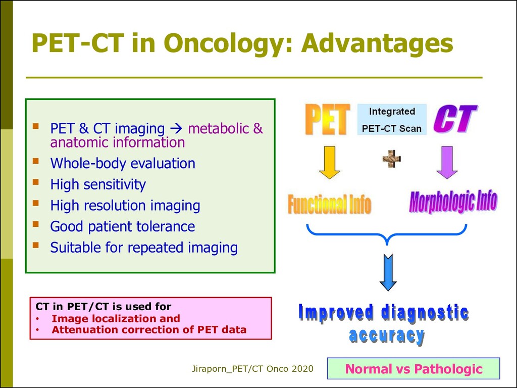
CT imaging metabolic & anatomic information Whole-body evaluation High sensitivity High resolution imaging Good patient tolerance Suitable for repeated imaging Normal vs Pathologic CT in PET/CT is used for • Image localization and • Attenuation correction of PET data
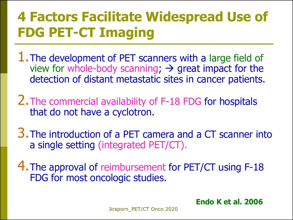
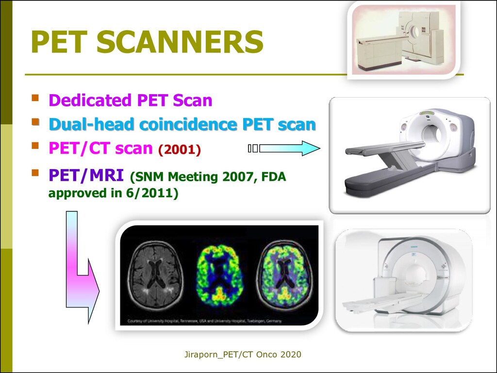
PET-CT Imaging 1.The development of PET scanners with a large field of view for whole-body scanning; great impact for the detection of distant metastatic sites in cancer patients. 2.The commercial availability of F-18 FDG for hospitals that do not have a cyclotron. 3.The introduction of a PET camera and a CT scanner into a single setting (integrated PET/CT). 4.The approval of reimbursement for PET/CT using F-18 FDG for most oncologic studies. Endo K et al. 2006
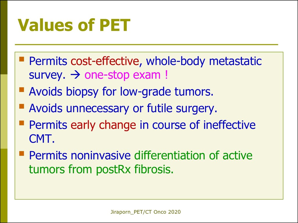
metastatic survey. one-stop exam ! Avoids biopsy for low-grade tumors. Avoids unnecessary or futile surgery. Permits early change in course of ineffective CMT. Permits noninvasive differentiation of active tumors from postRx fibrosis.
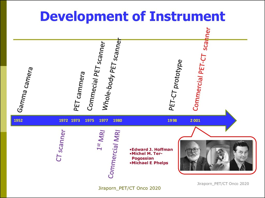
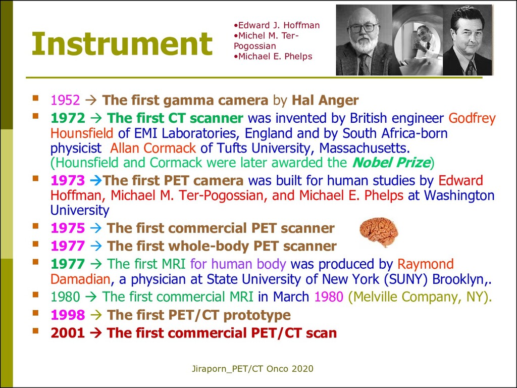
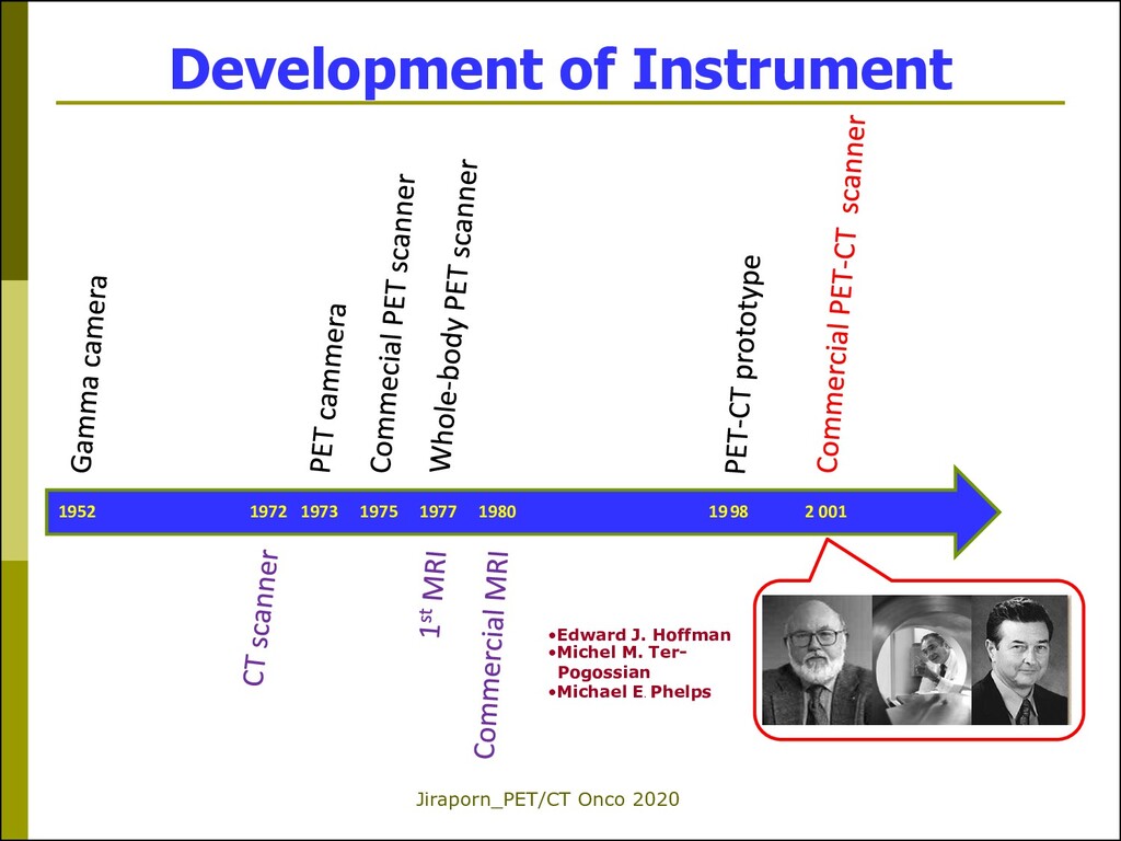
camera by Hal Anger 1972 The first CT scanner was invented by British engineer Godfrey Hounsfield of EMI Laboratories, England and by South Africa-born physicist Allan Cormack of Tufts University, Massachusetts. (Hounsfield and Cormack were later awarded the Nobel Prize) 1973 The first PET camera was built for human studies by Edward Hoffman, Michael M. Ter-Pogossian, and Michael E. Phelps at Washington University 1975 The first commercial PET scanner 1977 The first whole-body PET scanner 1977 The first MRI for human body was produced by Raymond Damadian, a physician at State University of New York (SUNY) Brooklyn,. 1980 The first commercial MRI in March 1980 (Melville Company, NY). 1998 The first PET/CT prototype 2001 The first commercial PET/CT scan •Edward J. Hoffman •Michel M. Ter- Pogossian •Michael E. Phelps
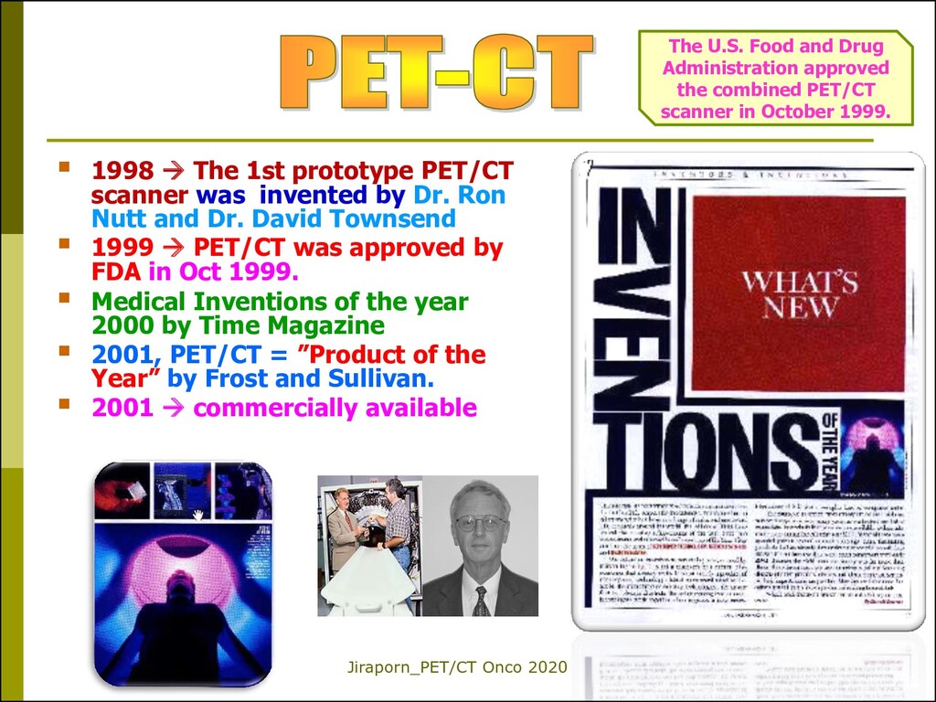
scanner was invented by Dr. Ron Nutt and Dr. David Townsend 1999 PET/CT was approved by FDA in Oct 1999. Medical Inventions of the year 2000 by Time Magazine 2001, PET/CT = ”Product of the Year” by Frost and Sullivan. 2001 commercially available The U.S. Food and Drug Administration approved the combined PET/CT scanner in October 1999.
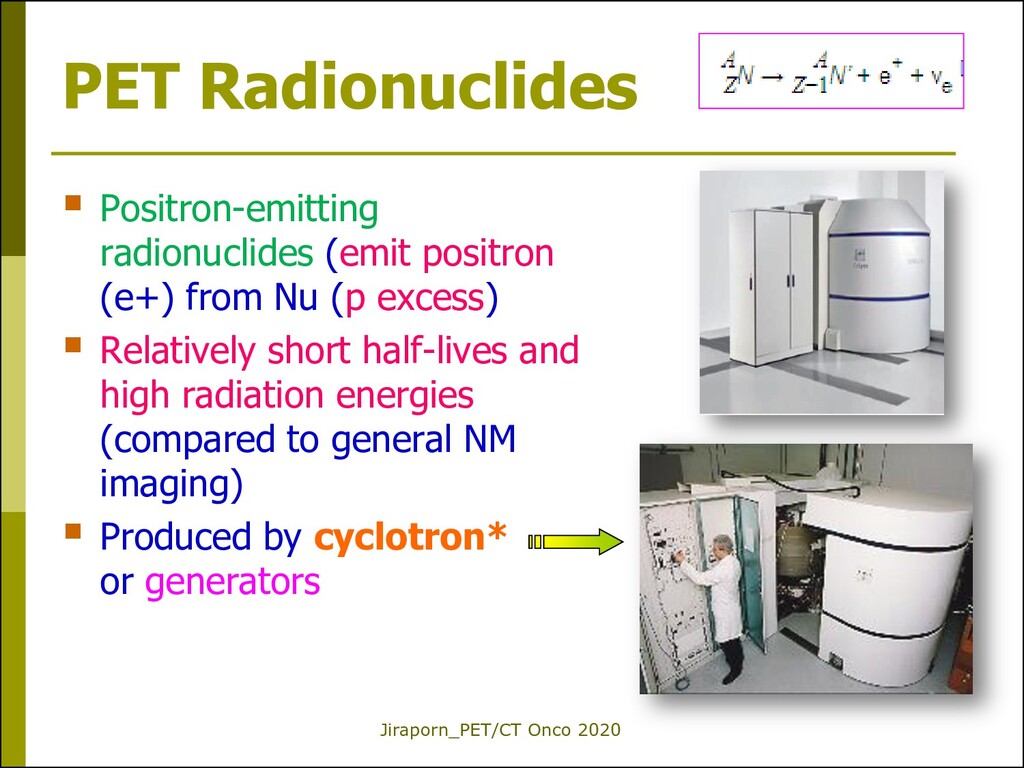
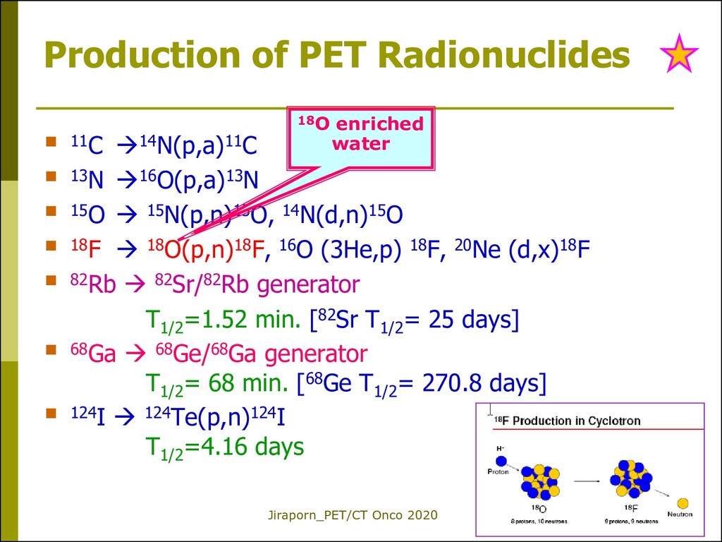
(e+) from Nu (p excess) Relatively short half-lives and high radiation energies (compared to general NM imaging) Produced by cyclotron* or generators
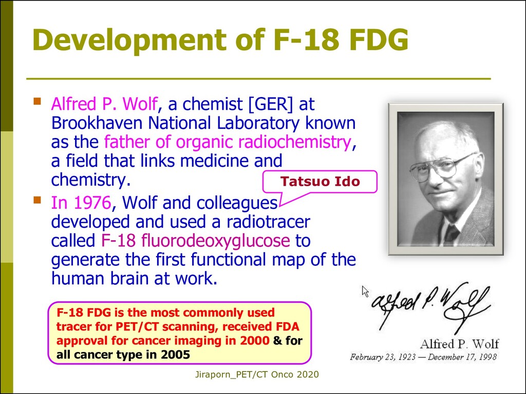
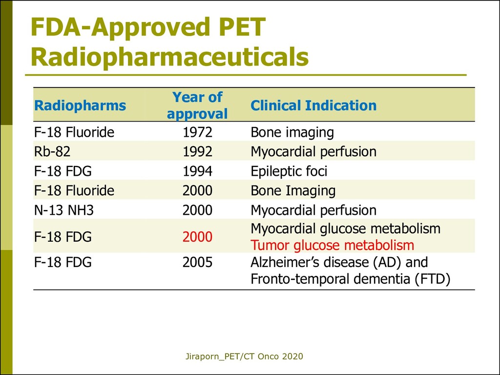
Wolf, a chemist [GER] at Brookhaven National Laboratory known as the father of organic radiochemistry, a field that links medicine and chemistry. In 1976, Wolf and colleagues developed and used a radiotracer called F-18 fluorodeoxyglucose to generate the first functional map of the human brain at work. Tatsuo Ido F-18 FDG is the most commonly used tracer for PET/CT scanning, received FDA approval for cancer imaging in 2000 & for all cancer type in 2005
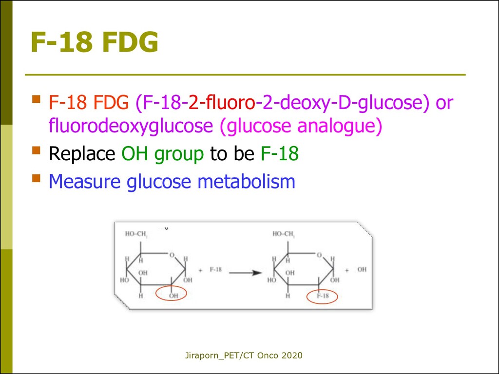
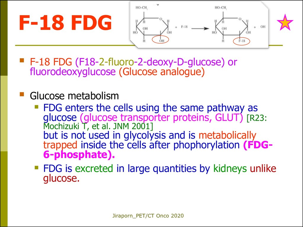
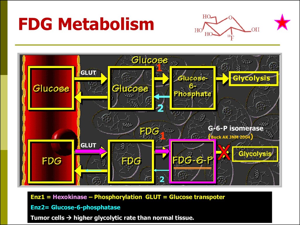
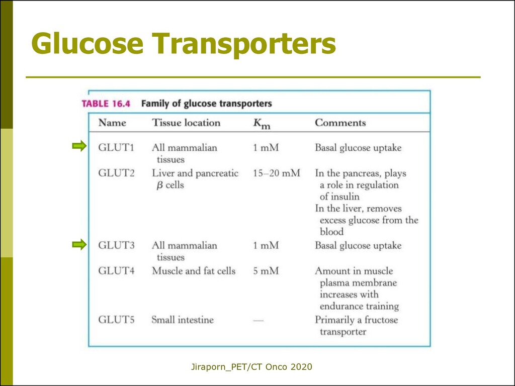
fluorodeoxyglucose (Glucose analogue) Glucose metabolism FDG enters the cells using the same pathway as glucose (glucose transporter proteins, GLUT) [R23: Mochizuki T, et al. JNM 2001] but is not used in glycolysis and is metabolically trapped inside the cells after phophorylation (FDG- 6-phosphate). FDG is excreted in large quantities by kidneys unlike glucose.
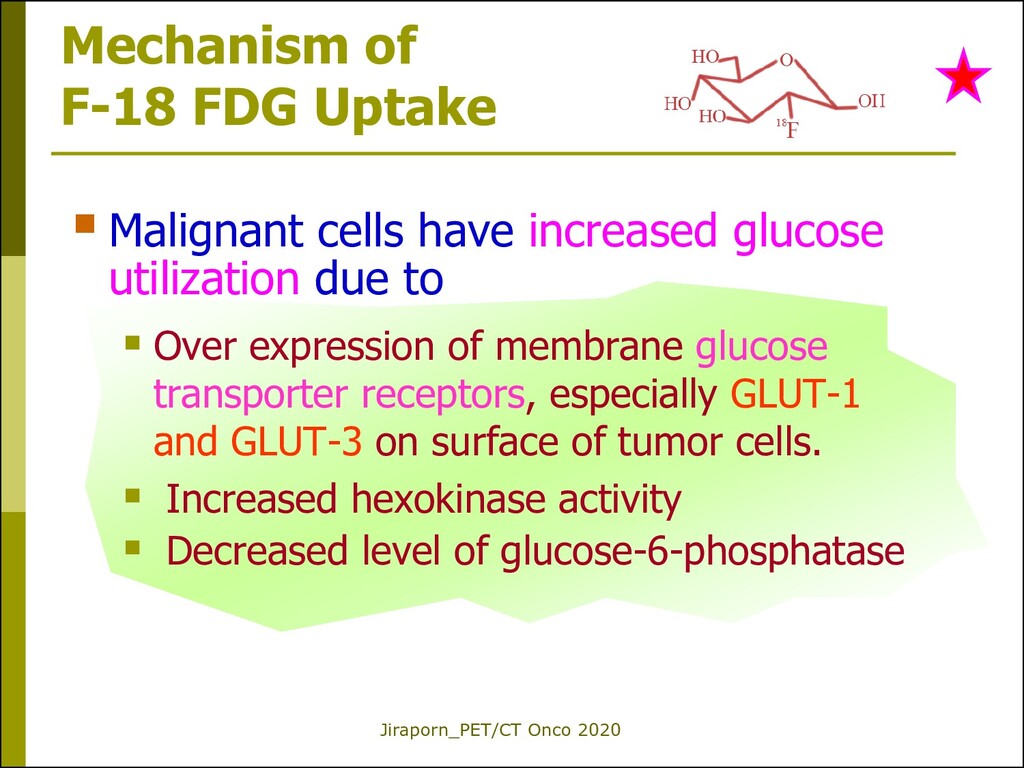
have increased glucose utilization due to Over expression of membrane glucose transporter receptors, especially GLUT-1 and GLUT-3 on surface of tumor cells. Increased hexokinase activity Decreased level of glucose-6-phosphatase
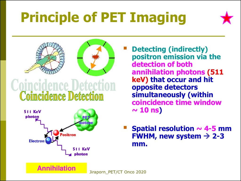
positron emission via the detection of both annihilation photons (511 keV) that occur and hit opposite detectors simultaneously (within coincidence time window ~ 10 ns) Spatial resolution ~ 4-5 mm FWHM, new system 2-3 mm. Annihilation
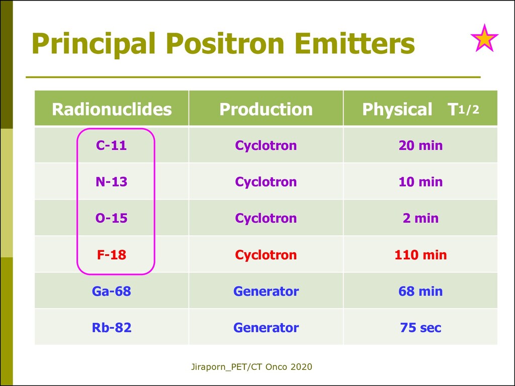
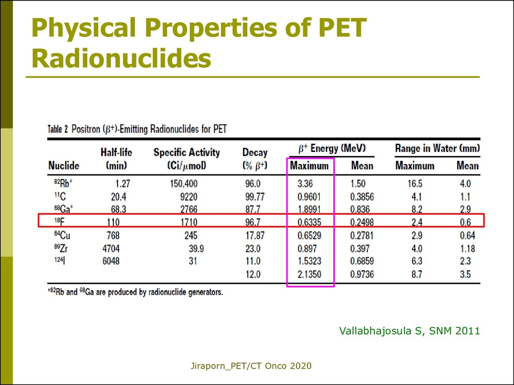
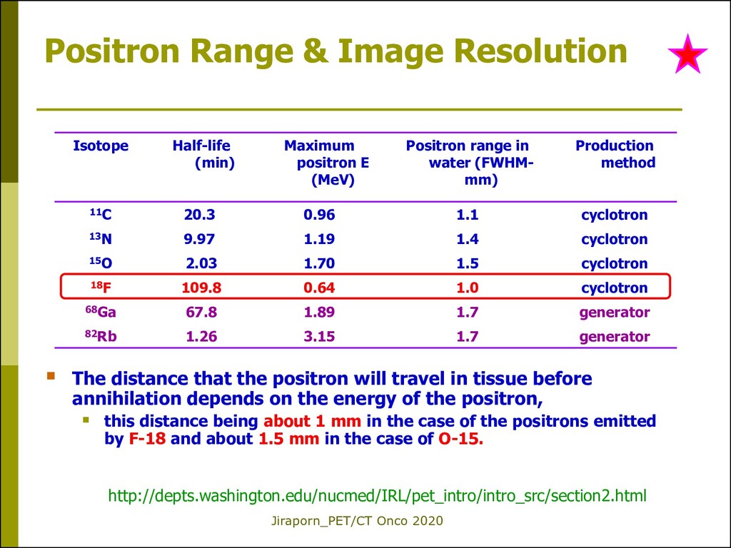
distance that the positron will travel in tissue before annihilation depends on the energy of the positron, this distance being about 1 mm in the case of the positrons emitted by F-18 and about 1.5 mm in the case of O-15. Isotope Half-life (min) Maximum positron E (MeV) Positron range in water (FWHM- mm) Production method 11C 20.3 0.96 1.1 cyclotron 13N 9.97 1.19 1.4 cyclotron 15O 2.03 1.70 1.5 cyclotron 18F 109.8 0.64 1.0 cyclotron 68Ga 67.8 1.89 1.7 generator 82Rb 1.26 3.15 1.7 generator http://depts.washington.edu/nucmed/IRL/pet_intro/intro_src/section2.html
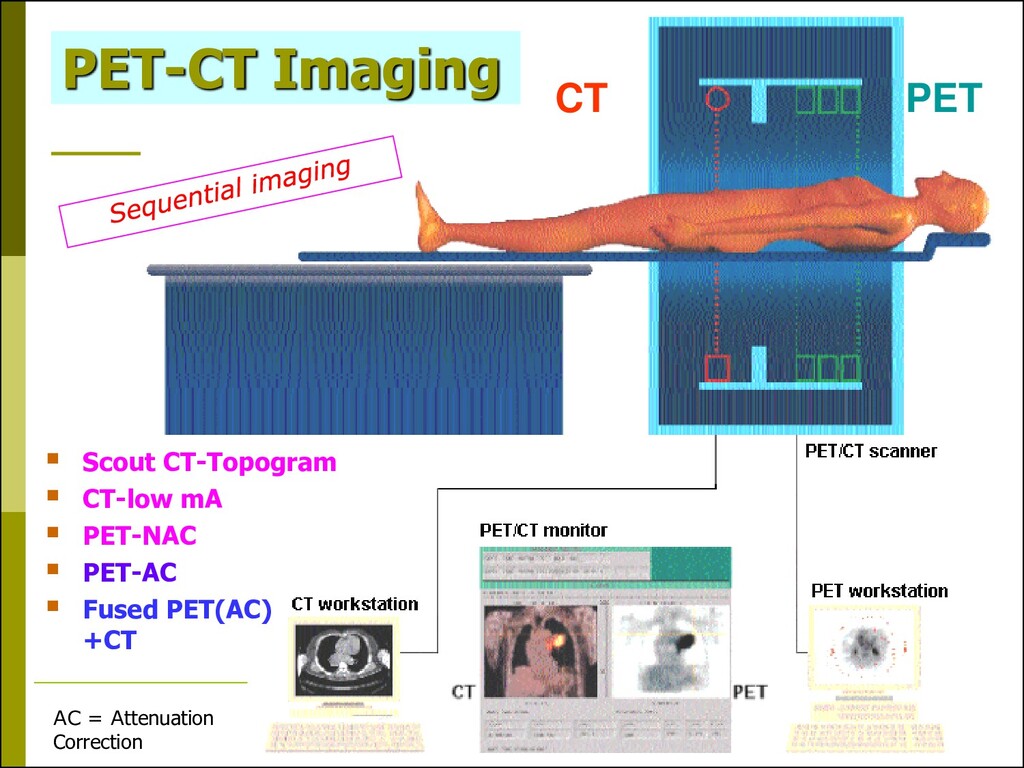
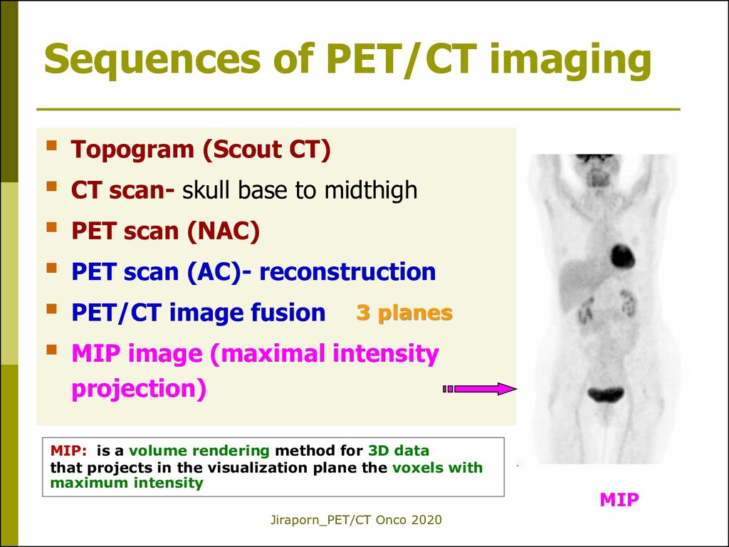
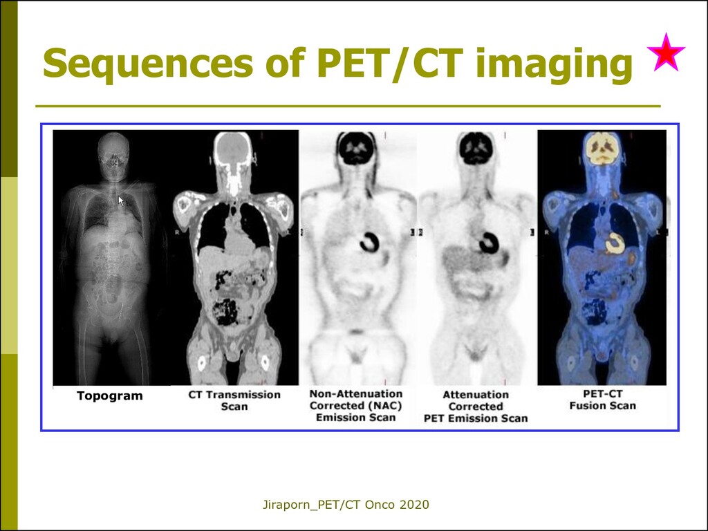
CT) CT scan- skull base to midthigh PET scan (NAC) PET scan (AC)- reconstruction PET/CT image fusion MIP image (maximal intensity projection) 3 planes MIP: is a volume rendering method for 3D data that projects in the visualization plane the voxels with maximum intensity MIP
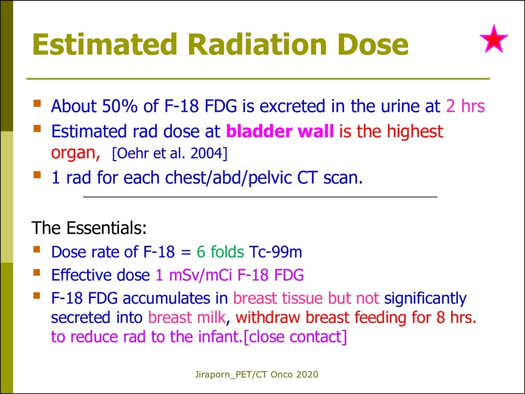
F-18 FDG is excreted in the urine at 2 hrs Estimated rad dose at bladder wall is the highest organ,. [Oehr et al. 2004] 1 rad for each chest/abd/pelvic CT scan. The Essentials: Dose rate of F-18 = 6 folds Tc-99m Effective dose 1 mSv/mCi F-18 FDG F-18 FDG accumulates in breast tissue but not significantly secreted into breast milk, withdraw breast feeding for 8 hrs. to reduce rad to the infant.[close contact]
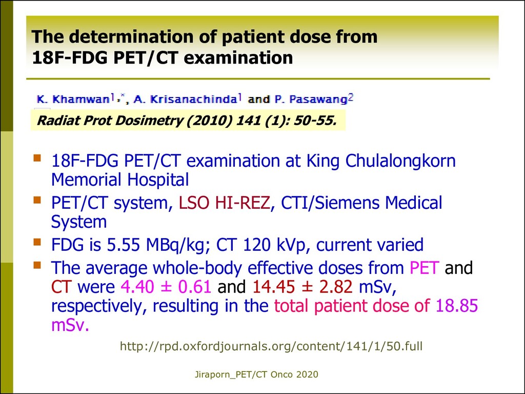
Memorial Hospital PET/CT system, LSO HI-REZ, CTI/Siemens Medical System FDG is 5.55 MBq/kg; CT 120 kVp, current varied The average whole-body effective doses from PET and CT were 4.40 ± 0.61 and 14.45 ± 2.82 mSv, respectively, resulting in the total patient dose of 18.85 mSv. http://rpd.oxfordjournals.org/content/141/1/50.full Radiat Prot Dosimetry (2010) 141 (1): 50-55. The determination of patient dose from 18F-FDG PET/CT examination
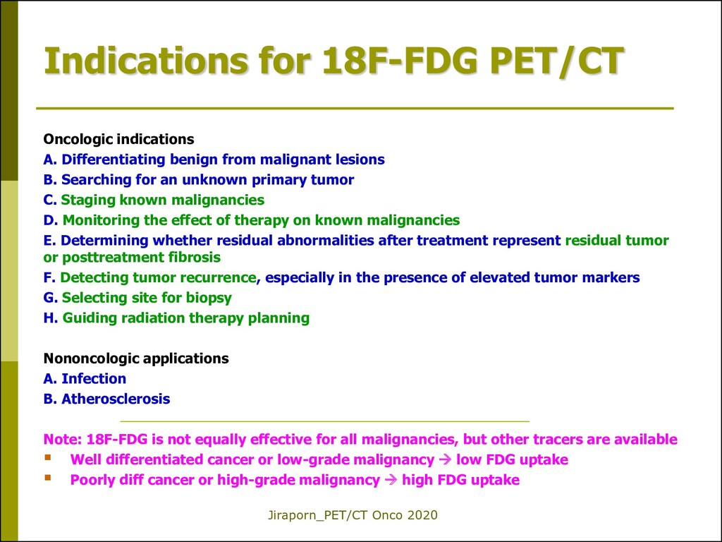
Differentiating benign from malignant lesions B. Searching for an unknown primary tumor C. Staging known malignancies D. Monitoring the effect of therapy on known malignancies E. Determining whether residual abnormalities after treatment represent residual tumor or posttreatment fibrosis F. Detecting tumor recurrence, especially in the presence of elevated tumor markers G. Selecting site for biopsy H. Guiding radiation therapy planning Nononcologic applications A. Infection B. Atherosclerosis Note: 18F-FDG is not equally effective for all malignancies, but other tracers are available Well differentiated cancer or low-grade malignancy low FDG uptake Poorly diff cancer or high-grade malignancy high FDG uptake
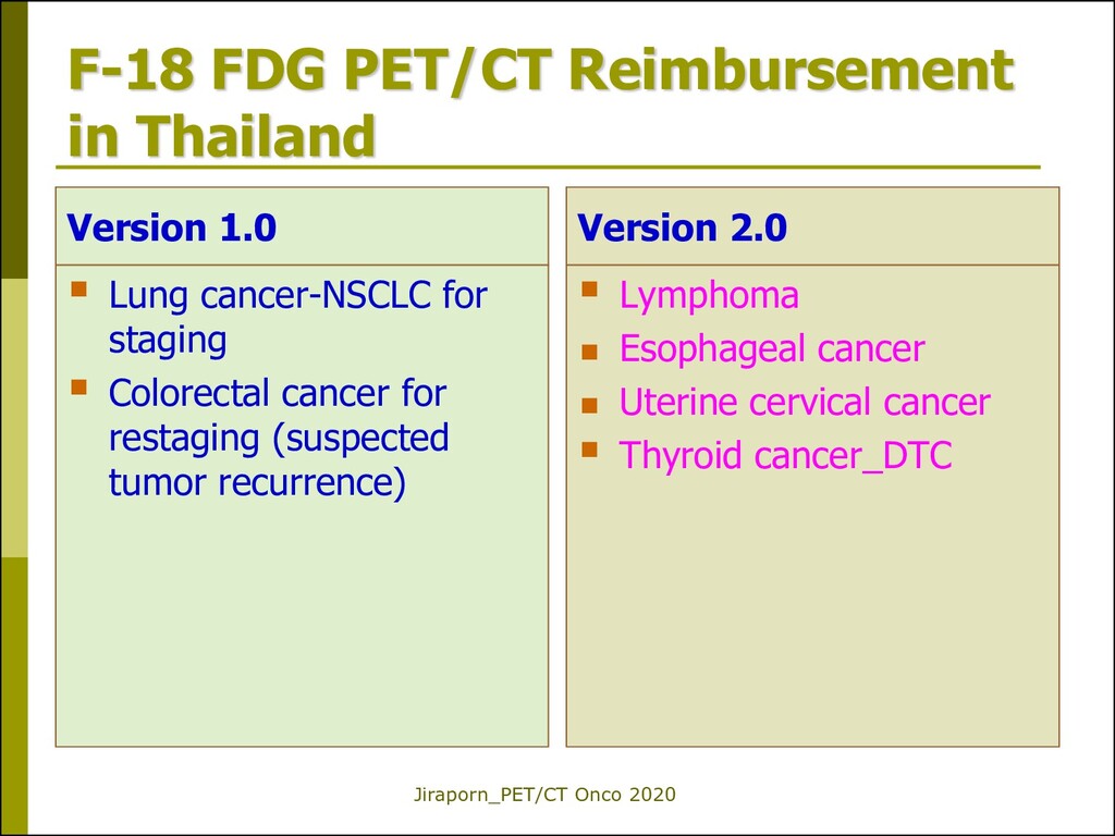
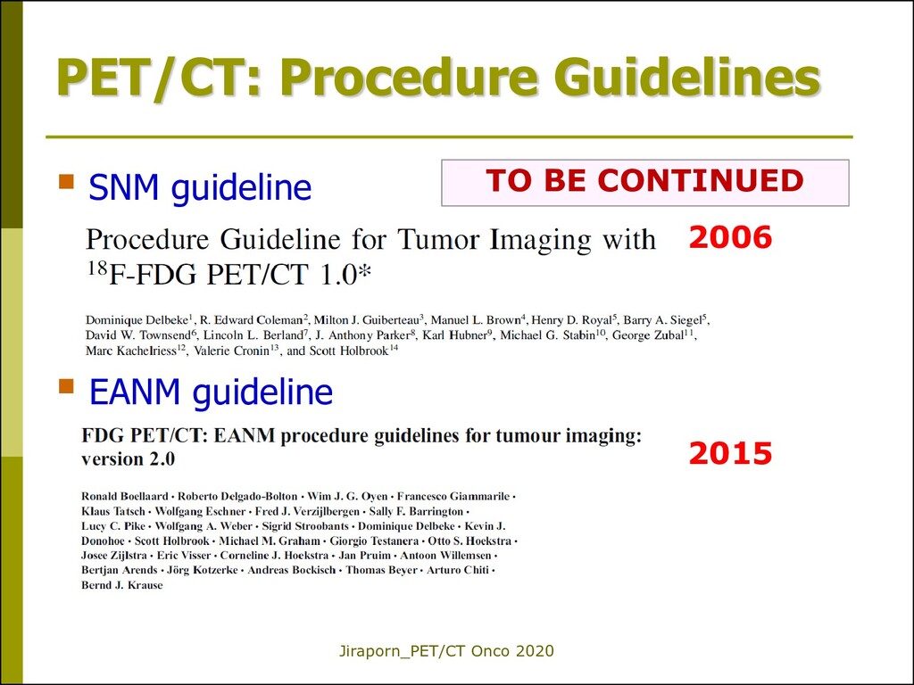
1.0 Lung cancer-NSCLC for staging Colorectal cancer for restaging (suspected tumor recurrence) Version 2.0 Lymphoma Esophageal cancer Uterine cervical cancer Thyroid cancer_DTC
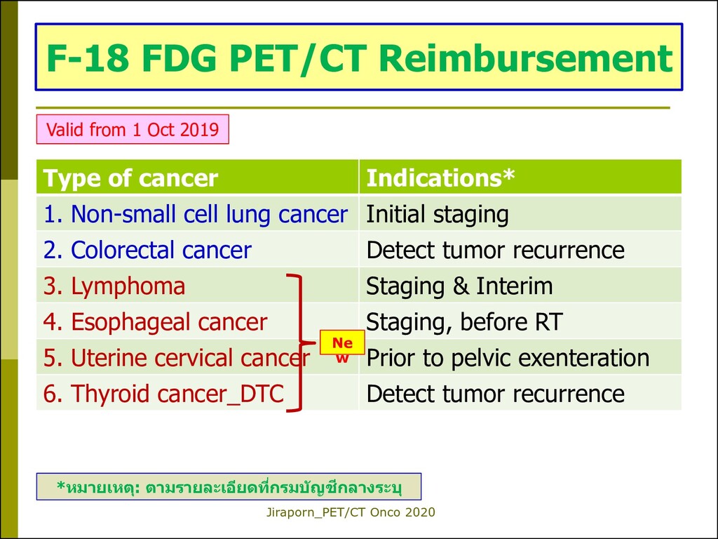
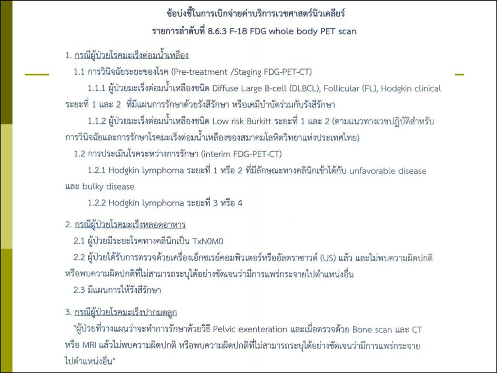
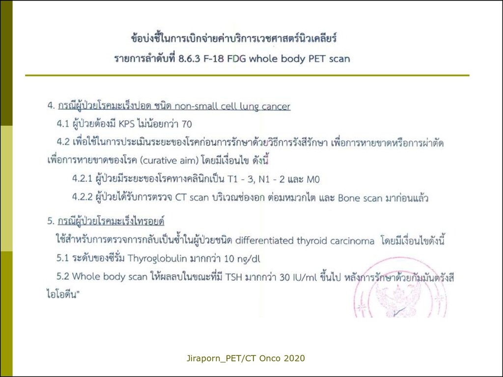
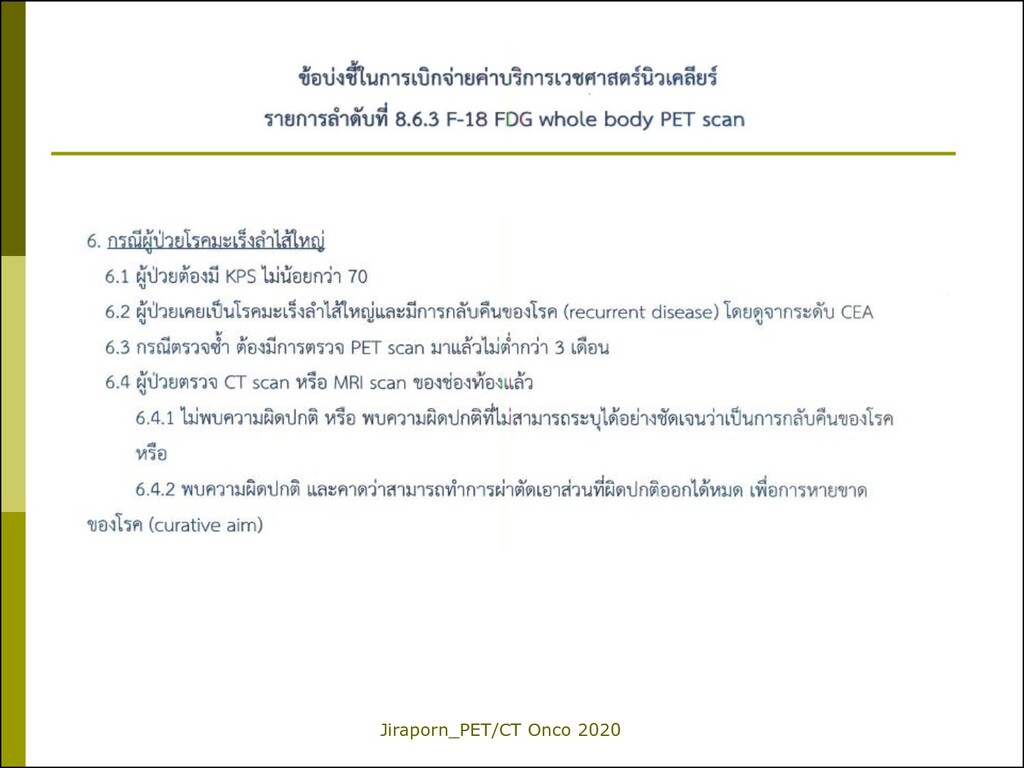
lung cancer Initial staging 2. Colorectal cancer Detect tumor recurrence 3. Lymphoma Staging & Interim 4. Esophageal cancer Staging, before RT 5. Uterine cervical cancer Prior to pelvic exenteration 6. Thyroid cancer_DTC Detect tumor recurrence Ne w F-18 FDG PET/CT Reimbursement Valid from 1 Oct 2019 *หมายเหตุ: ตามรายละเอียดที่กรมบัญชีกลางระบุ