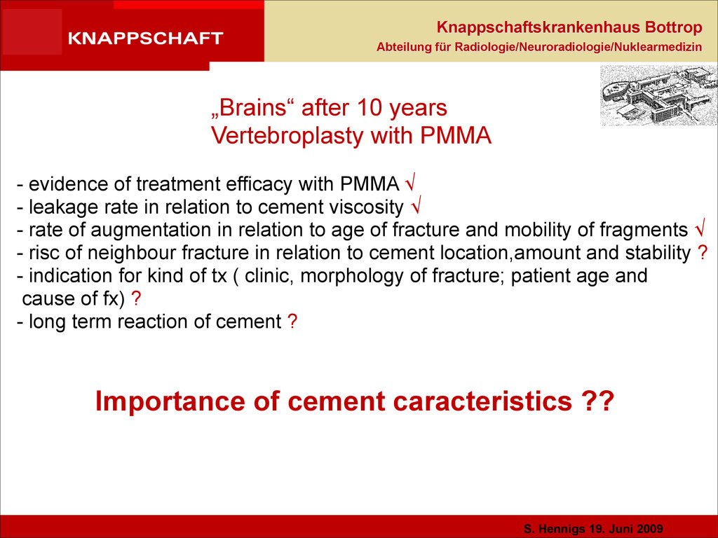
„Brains“ after 10 years Vertebroplasty with PMMA - evidence of treatment efficacy with PMMA √ - leakage rate in relation to cement viscosity √ - rate of augmentation in relation to age of fracture and mobility of fragments √ - risc of neighbour fracture in relation to cement location,amount and stability ? - indication for kind of tx ( clinic, morphology of fracture; patient age and cause of fx) ? - long term reaction of cement ? ! Importance of cement caracteristics ??
PMMA cement - Inert - high temperature - no osteoconduction or -induction - no bony integration - creation of necrosis and soft tissue interface - no long term results (critical for young patients)
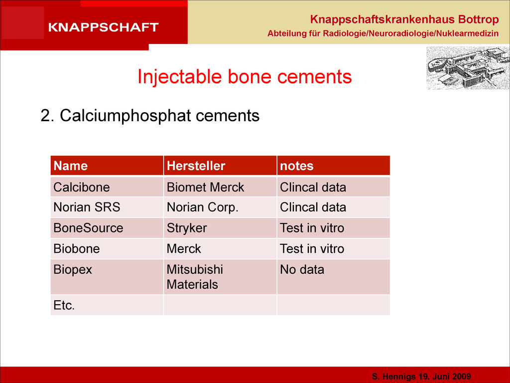
Injectable bone cements 2. Calciumphosphat cements Name Hersteller notes Calcibone Biomet Merck Clincal data Norian SRS Norian Corp. Clincal data BoneSource Stryker Test in vitro Biobone Merck Test in vitro Biopex Mitsubishi Materials No data Etc.
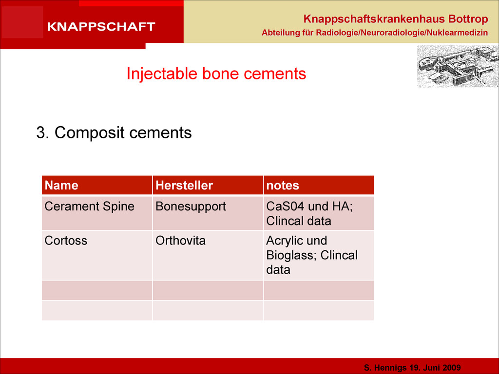
Injectable bone cements 3. Composit cements Name Hersteller notes Cerament Spine Bonesupport CaS04 und HA; Clincal data Cortoss Orthovita Acrylic und Bioglass; Clincal data
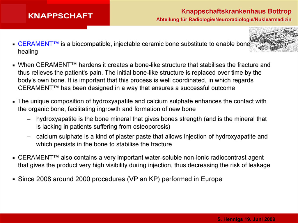
▪ CERAMENT™ is a biocompatible, injectable ceramic bone substitute to enable bone healing ▪ When CERAMENT™ hardens it creates a bone-like structure that stabilises the fracture and thus relieves the patient's pain. The initial bone-like structure is replaced over time by the body's own bone. It is important that this process is well coordinated, in which regards CERAMENT™ has been designed in a way that ensures a successful outcome ▪ The unique composition of hydroxyapatite and calcium sulphate enhances the contact with the organic bone, facilitating ingrowth and formation of new bone – hydroxyapatite is the bone mineral that gives bones strength (and is the mineral that is lacking in patients suffering from osteoporosis) – calcium sulphate is a kind of plaster paste that allows injection of hydroxyapatite and which persists in the bone to stabilise the fracture ▪ CERAMENT™ also contains a very important water-soluble non-ionic radiocontrast agent that gives the product very high visibility during injection, thus decreasing the risk of leakage ▪ Since 2008 around 2000 procedures (VP an KP) performed in Europe !
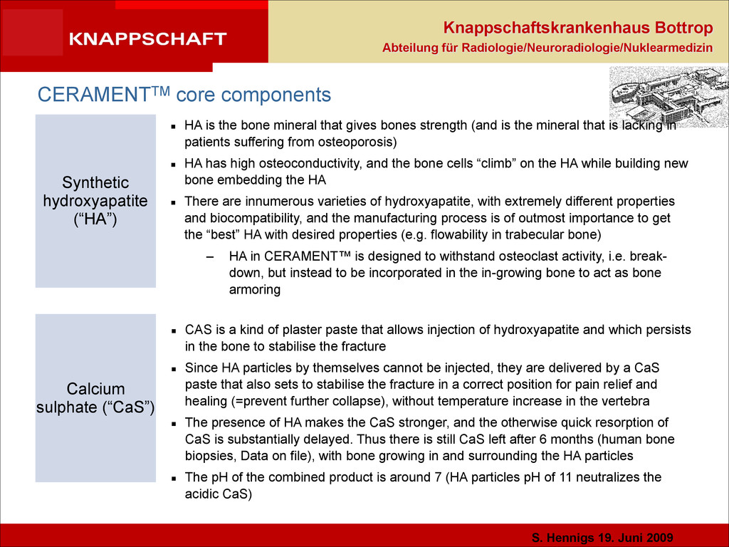
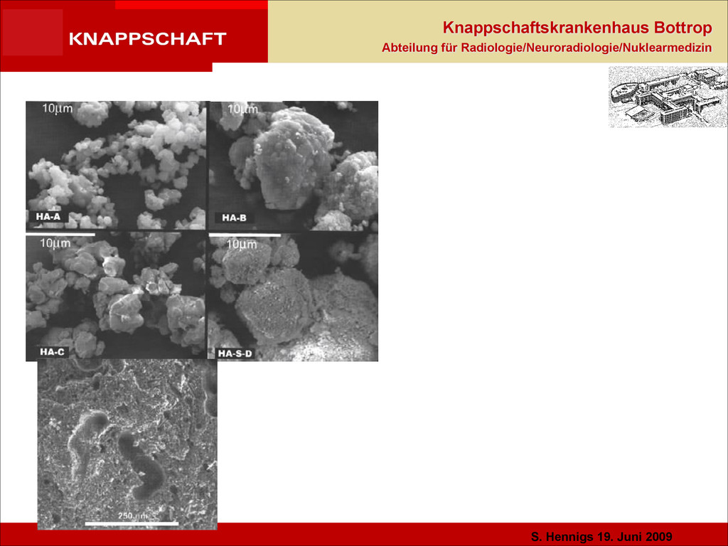
CERAMENTTM core components Synthetic hydroxyapatite (“HA”) Calcium sulphate (“CaS”) ▪ HA is the bone mineral that gives bones strength (and is the mineral that is lacking in patients suffering from osteoporosis) ▪ HA has high osteoconductivity, and the bone cells “climb” on the HA while building new bone embedding the HA ▪ There are innumerous varieties of hydroxyapatite, with extremely different properties and biocompatibility, and the manufacturing process is of outmost importance to get the “best” HA with desired properties (e.g. flowability in trabecular bone) – HA in CERAMENT™ is designed to withstand osteoclast activity, i.e. break- down, but instead to be incorporated in the in-growing bone to act as bone armoring ▪ CAS is a kind of plaster paste that allows injection of hydroxyapatite and which persists in the bone to stabilise the fracture ▪ Since HA particles by themselves cannot be injected, they are delivered by a CaS paste that also sets to stabilise the fracture in a correct position for pain relief and healing (=prevent further collapse), without temperature increase in the vertebra ▪ The presence of HA makes the CaS stronger, and the otherwise quick resorption of CaS is substantially delayed. Thus there is still CaS left after 6 months (human bone biopsies, Data on file), with bone growing in and surrounding the HA particles ▪ The pH of the combined product is around 7 (HA particles pH of 11 neutralizes the acidic CaS)
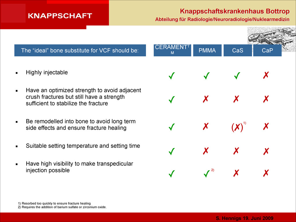
✓ ✗ ✗ ▪ Highly injectable ! ▪ Have an optimized strength to avoid adjacent crush fractures but still have a strength sufficient to stabilize the fracture ! ▪ Be remodelled into bone to avoid long term side effects and ensure fracture healing ! ▪ Suitable setting temperature and setting time ! ▪ Have high visibility to make transpedicular injection possible CERAMENTT M ✓ ✓ ✓ The “ideal” bone substitute for VCF should be: PMMA CaS CaP ✓ ✓ ✗ ✗ ✗ ✗ ✗ ✓ ✗ ✗ ✗ (✗) 1) Resorbed too quickly to ensure fracture healing 2) Requires the addition of barium sulfate or zirconium oxide. 2) 1) ✗ ✓
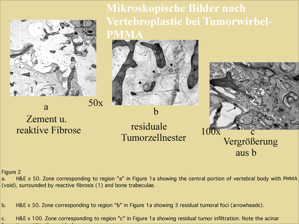
"a" in Figure 1a showing the central portion of vertebral body with PMMA (void), surrounded by reactive fibrosis (1) and bone trabeculae.! ! ! b.! H&E x 50. Zone corresponding to region "b" in Figure 1a showing 3 residual tumoral foci (arrowheads). ! ! c.! H&E x 100. Zone corresponding to region "c" in Figure 1a showing residual tumor infiltration. Note the acinar a b c Mikroskopische Bilder nach Vertebroplastie bei Tumorwirbel- PMMA Zement u. reaktive Fibrose 50x 100x residuale Tumorzellnester Vergrößerung aus b
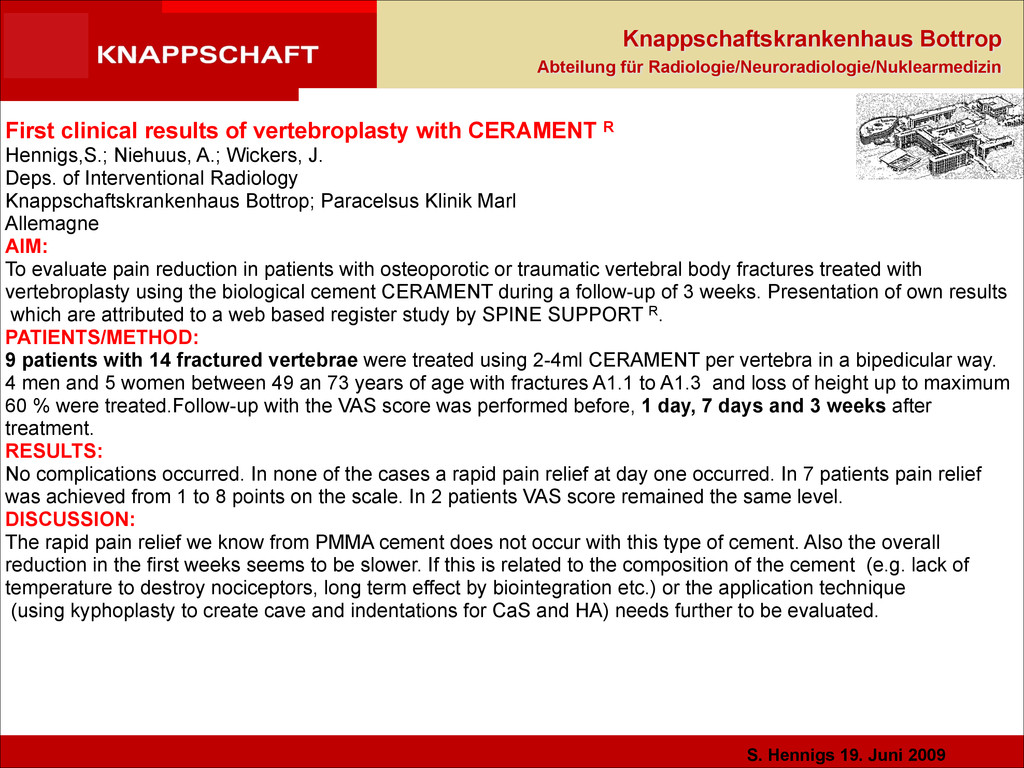
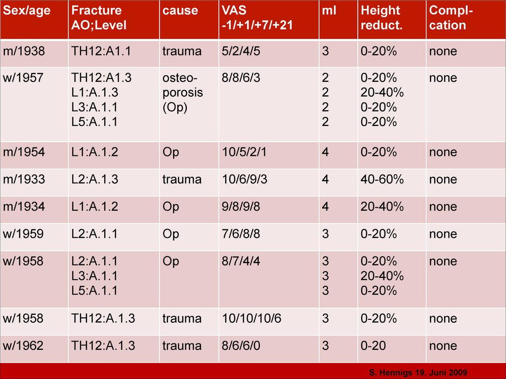
! ! ! ! ! ! First clinical results of vertebroplasty with CERAMENT R Hennigs,S.; Niehuus, A.; Wickers, J. Deps. of Interventional Radiology Knappschaftskrankenhaus Bottrop; Paracelsus Klinik Marl Allemagne AIM: To evaluate pain reduction in patients with osteoporotic or traumatic vertebral body fractures treated with vertebroplasty using the biological cement CERAMENT during a follow-up of 3 weeks. Presentation of own results which are attributed to a web based register study by SPINE SUPPORT R. PATIENTS/METHOD: 9 patients with 14 fractured vertebrae were treated using 2-4ml CERAMENT per vertebra in a bipedicular way. 4 men and 5 women between 49 an 73 years of age with fractures A1.1 to A1.3 and loss of height up to maximum 60 % were treated.Follow-up with the VAS score was performed before, 1 day, 7 days and 3 weeks after treatment. RESULTS: No complications occurred. In none of the cases a rapid pain relief at day one occurred. In 7 patients pain relief was achieved from 1 to 8 points on the scale. In 2 patients VAS score remained the same level. DISCUSSION: The rapid pain relief we know from PMMA cement does not occur with this type of cement. Also the overall reduction in the first weeks seems to be slower. If this is related to the composition of the cement (e.g. lack of temperature to destroy nociceptors, long term effect by biointegration etc.) or the application technique (using kyphoplasty to create cave and indentations for CaS and HA) needs further to be evaluated.
CERAMENT™|SPINE SUPPORT e-Registry by end of August 2010 http://www.ceramentspine.com 106 patients; 189 levels treated; follow-up day -1/1/7 analysed; long term follow-up to be done
CERAMENT, AN OSTEOCONDUCTIVE MATERIAL IN KYPHOPLASTIC PROCEDURES: RESULTS AT 1 YEAR Dr. Mario Dragani*; TSRM: Antonietta Occhiocupo*, Sandro Fantini* Dr. Stefano Marcia** *Dept. of Radiology, Hospital “Spirito Santo”, Pescara, Italy; **Dept. of Radiology O.C. San Giovanni di Dio , University of Cagliari, Italy ! ! ! ! Presented as abstract at GRIBOI, Boston, April, 2011
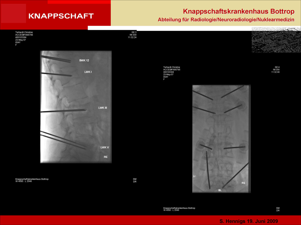
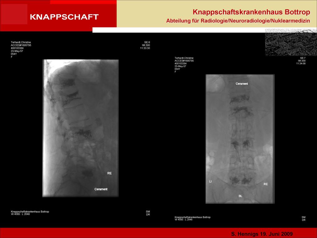
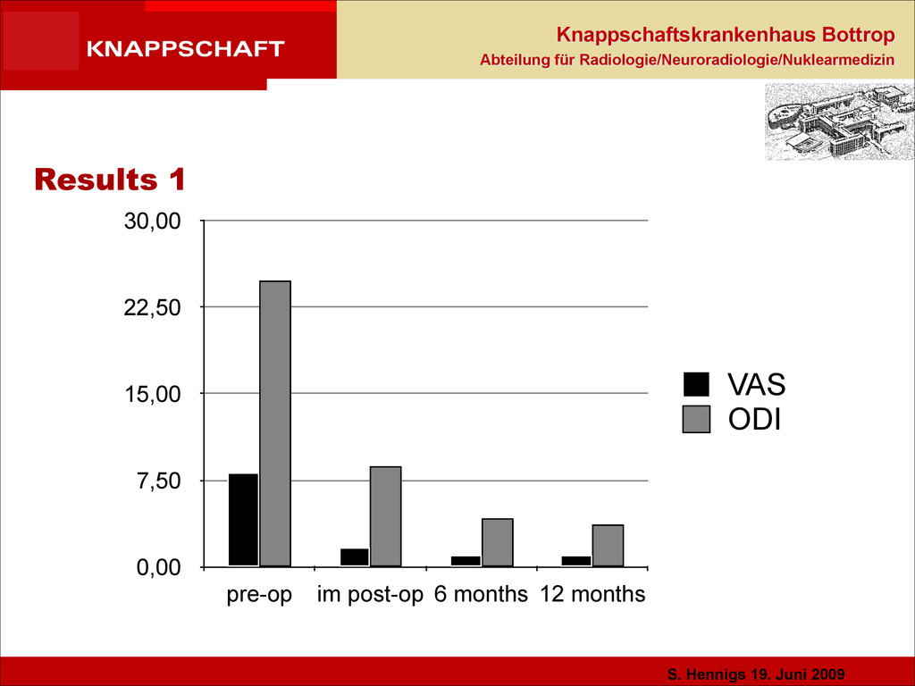
Materials and Methods •80 patients, osteoporotic or traumatic fractures, in sub acute phase with pain and edema. •64 female, 16 male, mean age 54 y. ! •Kyphoplasty - 53 dorsal and 127 lumbar vertebral bodies were treated. •Average injected quantity of Cerament was 2,5 cc/vb ! •Pre and post-operative exams: VAS and ODI score, RX plain film, CT. •Examinations: Pre-op, immediately after the procedure, the day after and at 6 and 12 months.
Osteoporotic vertebral compression fractures augmentation by injectable partly resorbable ceramic bone substitute (Cerament™|SPINE SUPPORT): a prospective nonrandomized study ! Salvatore Masala & Giovanni Nano & Stefano Marcia & Mario Muto & Francesco Paolo Maria Fucci & Giovanni Simonetti Interventional neuroradiology ! Abstract ! Introduction The aim of this study is to evaluate the longterm stabilizing–healing effectiveness and influence on adjacent intact vertebral bodies of a new injectable partly resorbable calcium sulfate (60 wt.%)/hydroxyapatite (40 wt.%) bone substitute employed in vertebral augmentation of osteoporotic collapses. Methods From April 2009 to April 2011, 80 patients underwent vertebral augmentation. Patients enrolling criteria were age >20 years and symptomatic osteoporotic vertebral collapse from low-energy trauma encompassed between levels T5 to L5. Preoperative and postoperative imaging studies consisted of computed tomography, plain X-ray, dual X-ray absorptiometry scanning, and magnetic resonance. Pain intensity has been evaluated by an 11-point visual analog scale (VAS) and physical and quality of life compromise assessments have been evaluated by Oswestry Disability Questionnaire (ODI). All procedures have been performed fluoroscopically guided by left unilateral approach under local anesthesia and mild sedation. Results VAS-based pain trend over the 12-month follow-up has shown a statistically significant (p<0.001) decrease, starting from 7.68 (SD 1.83) preoperatively with an immediate first day decrease at 3.51 (SD 2.16) and 0.96 (SD 0.93) at 12 months. ODI score dropped significantly from 54.78% to 20.12% at 6 months. No device-related complication has been reported. In no case a new incidental adjacent fracture has been reported. Conclusion :Data show how this injectable partly resorbable ceramic cement could be a nontoxic and lower stiffness alternative to polymethylmethacrylate for immediate and long-term stabilization of osteoporotic collapsed vertebral bodies.
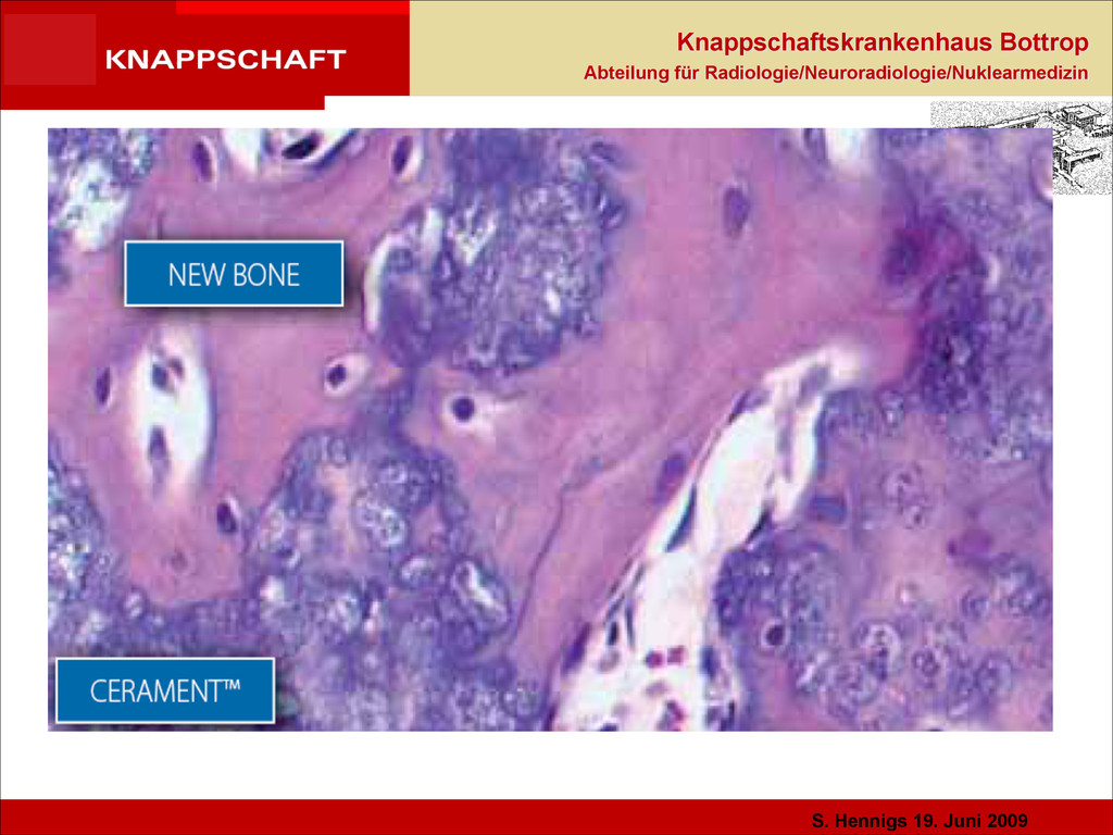
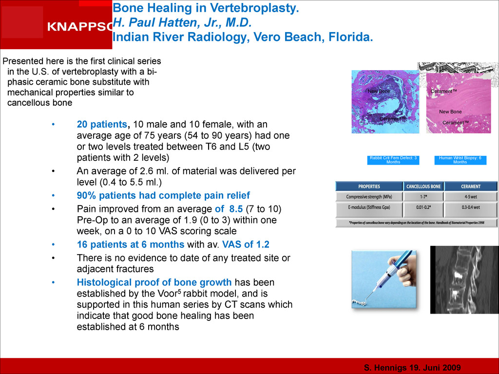
Bone Healing in Vertebroplasty. H. Paul Hatten, Jr., M.D. Indian River Radiology, Vero Beach, Florida. Human Wrist Biopsy: 6 Months Cerament™ Cerament™ New Bone Rabbit Crit Fem Defect: 3 Months Cerament™ New Bone Presented here is the first clinical series in the U.S. of vertebroplasty with a bi- phasic ceramic bone substitute with mechanical properties similar to cancellous bone • 20 patients, 10 male and 10 female, with an average age of 75 years (54 to 90 years) had one or two levels treated between T6 and L5 (two patients with 2 levels) • An average of 2.6 ml. of material was delivered per level (0.4 to 5.5 ml.) • 90% patients had complete pain relief • Pain improved from an average of 8.5 (7 to 10) Pre-Op to an average of 1.9 (0 to 3) within one week, on a 0 to 10 VAS scoring scale • 16 patients at 6 months with av. VAS of 1.2 • There is no evidence to date of any treated site or adjacent fractures • Histological proof of bone growth has been established by the Voor5 rabbit model, and is supported in this human series by CT scans which indicate that good bone healing has been established at 6 months
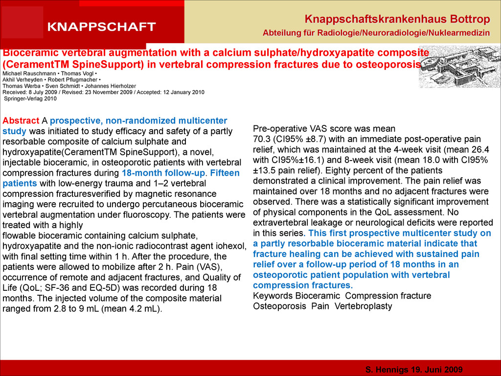
Bioceramic vertebral augmentation with a calcium sulphate/hydroxyapatite composite (CeramentTM SpineSupport) in vertebral compression fractures due to osteoporosis Michael Rauschmann • Thomas Vogl • Akhil Verheyden • Robert Pflugmacher • Thomas Werba • Sven Schmidt • Johannes Hierholzer Received: 8 July 2009 / Revised: 23 November 2009 / Accepted: 12 January 2010 Springer-Verlag 2010 Abstract A prospective, non-randomized multicenter study was initiated to study efficacy and safety of a partly resorbable composite of calcium sulphate and hydroxyapatite(CeramentTM SpineSupport), a novel, injectable bioceramic, in osteoporotic patients with vertebral compression fractures during 18-month follow-up. Fifteen patients with low-energy trauma and 1–2 vertebral compression fracturesverified by magnetic resonance imaging were recruited to undergo percutaneous bioceramic vertebral augmentation under fluoroscopy. The patients were treated with a highly flowable bioceramic containing calcium sulphate, hydroxyapatite and the non-ionic radiocontrast agent iohexol, with final setting time within 1 h. After the procedure, the patients were allowed to mobilize after 2 h. Pain (VAS), occurrence of remote and adjacent fractures, and Quality of Life (QoL; SF-36 and EQ-5D) was recorded during 18 months. The injected volume of the composite material ranged from 2.8 to 9 mL (mean 4.2 mL). Pre-operative VAS score was mean 70.3 (CI95% ±8.7) with an immediate post-operative pain relief, which was maintained at the 4-week visit (mean 26.4 with CI95%±16.1) and 8-week visit (mean 18.0 with CI95% ±13.5 pain relief). Eighty percent of the patients demonstrated a clinical improvement. The pain relief was maintained over 18 months and no adjacent fractures were observed. There was a statistically significant improvement of physical components in the QoL assessment. No extravertebral leakage or neurological deficits were reported in this series. This first prospective multicenter study on a partly resorbable bioceramic material indicate that fracture healing can be achieved with sustained pain relief over a follow-up period of 18 months in an osteoporotic patient population with vertebral compression fractures. Keywords Bioceramic Compression fracture Osteoporosis Pain Vertebroplasty
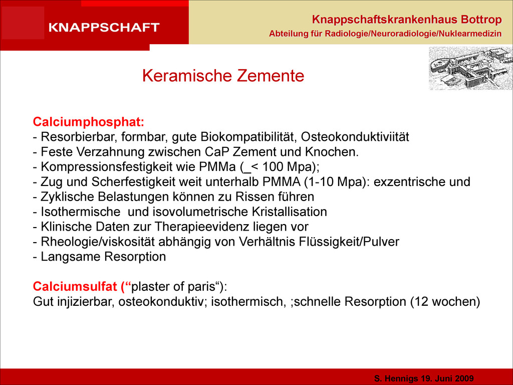
Keramische Zemente Calciumphosphat: - Resorbierbar, formbar, gute Biokompatibilität, Osteokonduktiviität - Feste Verzahnung zwischen CaP Zement und Knochen. - Kompressionsfestigkeit wie PMMa (_< 100 Mpa); - Zug und Scherfestigkeit weit unterhalb PMMA (1-10 Mpa): exzentrische und - Zyklische Belastungen können zu Rissen führen - Isothermische und isovolumetrische Kristallisation - Klinische Daten zur Therapieevidenz liegen vor - Rheologie/viskosität abhängig von Verhältnis Flüssigkeit/Pulver - Langsame Resorption ! Calciumsulfat (“plaster of paris“): Gut injizierbar, osteokonduktiv; isothermisch, ;schnelle Resorption (12 wochen)
Keramische Zemente Kompositzement: Cerament - Calciumsulfat 60% - Hydroxylapatit 40% - Jod als Kontrastmedium - Der Zusatz von HA als schwer resorbierbares Knochenersatzmaterial soll nach Resorption von CS dem osteoporotischen WK weiterhin Stabilität verleihen. - Klinische Daten vorhanden bei stabilen A1.1 bis A3 Frakturen mit klinisch signifikanter Besserung; Frage Stabilität des Zementes
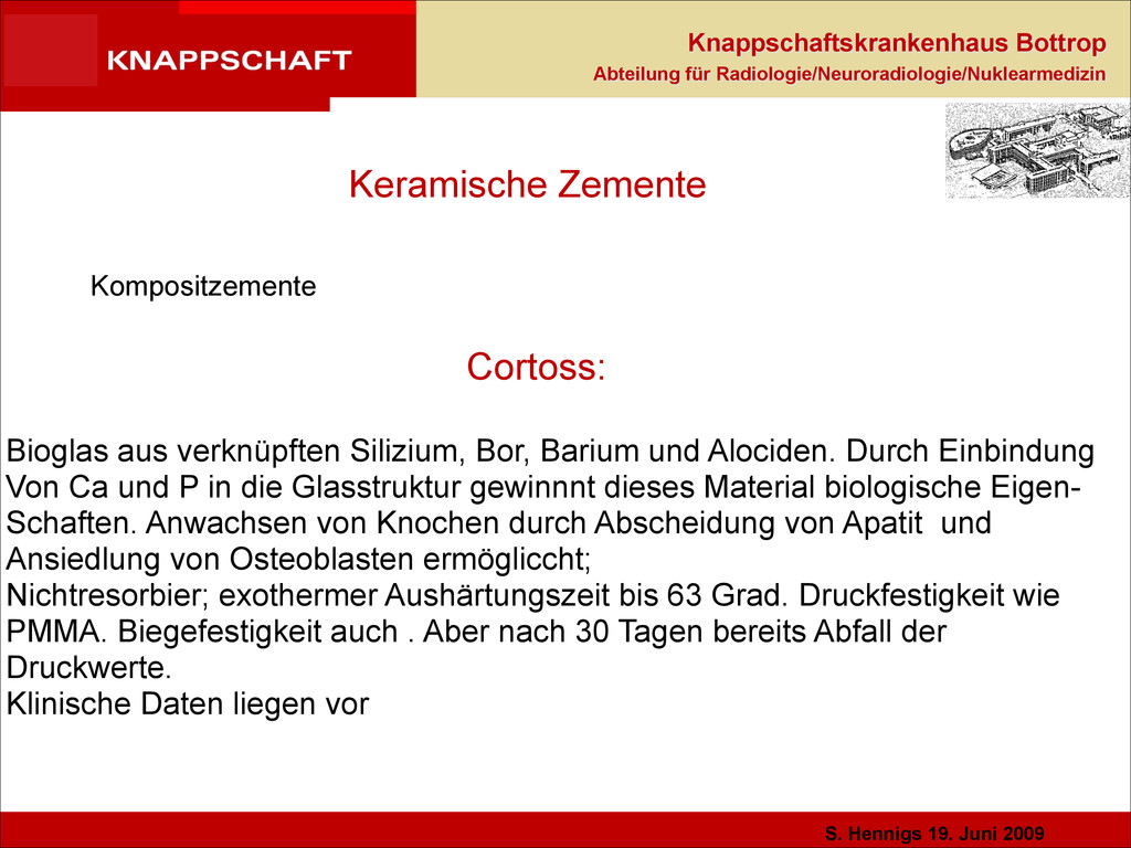
Keramische Zemente Kompositzemente Cortoss: ! Bioglas aus verknüpften Silizium, Bor, Barium und Alociden. Durch Einbindung Von Ca und P in die Glasstruktur gewinnnt dieses Material biologische Eigen- Schaften. Anwachsen von Knochen durch Abscheidung von Apatit und Ansiedlung von Osteoblasten ermögliccht; Nichtresorbier; exothermer Aushärtungszeit bis 63 Grad. Druckfestigkeit wie PMMA. Biegefestigkeit auch . Aber nach 30 Tagen bereits Abfall der Druckwerte. Klinische Daten liegen vor
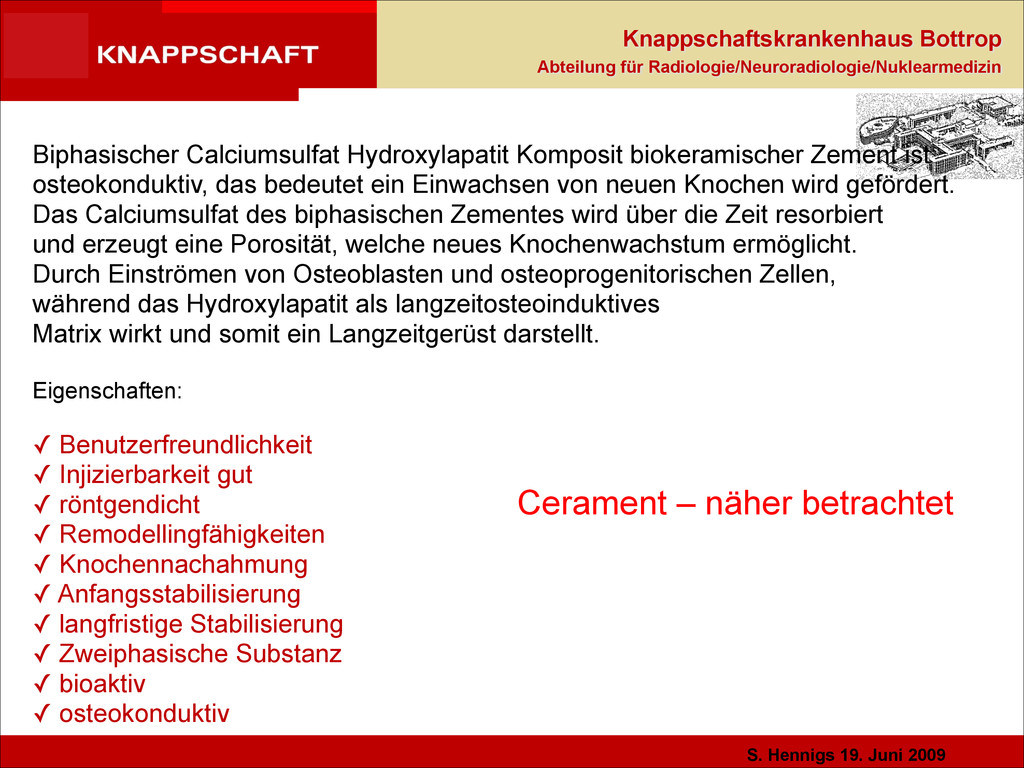
Cerament – näher betrachtet Biphasischer Calciumsulfat Hydroxylapatit Komposit biokeramischer Zement ist osteokonduktiv, das bedeutet ein Einwachsen von neuen Knochen wird gefördert. Das Calciumsulfat des biphasischen Zementes wird über die Zeit resorbiert und erzeugt eine Porosität, welche neues Knochenwachstum ermöglicht. Durch Einströmen von Osteoblasten und osteoprogenitorischen Zellen, während das Hydroxylapatit als langzeitosteoinduktives Matrix wirkt und somit ein Langzeitgerüst darstellt. ! Eigenschaften: ! ✓ Benutzerfreundlichkeit ✓ Injizierbarkeit gut ✓ röntgendicht ✓ Remodellingfähigkeiten ✓ Knochennachahmung ✓ Anfangsstabilisierung ✓ langfristige Stabilisierung ✓ Zweiphasische Substanz ✓ bioaktiv ✓ osteokonduktiv
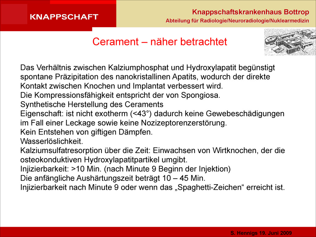
Cerament – näher betrachtet Das Verhältnis zwischen Kalziumphosphat und Hydroxylapatit begünstigt spontane Präzipitation des nanokristallinen Apatits, wodurch der direkte Kontakt zwischen Knochen und Implantat verbessert wird. Die Kompressionsfähigkeit entspricht der von Spongiosa. Synthetische Herstellung des Ceraments Eigenschaft: ist nicht exotherm (<43°) dadurch keine Gewebeschädigungen im Fall einer Leckage sowie keine Nozizeptorenzerstörung. Kein Entstehen von giftigen Dämpfen. Wasserlöslichkeit. Kalziumsulfatresorption über die Zeit: Einwachsen von Wirtknochen, der die osteokonduktiven Hydroxylapatitpartikel umgibt. Injizierbarkeit: >10 Min. (nach Minute 9 Beginn der Injektion) Die anfängliche Aushärtungszeit beträgt 10 – 45 Min. Injizierbarkeit nach Minute 9 oder wenn das „Spaghetti-Zeichen“ erreicht ist.
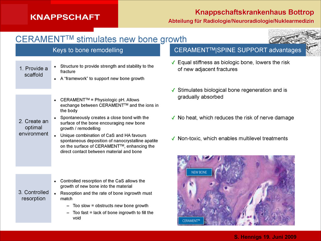
CERAMENTTM stimulates new bone growth ✓ Equal stiffness as biologic bone, lowers the risk of new adjacent fractures ! ✓ Stimulates biological bone regeneration and is gradually absorbed ! ✓ No heat, which reduces the risk of nerve damage ! ✓ Non-toxic, which enables multilevel treatments Keys to bone remodelling CERAMENTTM|SPINE SUPPORT advantages ▪ Structure to provide strength and stability to the fracture ▪ A “framework” to support new bone growth 1. Provide a scaffold 2. Create an optimal environment 3. Controlled resorption ▪ CERAMENTTM = Physiologic pH. Allows exchange between CERAMENTTM and the ions in the body ▪ Spontaneously creates a close bond with the surface of the bone encouraging new bone growth / remodelling ▪ Unique combination of CaS and HA favours spontaneous deposition of nanocrystalline apatite on the surface of CERAMENTTM, enhancing the direct contact between material and bone ▪ Controlled resorption of the CaS allows the growth of new bone into the material ▪ Resorption and the rate of bone ingrowth must match – Too slow = obstructs new bone growth – Too fast = lack of bone ingrowth to fill the void
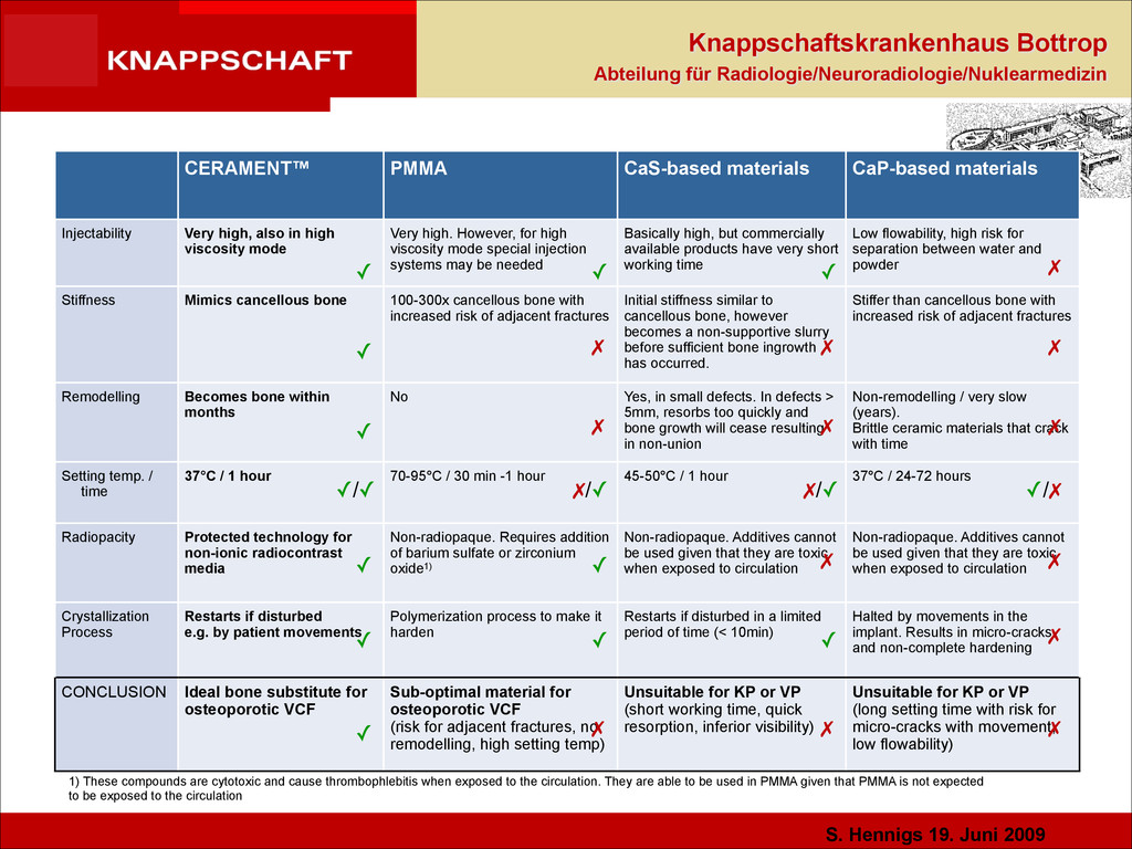
CERAMENT™ PMMA CaS-based materials CaP-based materials Injectability Very high, also in high viscosity mode Very high. However, for high viscosity mode special injection systems may be needed Basically high, but commercially available products have very short working time Low flowability, high risk for separation between water and powder Stiffness Mimics cancellous bone 100-300x cancellous bone with increased risk of adjacent fractures Initial stiffness similar to cancellous bone, however becomes a non-supportive slurry before sufficient bone ingrowth has occurred. Stiffer than cancellous bone with increased risk of adjacent fractures Remodelling Becomes bone within months No Yes, in small defects. In defects > 5mm, resorbs too quickly and bone growth will cease resulting in non-union Non-remodelling / very slow (years). Brittle ceramic materials that crack with time Setting temp. / time 37°C / 1 hour 70-95°C / 30 min -1 hour 45-50°C / 1 hour 37°C / 24-72 hours Radiopacity Protected technology for non-ionic radiocontrast media Non-radiopaque. Requires addition of barium sulfate or zirconium oxide1) Non-radiopaque. Additives cannot be used given that they are toxic when exposed to circulation Non-radiopaque. Additives cannot be used given that they are toxic when exposed to circulation Crystallization Process Restarts if disturbed e.g. by patient movements Polymerization process to make it harden Restarts if disturbed in a limited period of time (< 10min) Halted by movements in the implant. Results in micro-cracks and non-complete hardening CONCLUSION Ideal bone substitute for osteoporotic VCF Sub-optimal material for osteoporotic VCF (risk for adjacent fractures, no remodelling, high setting temp) Unsuitable for KP or VP (short working time, quick resorption, inferior visibility) Unsuitable for KP or VP (long setting time with risk for micro-cracks with movement, low flowability) 1) These compounds are cytotoxic and cause thrombophlebitis when exposed to the circulation. They are able to be used in PMMA given that PMMA is not expected to be exposed to the circulation ✓ ✗ ✓ ✓ ✓/✓ ✓ ✓ ✓ ✓ ✗ ✗/✓ ✓ ✓ ✗ ✓ ✓ ✗ ✗ ✗/✓ ✗ ✗ ✗ ✗ ✗ ✗ ✗ ✗ ✓/✗
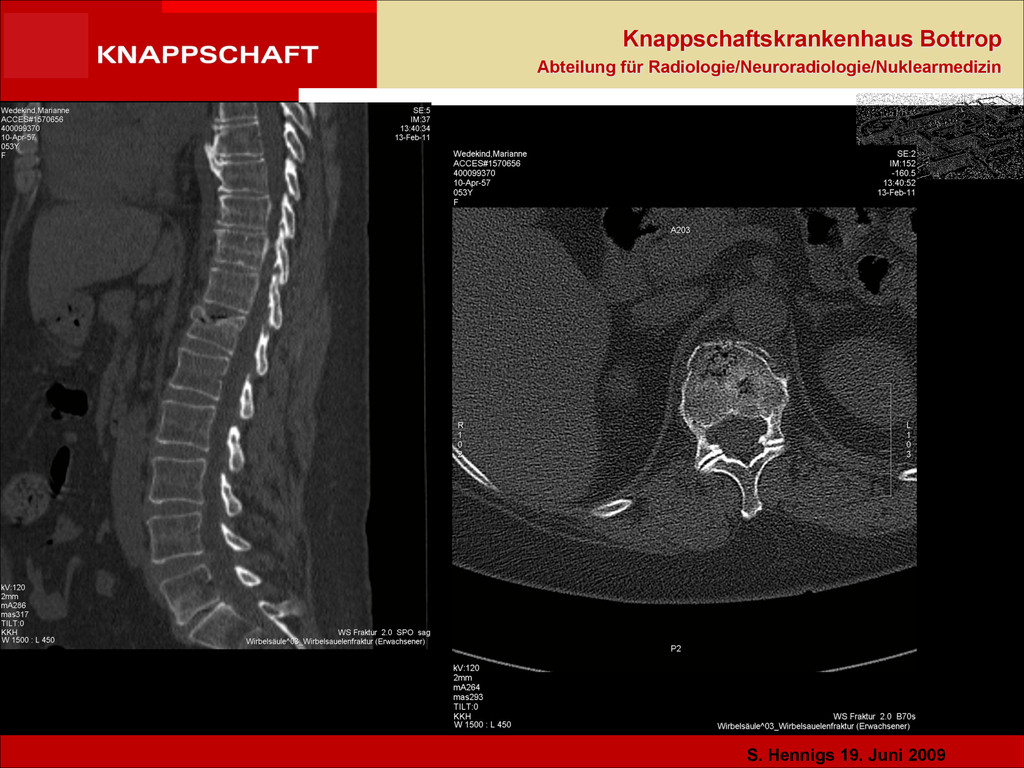
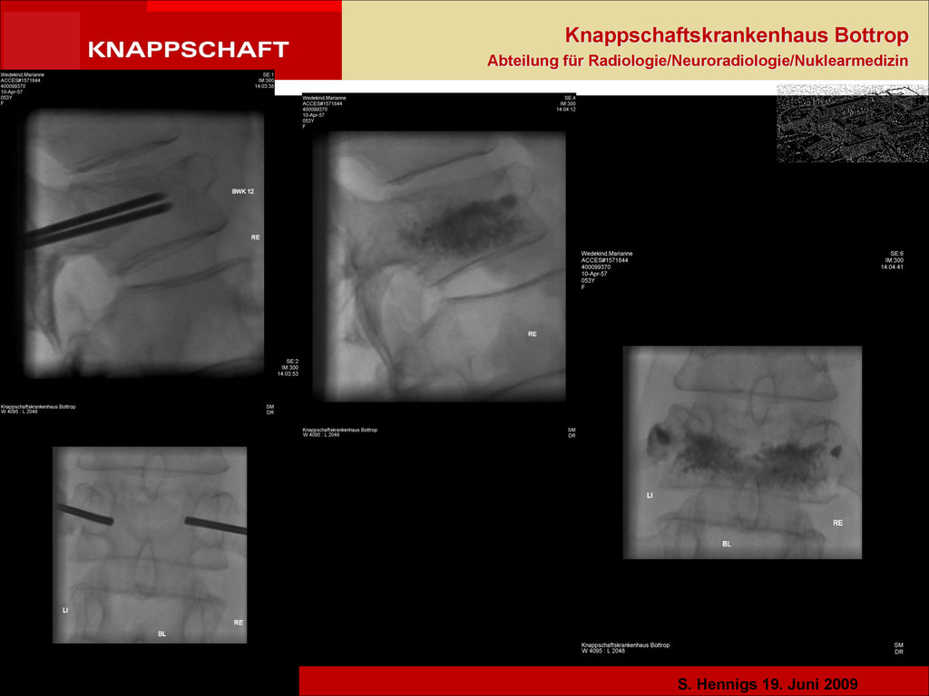
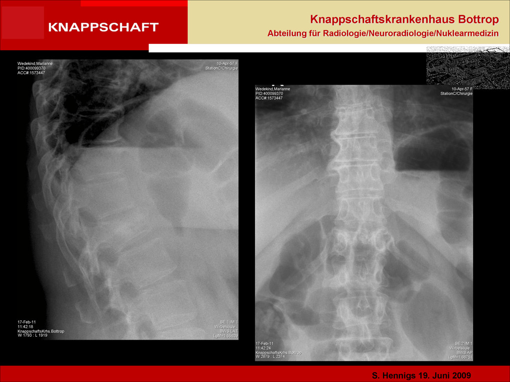
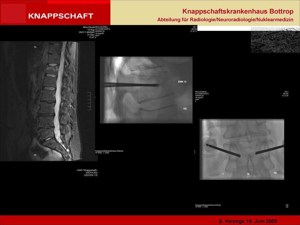
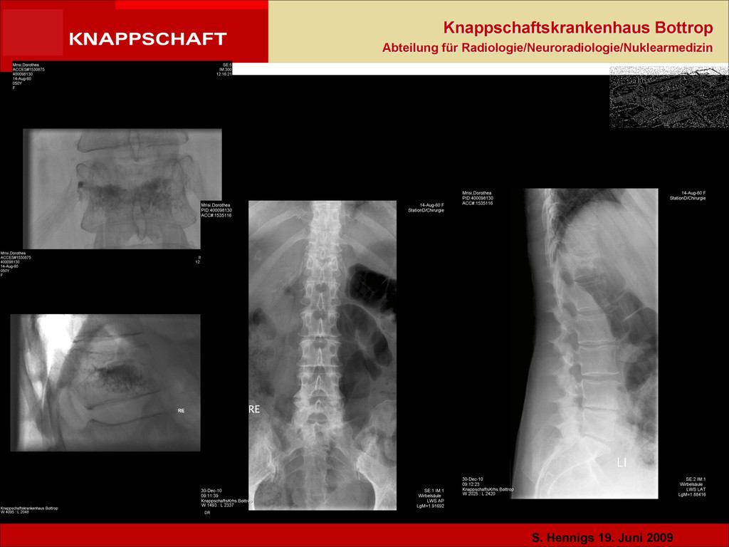
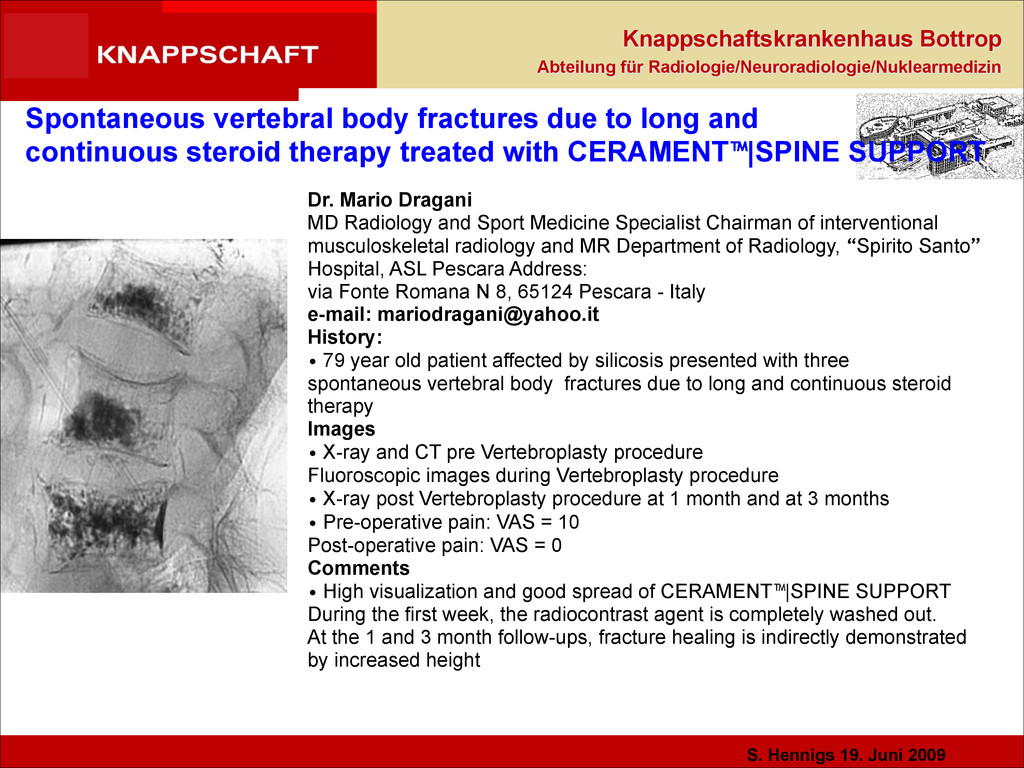
Spontaneous vertebral body fractures due to long and continuous steroid therapy treated with CERAMENT™|SPINE SUPPORT Dr. Mario Dragani MD Radiology and Sport Medicine Specialist Chairman of interventional musculoskeletal radiology and MR Department of Radiology, “Spirito Santo” Hospital, ASL Pescara Address: via Fonte Romana N 8, 65124 Pescara - Italy e-mail: [email protected] History: • 79 year old patient affected by silicosis presented with three spontaneous vertebral body fractures due to long and continuous steroid therapy Images • X-ray and CT pre Vertebroplasty procedure Fluoroscopic images during Vertebroplasty procedure • X-ray post Vertebroplasty procedure at 1 month and at 3 months • Pre-operative pain: VAS = 10 Post-operative pain: VAS = 0 Comments • High visualization and good spread of CERAMENT™|SPINE SUPPORT During the first week, the radiocontrast agent is completely washed out. At the 1 and 3 month follow-ups, fracture healing is indirectly demonstrated by increased height
ORIGINAL ARTICLE Bioceramic vertebral augmentation with a calcium sulphate/ hydroxyapatite composite (CeramentTM SpineSupport) in vertebral compression fractures due to osteoporosis Michael Rauschmann • Thomas Vogl • Akhil Verheyden • Robert Pflugmacher • Thomas Werba • Sven Schmidt • Johannes Hierholzer Received: 8 July 2009 / Revised: 23 November 2009 / Accepted: 12 January 2010 Springer-Verlag 2010 Abstract A prospective, non-randomized multicenter study was initiated to study efficacy and safety of a partly resorbable composite of calcium sulphate and hydroxyapatite (CeramentTM SpineSupport), a novel, injectable bioceramic, in osteoporotic patients with vertebral compression fractures during 18-month follow-up. Fifteen patients with low-energy trauma and 1–2 vertebral compression fractures verified by magnetic resonance imaging were recruited to undergo percutaneous bioceramic vertebral augmentation under fluoroscopy. The patients were treated with a highly flowable bioceramic containing calcium sulphate, hydroxyapatite and the non-ionic radiocontrast agent iohexol, with final setting time within 1 h. After the procedure, the patients were allowed to mobilize after 2 h. Pain (VAS), occurrence of remote and adjacent fractures, and Quality of Life (QoL; SF-36 and EQ-5D) was recorded during 18 months. The injected volume of the composite material ranged from 2.8 to 9 mL (mean 4.2 mL). Pre-operative VAS score was mean 70.3 (CI95% ±8.7) with an immediate post-operative pain relief, which was maintained at the 4-week visit (mean 26.4 with CI95%±16.1) and 8-week visit (mean 18.0 with CI95% ±13.5 pain relief). Eighty percent of the patients demonstrated a clinical improvement. The pain relief was maintained over 18 months and no adjacent fractures were observed. There was a statistically significant improvement of physical components in the QoL assessment. No extravertebral leakage or neurological deficits were reported in this series. This first prospective multicenter study on a partly resorbable bioceramic material indicate that fracture healing can be achieved with sustained pain relief over a follow-up period of 18 months in an osteoporotic vertebral fractures.
Bone Healing in Vertebroplasty. H. Paul Hatten, Jr., M.D. Indian River Radiology, Vero Beach, Florida. • PMMA bone cement has been the material of choice to date, but adjacent segmental fractures have been as high as 20% within the first year leading to further intervention.3 • A cement of altered modulus that represents the properties of trabecular bone may reduce this risk. 4 • Presented here is the first clinical series in the U.S. of vertebroplasty with a biphasic ceramic bone substitute with mechanical properties similar to cancellous bone • There is no evidence to date of any treated site or adjacent fractures • Histological proof of bone growth has been established by the Voor5 rabbit model, and is supported in this human series by CT scans which indicate that good bone healing has been established at 6 months bone.