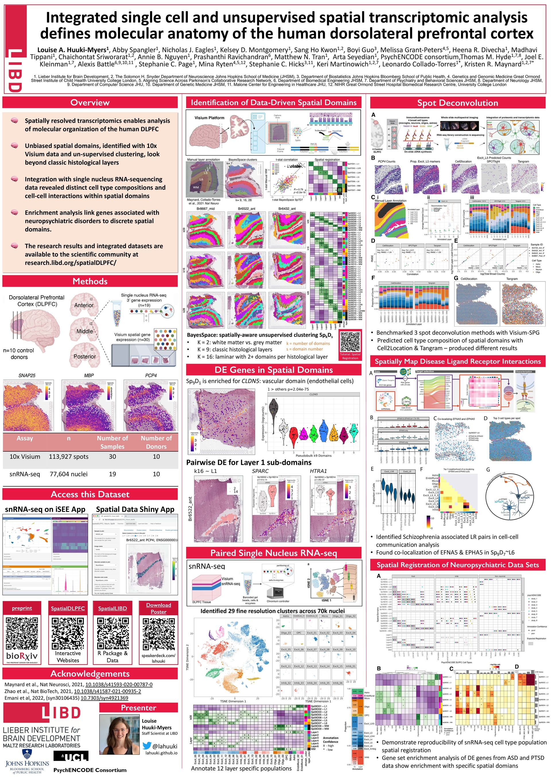
anatomy of the human dorsolateral prefrontal cortex Louise A. Huuki-Myers1, Abby Spangler1, Nicholas J. Eagles1, Kelsey D. Montgomery1, Sang Ho Kwon1,2, Boyi Guo3, Melissa Grant-Peters4,5, Heena R. Divecha1, Madhavi Tippani1, Chaichontat Sriworarat1,2, Annie B. Nguyen1, Prashanthi Ravichandran6, Matthew N. Tran1, Arta Seyedian1, PsychENCODE consortium,Thomas M. Hyde1,7,8, Joel E. Kleinman1,7, Alexis Battle6,9,10,11 , Stephanie C. Page1, Mina Ryten4,5,12, Stephanie C. Hicks3,11, Keri Martinowich1,2,7, Leonardo Collado-Torres1*, Kristen R. Maynard1,2,7* 1. Lieber Institute for Brain Development, 2. The Solomon H. Snyder Department of Neuroscience Johns Hopkins School of Medicine (JHSM), 3. Department of Biostatistics Johns Hopkins Bloomberg School of Public Health, 4. Genetics and Genomic Medicine Great Ormond Street Institute of Child Health University College London, 5. Aligning Science Across Parkinson’s Collaborative Research Network, 6. Department of Biomedical Engineering JHSM, 7. Department of Psychiatry and Behavioral Sciences JHSM, 8. Department of Neurology JHSM, 9. Department of Computer Science JHU, 10. Department of Genetic Medicine JHSM, 11. Malone Center for Engineering in Healthcare JHU, 12. NIHR Great Ormond Street Hospital Biomedical Research Centre, University College London Overview Methods Assay n Number of Samples Number of Donors 10x Visium 113,927 spots 30 10 snRNA-seq 77,604 nuclei 19 10 ! Spatially resolved transcriptomics enables analysis of molecular organization of the human DLPFC ! Unbiased spatial domains, identified with 10x Visium data and un-supervised clustering, look beyond classic histological layers ! Integration with single nucleus RNA-sequencing data revealed distinct cell type compositions and cell-cell interactions within spatial domains ! Enrichment analysis link genes associated with neuropsychiatric disorders to discrete spatial domains. ! The research results and integrated datasets are available to the scientific community at research.libd.org/spatialDLPFC/ Access this Dataset Spatial Data Shiny App snRNA-seq on iSEE App preprint SpatialDLPFC Interactive Websites SpatialLIBD R Package & Data Download Poster speakerdeck.com/ lahuuki Acknowledgements Maynard et al., Nat Neurosci, 2021, 10.1038/s41593-020-00787-0 Zhao et al., Nat BioTech, 2021, 10.1038/s41587-021-00935-2 Emani et al, 2022, (syn30106435) 10.7303/syn4921369 Presenter Louise Huuki-Myers Staff Scientist at LIBD @lahuuki lahuuki.github.io PsychENCODE Consortium snRNA-seq Identification of Data-Driven Spatial Domains BayesSpace: spatially-aware unsupervised clustering Spk Ds • K = 2: white matter vs. grey matter • K = 9: classic histological layers • K = 16: laminar with 2+ domains per histological layer k = number of domains s = domain number DE Genes in Spatial Domains Sp9 D1 is enriched for CLDN5: vascular domain (endothelial cells) Pairwise DE for Layer 1 sub-domains 1 2 logcounts min > 0 CLDN5 1 2 3 logcounts min > 0 1 2 3 logcounts min > 0 Identified 29 fine resolution clusters across 70k nuclei Annotation Confidence X - high * - low Spot Deconvolution Spatially Map Disease Ligand Receptor Interactions Spatial Registration of Neuropsychiatric Data Sets • Benchmarked 3 spot deconvolution methods with Visium-SPG • Predicted cell type composition of spatial domains with Cell2Location & Tangram – produced different results • Identified Schizophrenia associated LR pairs in cell-cell communication analysis • Found co-localization of EFNA5 & EPHA5 in Sp9 D7 ~L6 • Demonstrate reproducibility of snRNA-seq cell type population spatial registration • Gene set enrichment analysis of DE genes from ASD and PTSD data show enrichment with specific spatial domians Annotate 12 layer specific populations Paired Single Nucleus RNA-seq Tutorial: Spatial Registration