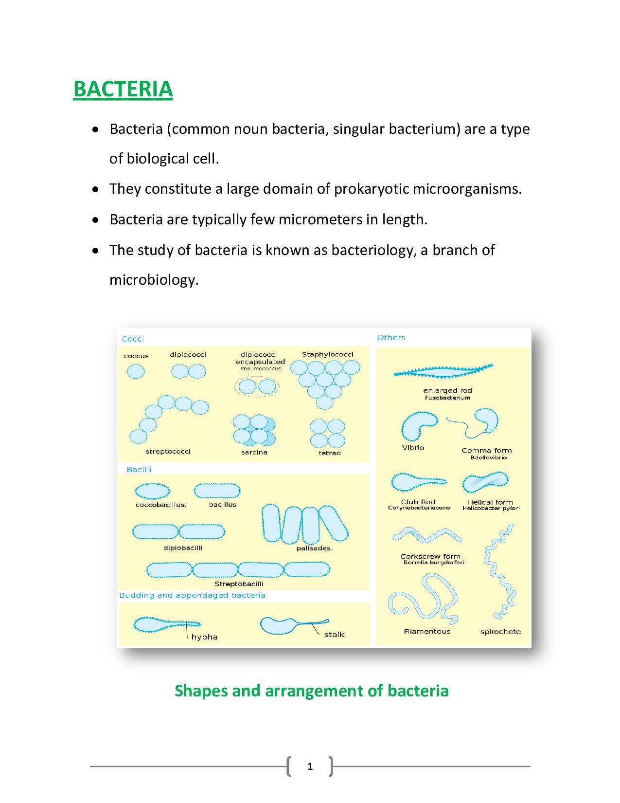
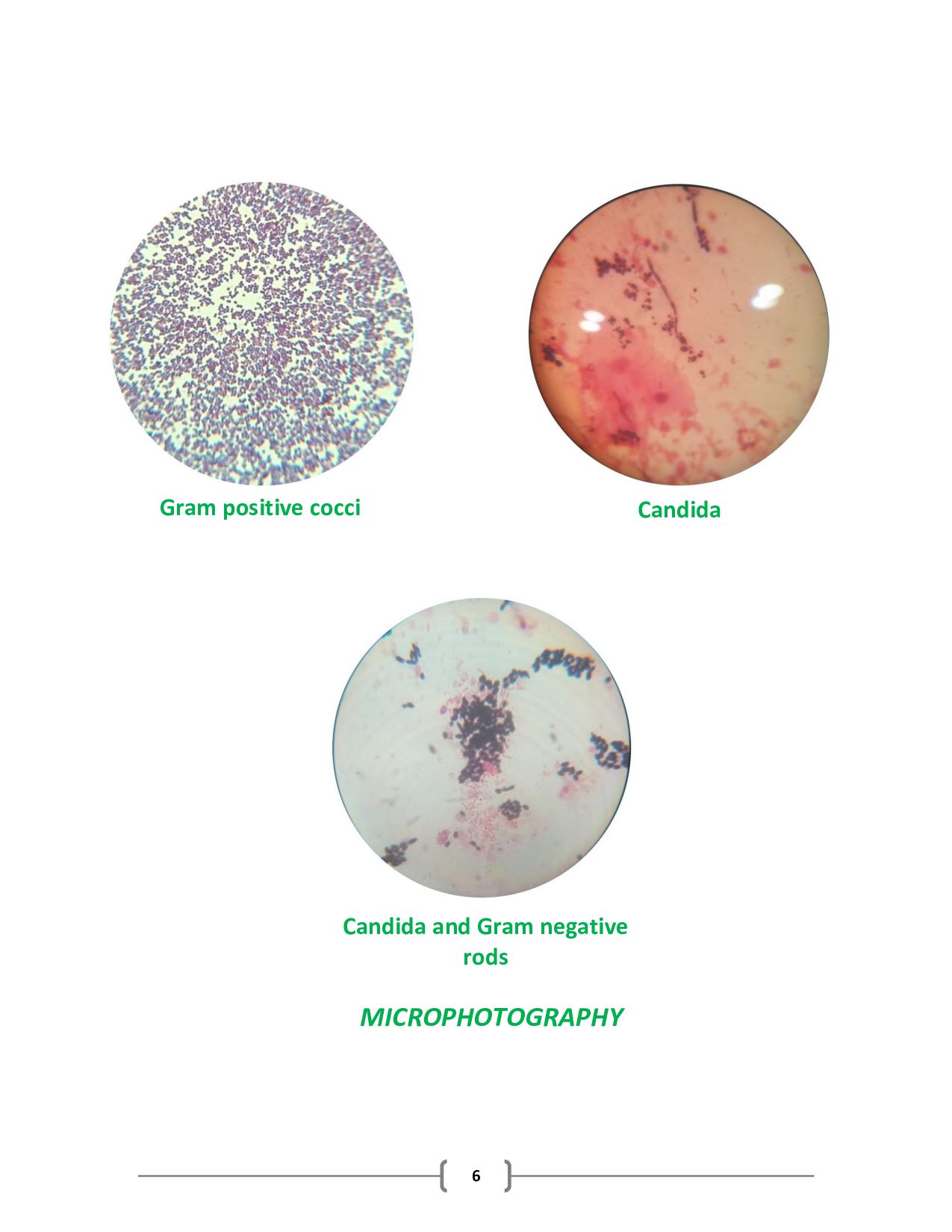
a type of biological cell. They constitute a large domain of prokaryotic microorganisms. Bacteria are typically few micrometers in length. The study of bacteria is known as bacteriology, a branch of microbiology. Shapes and arrangement of bacteria
identify the bacteria. The gram staining technique was developed by Danish bacteriologist Hans Christian Gram. The gram stain technique is based on the differential structure of the cellular membranes and cell walls of the two groups. The two major groups of bacteria can be divided into gram-positive and gram-negative. This technique utilizes four steps staining procedure with two different dyes to identify unknown species of bacteria. REAGENTS 1. Primary stain - Crystal violet 2. Mordant - Gram iodine 3. Gram decolorizer – Ethanol 4. Counter stain – Gram safranin
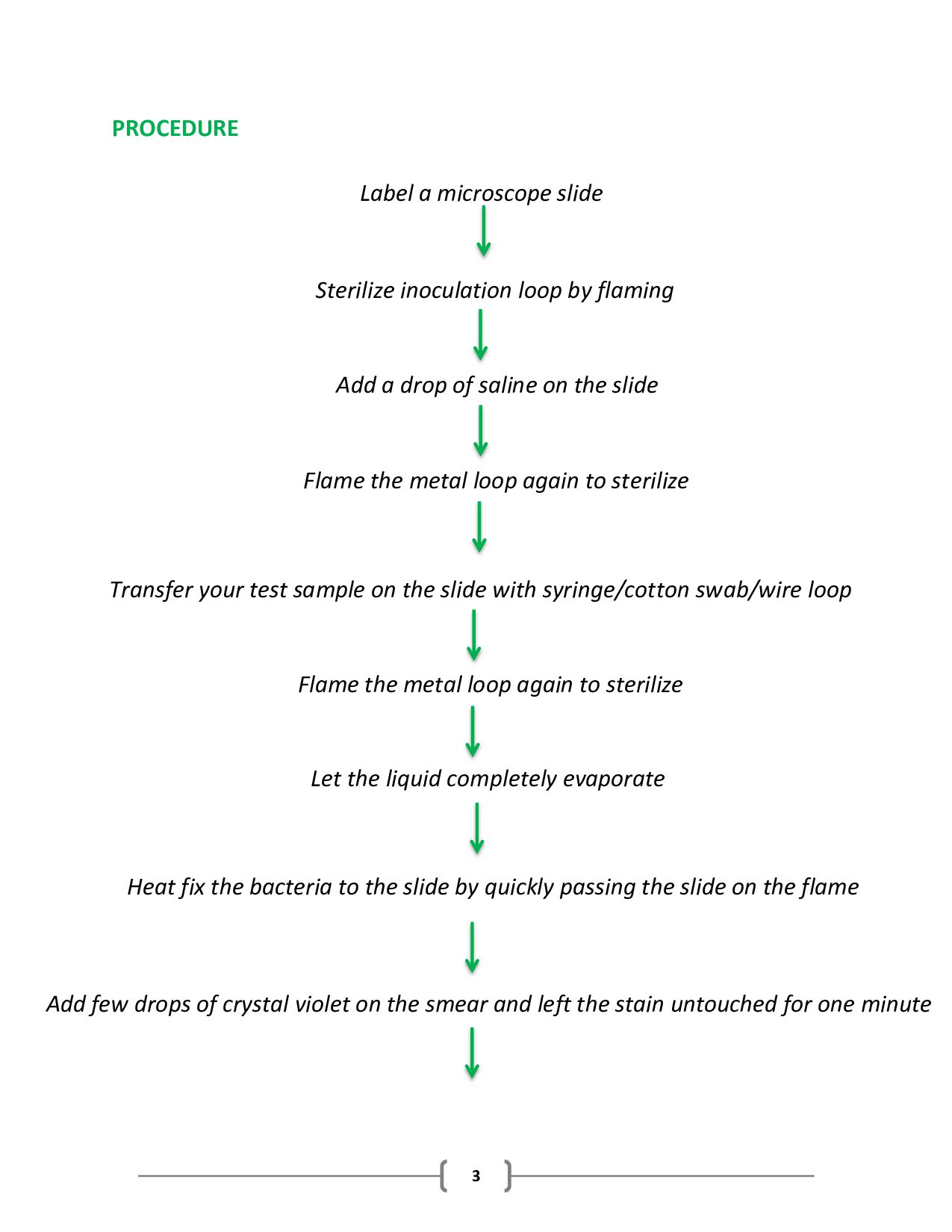
flaming Add a drop of saline on the slide Flame the metal loop again to sterilize Transfer your test sample on the slide with syringe/cotton swab/wire loop Flame the metal loop again to sterilize Let the liquid completely evaporate Heat fix the bacteria to the slide by quickly passing the slide on the flame Add few drops of crystal violet on the smear and left the stain untouched for one minute
of gram iodine on the smear and left the stain untouched for one minute Rinse the slide with tap water Add few drops of decolorizer & let the decolorizer flow over the bacteria at a 45° angle until the flow clear Rinse the slide with tap water Add few drops of safranin on the smear and left the stain untouched for one minute Rinse the slide with tap water Allow the slide to dry it completely Observe the slide under a microscope by adding oil on the slide
of peptidoglycan that retains the primary dye, crystal violet, following the application of the mordant, iodine. The iodine and crystal violet form a complex within the peptidoglycan. When decolorizer is applied to the cells, the Crystal violet-Iodine complex remains within the cell, making it appear dark purple to blue. The gram-negative organisms do not contain a thick cross-linked layer of peptidoglycan. The peptidoglycan is loosely distributed between the inner cell and the outer cell membranes. Following the application of the crystal violet and iodine, the Crystal violet-Iodine complexes are not trapped within the peptidoglycan. Application of the acid-alcohol decolorizer dehydrates the outer cellular membrane, leaving holes in the membrane and effectively washing or removing the CV-I complex from the cells. The cells appear colorless. To make the colorless cells visible, a secondary stain, safranin, is applied, leaving the gram-negative cells pink.
or on the medium is known as a culture. The substances & environment, which is provided to the microorganisms for their growth is called as media. Culture media are contained in test tubes, flasks, or Petri dishes. Media are extremely varied in nutrient content & consistency & can be specially formulated for a particular purpose. TYPES OF MEDIA Media can be classified on the three primary levels: 1. Physically 2. Chemically 3. Functionally
defined as water-based solutions that do not solidify at temperatures above freezing & that tends to flow freely when the container is tilted. These media termed broths, milks, or infusion, are made by dissolving various solutes in distilled water. Growth occurs throughout the container & can then present a dispersed cloudy or particulate appearance. Examples: Luria broth, Nutrient broth. 2- SEMISOLID MEDIA It exhibit clot-like consistency. It contain solidifying agent (agar or gelatin) that thickens the media but does not produce a firm substrate. This media is used to determine the motility of bacteria & to localize a reaction at a specific site. Examples: Both motility test medium & Sulfide Indole Motility (SIM) contain a small amount of agar. The medium is
the pattern of growth around the stab line. 3- SOLID MEDIA It provides a firm surface on which cells can form discrete colonies & are advantages for isolating & sub- culturing bacteria & fungi. They come in two forms: Liquefiable, Non-liquefiable. Liquefiable solid media (also known as reversible solid media) contains solidifying agent i.e. agar which is thermoplastic. Agar is solid at room temperature and it melts or liquefies at 100 °C. Non-liquefiable solid media have less versatile applications than agar media because they are not thermoplastic. This type of media include materials such as rice grains (used to grow fungi), cooked meat media (good for anaerobes), potato slices etc. All of these media start out solid and remain solid after heat sterilization.
compositions are chemically defined are termed as synthetic media. These media contain highly pure organic & inorganic compounds that vary little from one source to another & have a molecular content specified by means of an exact formula. Examples: Glucose-salt agar, Inorganic synthetic broth. 2- NON-SYNTHETIC MEDIA Media whose compositions are not chemically defined are termed as non-synthetic media. It contains at least one ingredient that is not chemically definable. Most of these substances are extracts of animals, plants, or yeasts including such materials as ground-up cells, tissues, secretions, milk, yeast extract, soybean digests, and peptone. Examples: Nutrient broth, Blood agar, EMB agar.
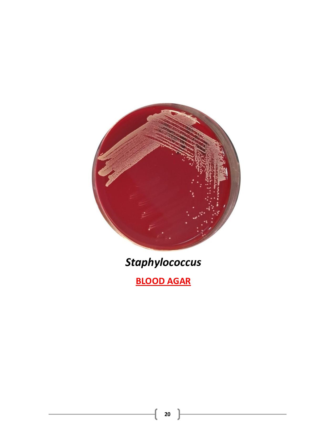
are designed to grow as broad a spectrum of microbes as possible. They are nonsynthetic & contain a mixture of nutrients that could support the growth of pathogens & non pathogens alike. Examples: Nutrient agar, Nutrient broth, Brain-heart infusion, Trypticase soy agar. 2- ENRICHED MEDIA It contains complex organic substances (blood, serum, hemoglobin or special growth factors) that allow certain species to grow. Examples: Streptococcus pneumoniae is cultured on blood agar, which is made by adding sterile sheep, horse or rabbit blood to a sterile agar base, Neisseria can grow on Thayer-Martin medium or chocolate agar, which is essentially cooked blood agar.
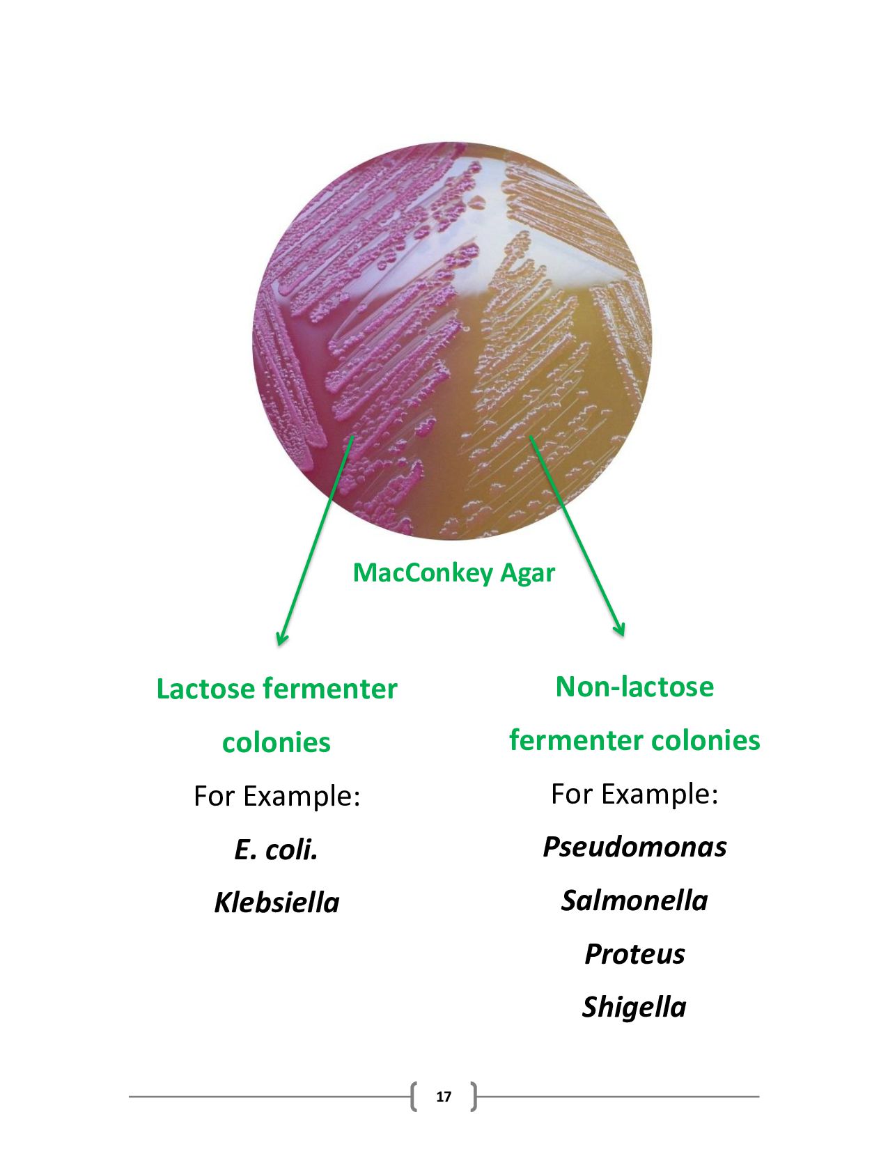
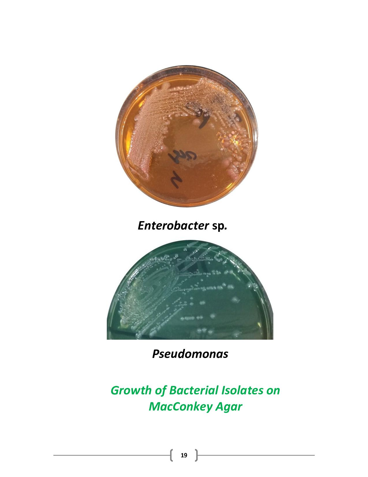
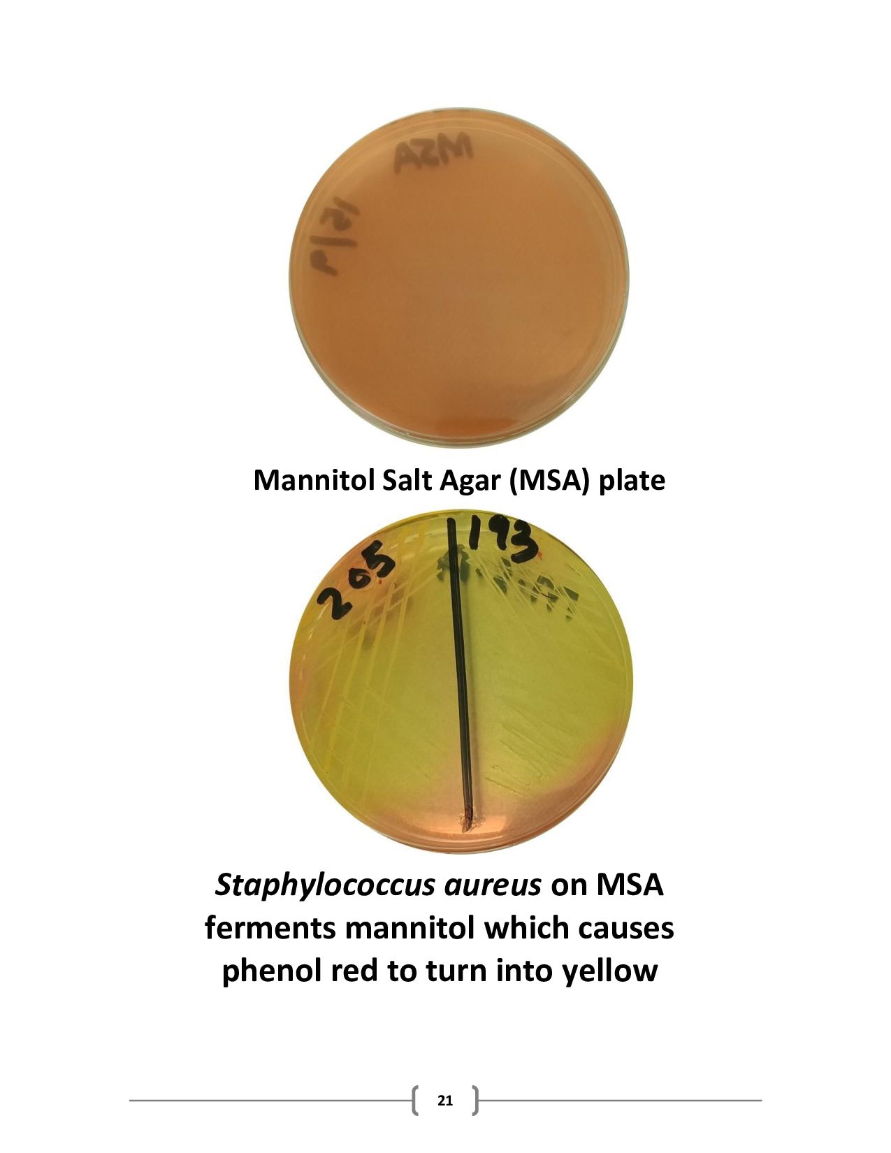
that inhibit the growth of certain microbes thereby encourage the growth of desired microbe. This type of media is very important in primary isolation of a specific type of microorganism from samples containing a highly mixed population. Examples: Mannitol Salt Agar (MSA), MacConkey agar, Eosin-Methylene Blue (EMB) agar. 4- DIFFERENTIAL MEDIA A differential media can grow several types of microorganisms, but it is designed to highlight differences among these microorganisms. Its ability of differentiation is due to the type of agents added. Examples: Sulfide Indole Motility (SIM) agar, Xylose Lysine Deoxycholate (XLD) agar, Triple Sugar Iron (TSI) agar.
(thioglycollic acid or cystine) that absorbs oxygen or slows the penetration of oxygen in a medium thus reducing its availability. These media are important for growing anaerobic bacteria or determining oxygen requirements. Example: Thioglycolate broth. Carbohydrate fermentation media contain sugars that can be fermented (converted to acids) & a pH indicator to show this reaction. Transport media is used to maintain & preserve specimens that have to be held for a period of time prior to clinical analysis or to sustain delicate species that die rapidly if not held under stable conditions. Example: Stuart’s & Amies transport media contain salts, buffers & absorbants to
substances but will not support growth. Assay media are used by technologists to test the effectiveness of antimicrobial drugs & by drugs manufacturers to assess the effect of disinfectants, antiseptics, cosmetics & preservatives on the growth of microorganisms. Enumeration media is used by industrial & environmental microbiologists to count the numbers of microorganisms in milk, water, food, soil & other samples.
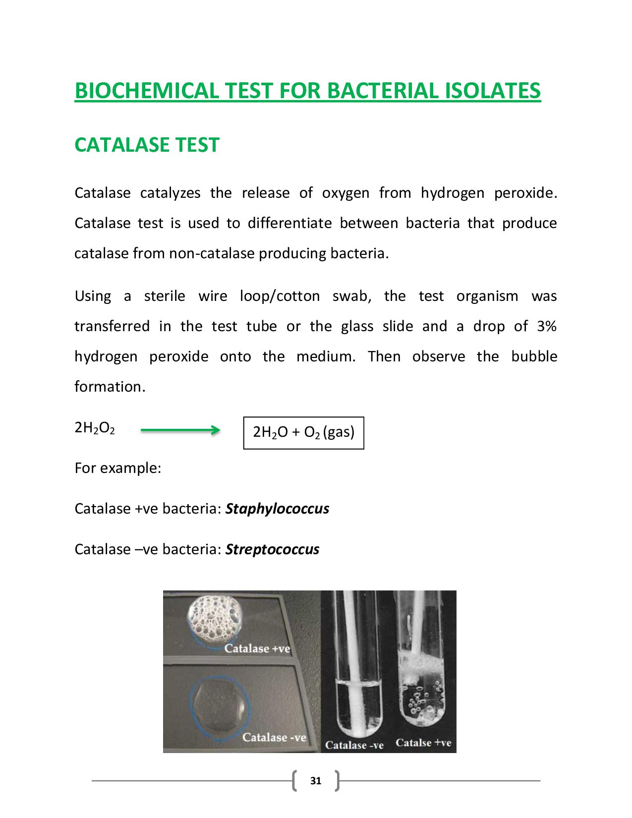
the release of oxygen from hydrogen peroxide. Catalase test is used to differentiate between bacteria that produce catalase from non-catalase producing bacteria. Using a sterile wire loop/cotton swab, the test organism was transferred in the test tube or the glass slide and a drop of 3% hydrogen peroxide onto the medium. Then observe the bubble formation. 2H2 O2 For example: Catalase +ve bacteria: Staphylococcus Catalase –ve bacteria: Streptococcus 2H2 O + O2 (gas)
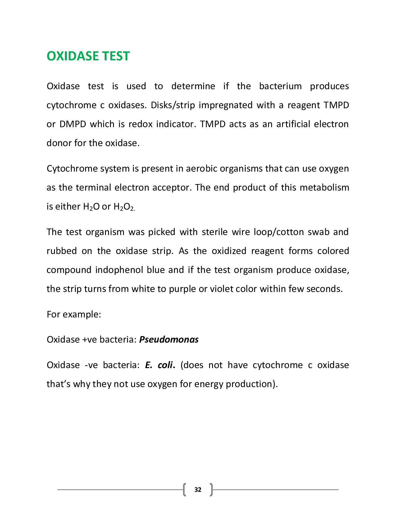
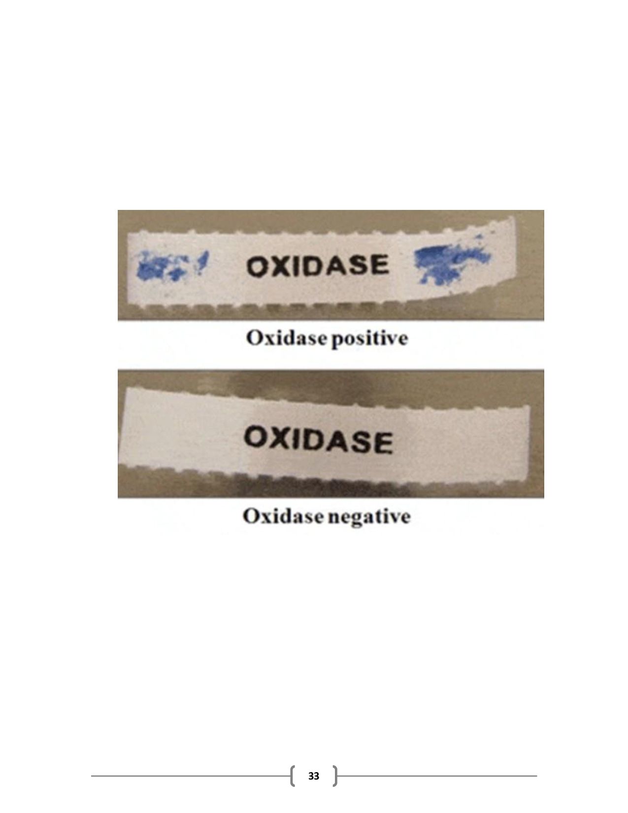
the bacterium produces cytochrome c oxidases. Disks/strip impregnated with a reagent TMPD or DMPD which is redox indicator. TMPD acts as an artificial electron donor for the oxidase. Cytochrome system is present in aerobic organisms that can use oxygen as the terminal electron acceptor. The end product of this metabolism is either H2 O or H2 O2. The test organism was picked with sterile wire loop/cotton swab and rubbed on the oxidase strip. As the oxidized reagent forms colored compound indophenol blue and if the test organism produce oxidase, the strip turns from white to purple or violet color within few seconds. For example: Oxidase +ve bacteria: Pseudomonas Oxidase -ve bacteria: E. coli. (does not have cytochrome c oxidase that’s why they not use oxygen for energy production).
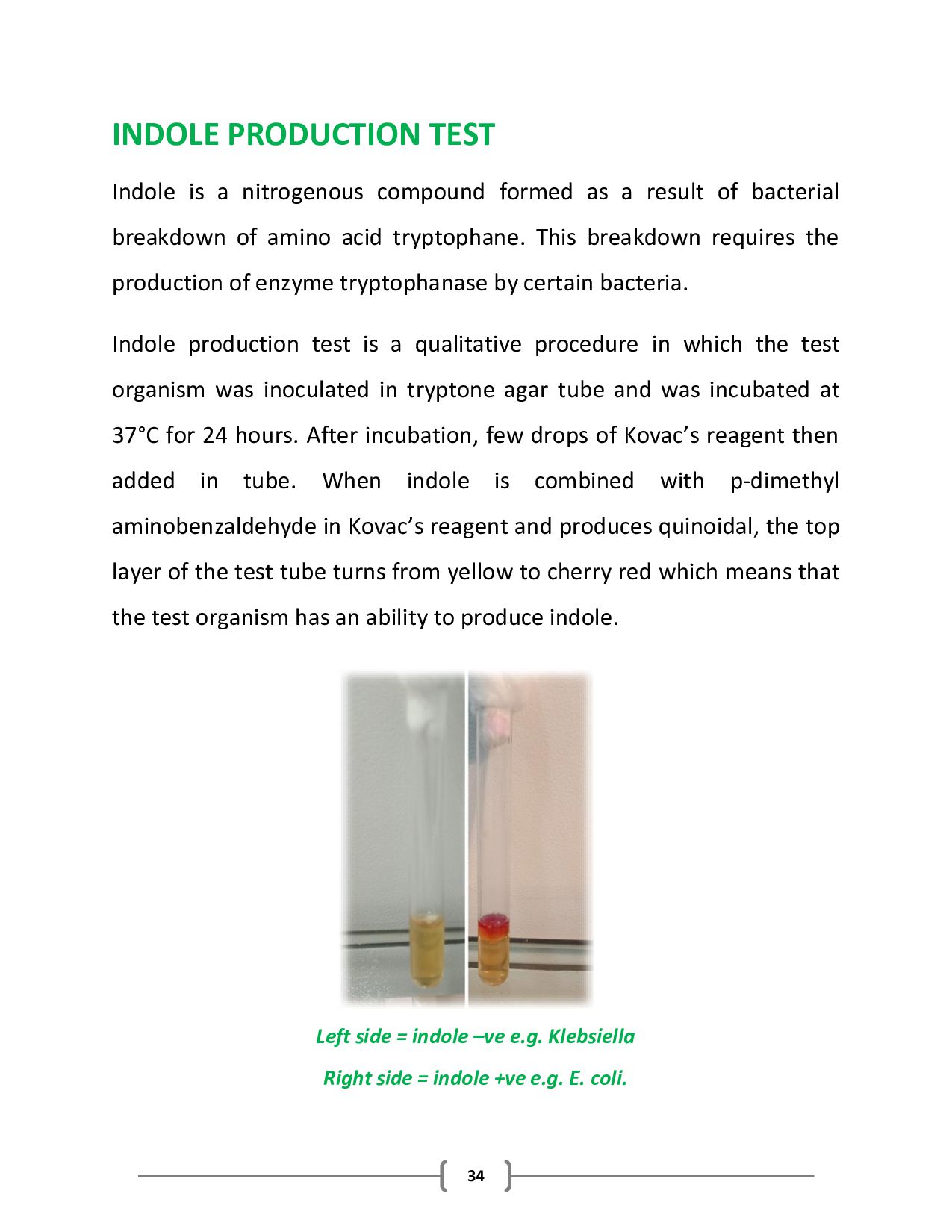
as a result of bacterial breakdown of amino acid tryptophane. This breakdown requires the production of enzyme tryptophanase by certain bacteria. Indole production test is a qualitative procedure in which the test organism was inoculated in tryptone agar tube and was incubated at 37°C for 24 hours. After incubation, few drops of Kovac’s reagent then added in tube. When indole is combined with p-dimethyl aminobenzaldehyde in Kovac’s reagent and produces quinoidal, the top layer of the test tube turns from yellow to cherry red which means that the test organism has an ability to produce indole. Left side = indole –ve e.g. Klebsiella Right side = indole +ve e.g. E. coli.
with test organism. The tube was incubated at 37°C for 24 hours. Bacteria that can grow on this medium use citrate and covert ammonium phosphate to ammonia and ammonium hydroxide, creating an alkaline pH. The pH change turns the bromothymol blue indicator from green to blue. Left side = citrate –ve e.g. E. coli. Right side = citrate +ve e.g. Klebsiella
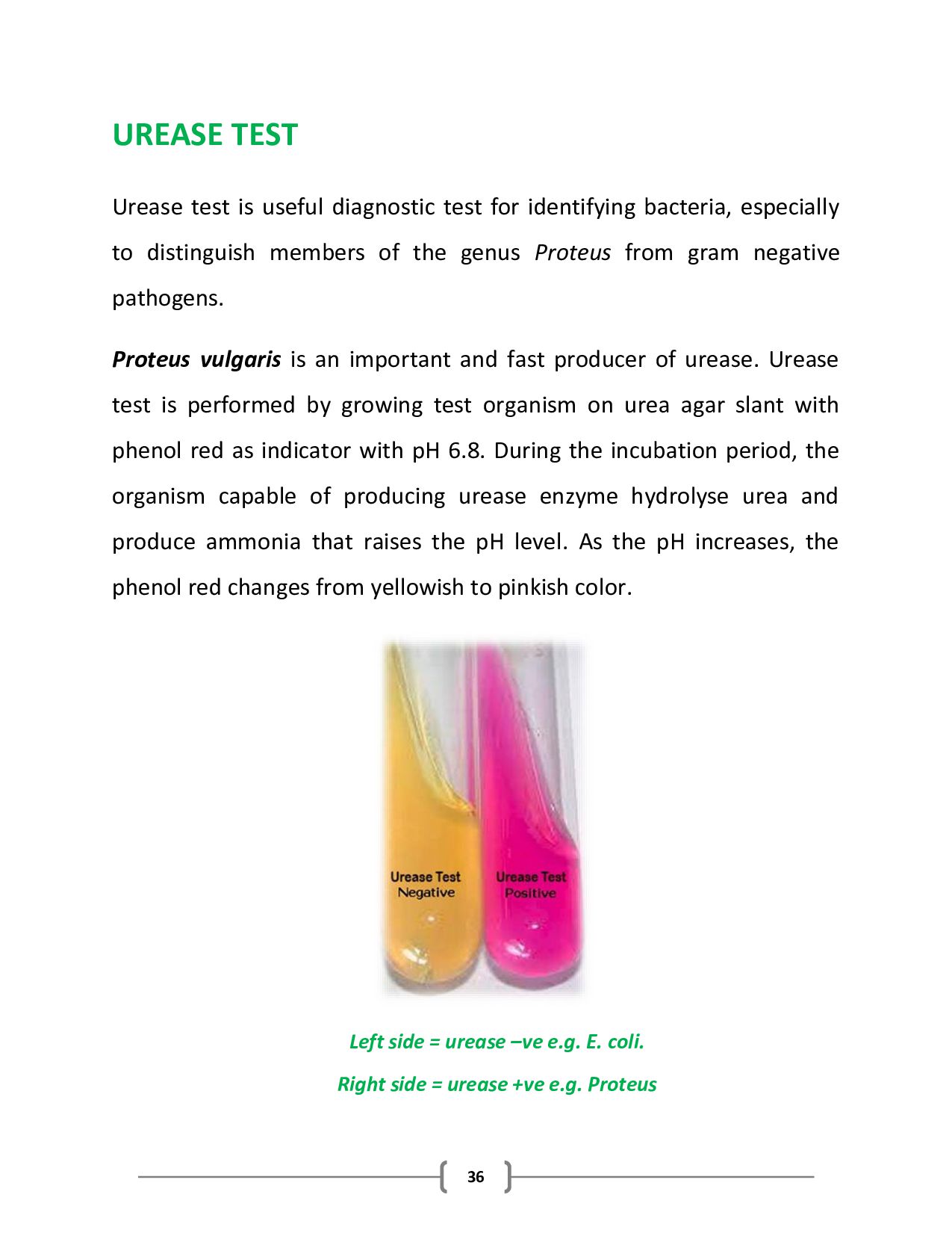
identifying bacteria, especially to distinguish members of the genus Proteus from gram negative pathogens. Proteus vulgaris is an important and fast producer of urease. Urease test is performed by growing test organism on urea agar slant with phenol red as indicator with pH 6.8. During the incubation period, the organism capable of producing urease enzyme hydrolyse urea and produce ammonia that raises the pH level. As the pH increases, the phenol red changes from yellowish to pinkish color. Left side = urease –ve e.g. E. coli. Right side = urease +ve e.g. Proteus
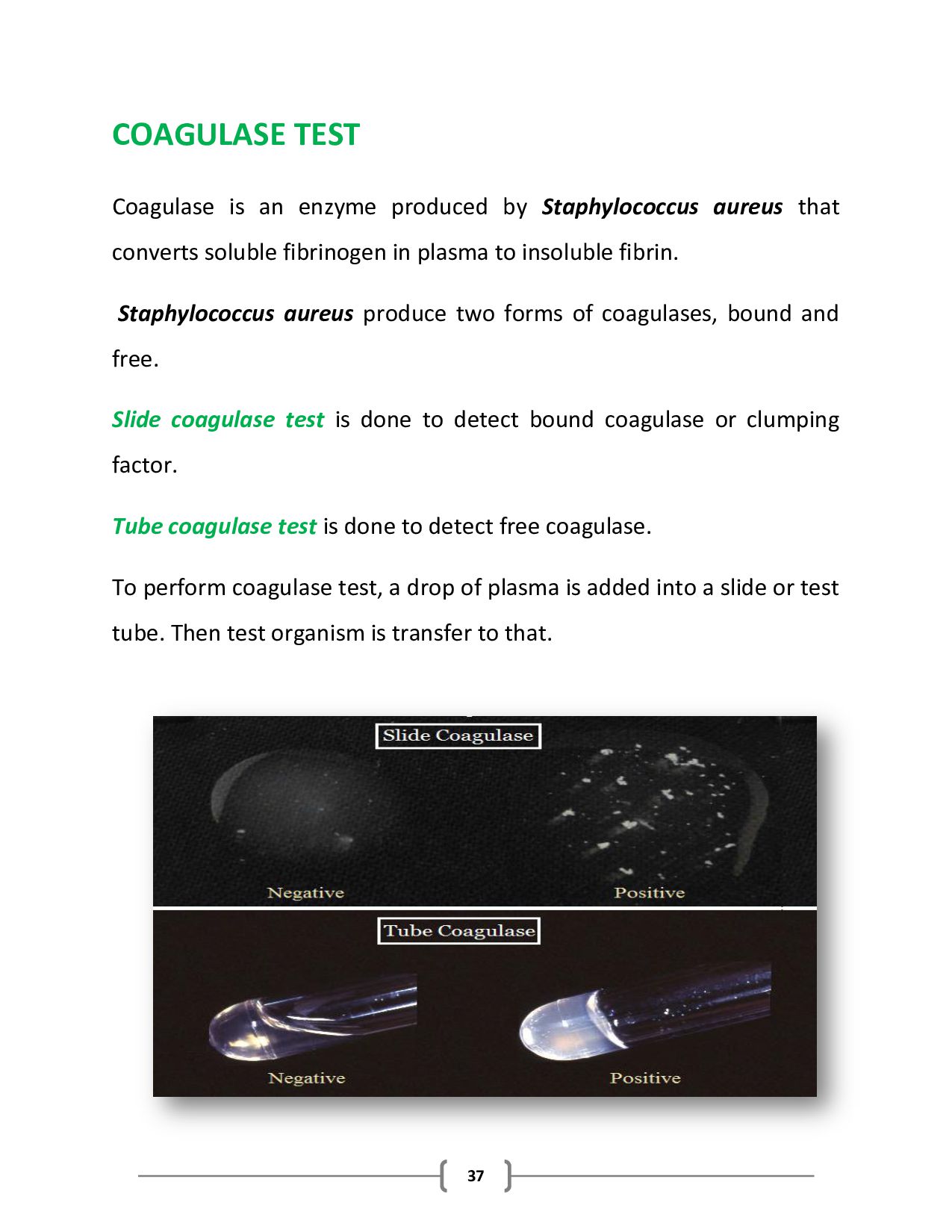
aureus that converts soluble fibrinogen in plasma to insoluble fibrin. Staphylococcus aureus produce two forms of coagulases, bound and free. Slide coagulase test is done to detect bound coagulase or clumping factor. Tube coagulase test is done to detect free coagulase. To perform coagulase test, a drop of plasma is added into a slide or test tube. Then test organism is transfer to that.
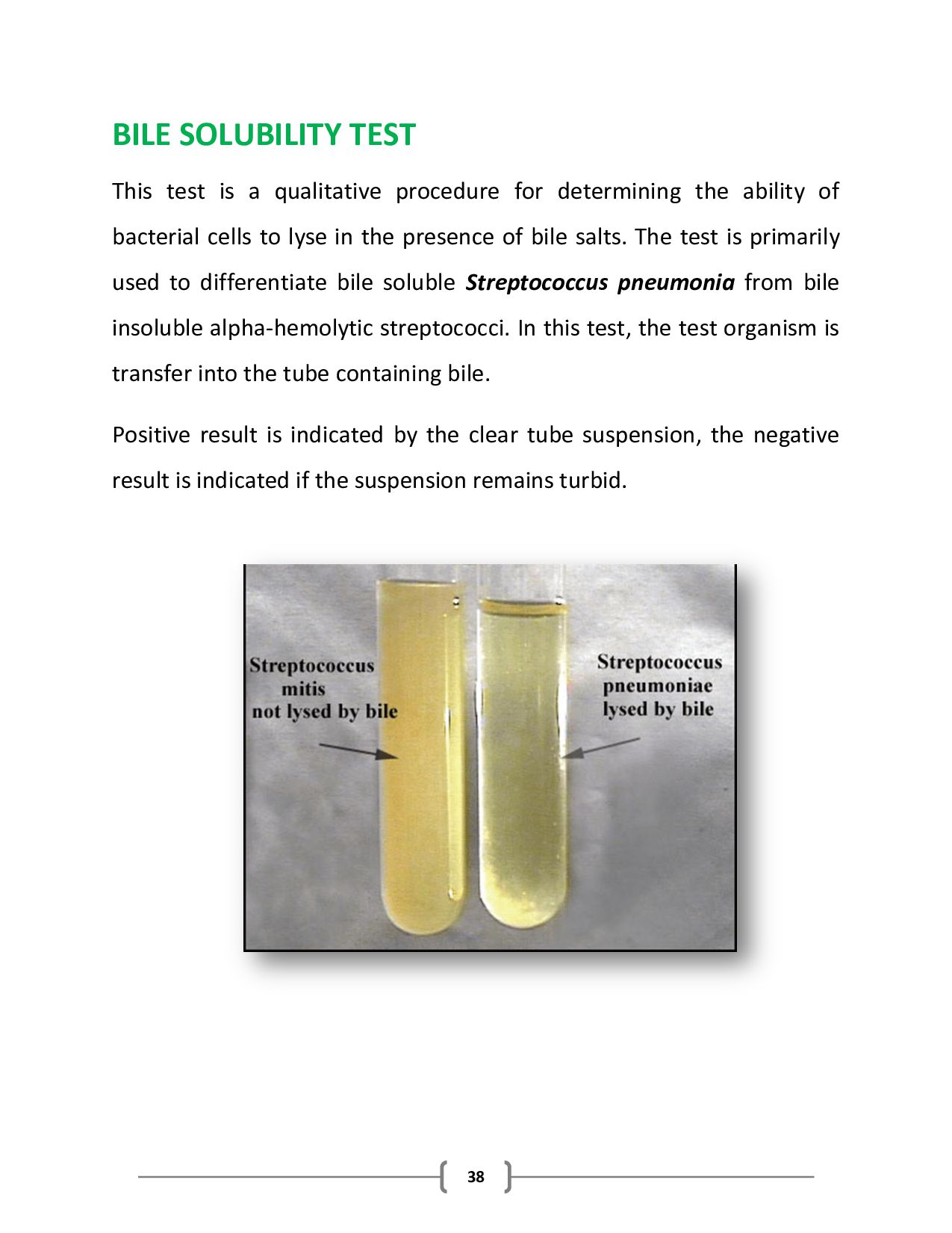
for determining the ability of bacterial cells to lyse in the presence of bile salts. The test is primarily used to differentiate bile soluble Streptococcus pneumonia from bile insoluble alpha-hemolytic streptococci. In this test, the test organism is transfer into the tube containing bile. Positive result is indicated by the clear tube suspension, the negative result is indicated if the suspension remains turbid.
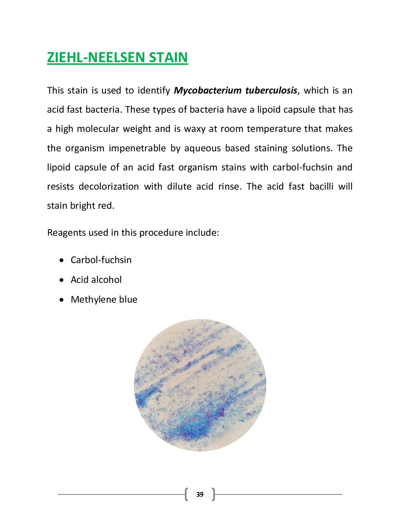
tuberculosis, which is an acid fast bacteria. These types of bacteria have a lipoid capsule that has a high molecular weight and is waxy at room temperature that makes the organism impenetrable by aqueous based staining solutions. The lipoid capsule of an acid fast organism stains with carbol-fuchsin and resists decolorization with dilute acid rinse. The acid fast bacilli will stain bright red. Reagents used in this procedure include: Carbol-fuchsin Acid alcohol Methylene blue
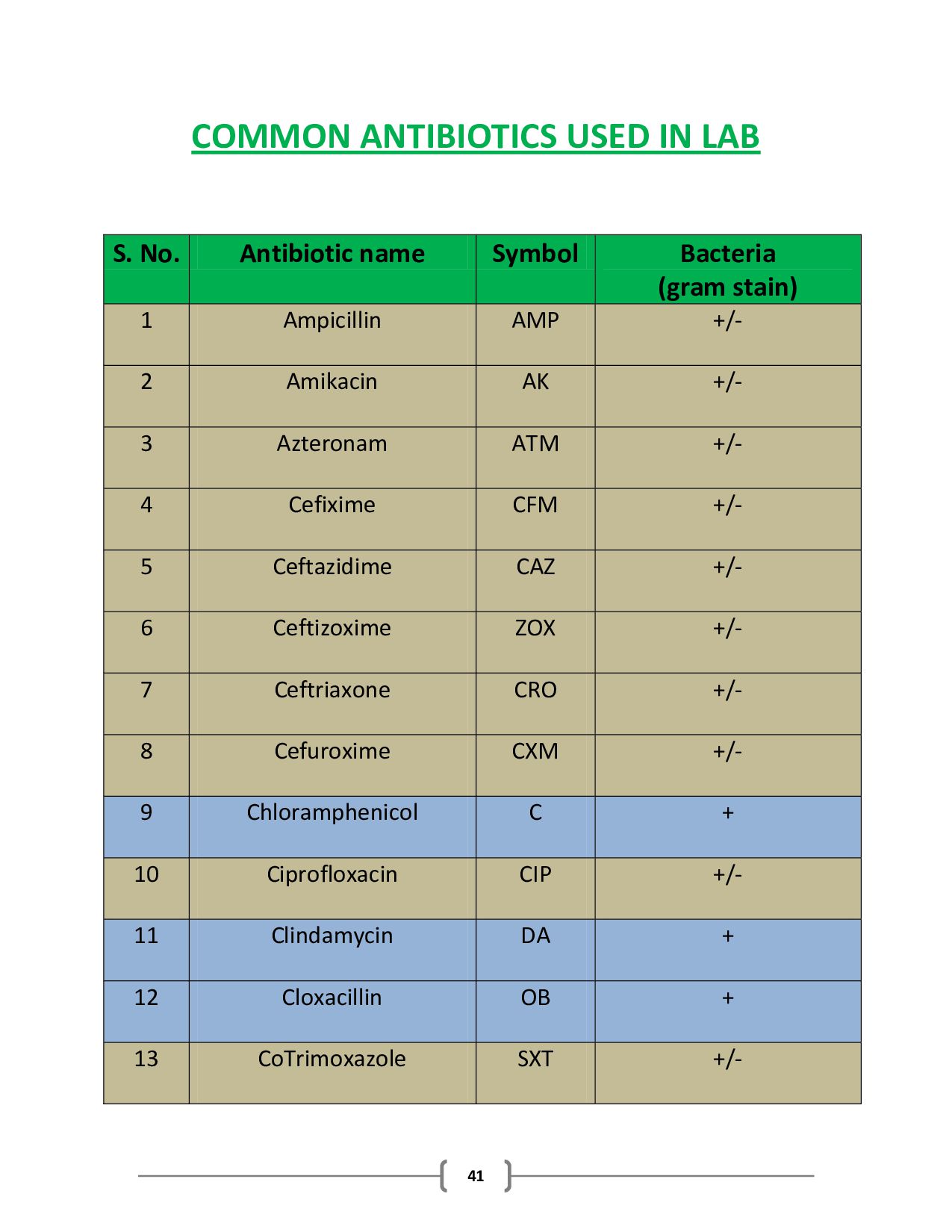
the measurement of the susceptibility of bacteria to antibiotics as bacteria may have resistance to some antibiotics. Commonly for qualitative antibiotic sensitivity testing, the disc diffusion method is used. In this method, antibiotic- impregnated discs are placed on agar plate (Mueller-Hinton Agar frequently use for this purpose) having bacterial growth. This is called as the Kirby-Bauer method. If the antibiotic inhibits microbial growth, a clear zone called zone of inhibition is seen around the antibiotic disc but, if the bacteria will grow around antibiotics to which they are resistant, no zone of inhibition will be observed around antibiotic disc. MHA plates with bacterial growth and antibiotic discs Zone of inhibition around antibiotic disc Bacterial growth around antibiotic disc
Imipenem IPM +/- 17 Levofloxcin LEV + 18 Linezolid LZD + 19 Meropenem MEM +/- 20 Nalidixic acid NA For urine only 21 Nitrofurantoin F For urine only 22 Norfloxacin NOR For urine only 23 Sulzone SCI +/- 24 Tigecycline TGC +/- 25 Tozabaclam TZP +/- 26 Urixcin UR For urine only 27 Vancomycin V +